A 21-year-old unmarried male, with a known history of opioid dependence, presented to our dermatology outpatient department following referral from the psychiatry de-addiction unit. He complained of multiple, painful, slowly progressive skin lesions over both forearms and upper arms for the past three months. These lesions had started as firm nodules that gradually enlarged, became erythematous, tender, and in some instances, ulcerated with seropurulent discharge. The distribution of the lesions was notably linear, following the course of superficial veno-lymphatics, in a classic sporotrichoid lymphangitic pattern.
The patient admitted to injecting heroin intravenously for over a year, often using multiple and unconventional sites due to difficulties accessing veins. His injection practices were inconsistent, frequently performed without sterile techniques. He reported no associated systemic symptoms such as fever, malaise, night sweats, joint pain, or weight loss. There was no recent travel or history of exposure to fish tanks, animals, or soil that might suggest an environmental fungal or atypical bacterial infection. He denied any other comorbidities and was not taking any medications other than those prescribed in the de-addiction program.
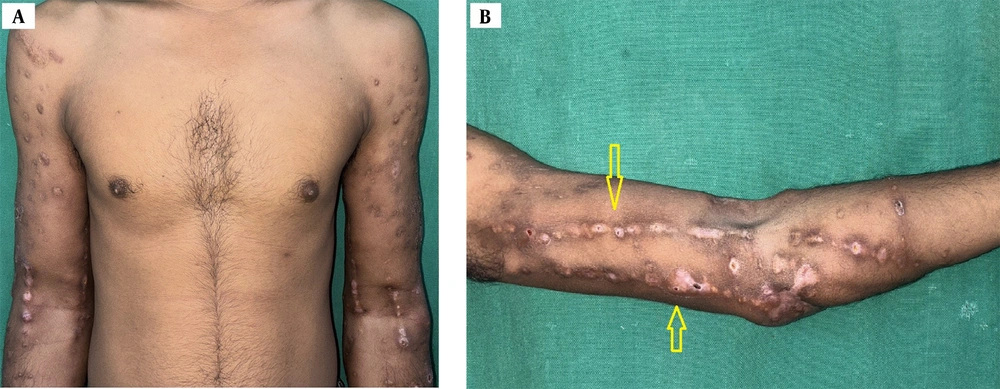
On clinical examination, both upper limbs exhibited multiple nodules ranging from 0.5 to 2 cm in diameter, with overlying erythema, tenderness, and crusting. A few lesions had broken down to form ulcers with seropurulent discharge. The nodules and ulcers were distributed in a linear fashion along the lymphatics of the forearms and arms (
Figure 1A and
B). There was evidence of chronic injection site scarring, skin thickening, and post-inflammatory hyperpigmentation. No regional lymphadenopathy or systemic signs were evident at the time of evaluation.
Clinical photograph showing multiple tender nodules, ulcers and scars over bilateral arms and forearms (A). Sporotrichoid (linear lymphocutaneous) distribution (yellow arrows) of erythematous and hyperpigmented nodules, abscesses and scars, with surrounding hyperpigmentation (B).
Based on the clinical history and examination findings, the differentials included deep mycoses (sporotrichosis, histoplasmosis, blastomycosis), atypical mycobacterial infections (fish tank granuloma/infection with environmental mycobacteria including rapid growers), nocardiosis, actinomycosis, and botryomycosis. Laboratory investigations, including complete blood count, liver, and renal function tests, were within normal limits. Serological tests for human immunodeficiency virus (HIV), hepatitis B surface antigen (HBsAg), and hepatitis C virus (HCV) were non-reactive. Given the clinical suspicion of an infectious etiology, a punch biopsy was obtained from an active nodule, and samples were sent for bacterial, fungal, and mycobacterial cultures.
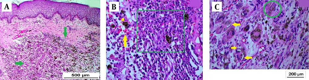
Hematoxylin and eosin-stained examination of the biopsy specimen revealed a dense dermal infiltrate composed of pyogranulomatous inflammation with areas of central suppuration (
Figure 2A). Numerous hemosiderin-laden histiocytes and foreign body-type giant cells were observed. The pigment was identified as hemosiderin rather than melanin, as evidenced by its golden-brown, refractile appearance and the presence of extravasated red blood cells, supporting hemosiderin deposition over melanin (
Figure 2B). Within the cytoplasm of histiocytes and giant cells, multiple refractile, needle-shaped, tubular, and occasionally curved unstained structures were seen (
Figure 2C). These structures raised suspicion for foreign material deposition.
Photomicrograph showing mid and deep dermal granulomatous infiltrate (green arrows; H and E, 4x) (A). Suppurative granuloma (green square), hemosiderin deposition (yellow star), red blood cell extravasation (yellow arrow; H and E, 20x) (B). Foreign body giant cells which have engulfed- curved, spicule-like refractile, unstained structures; seen as empty spaces (yellow arrows) and tubular/quadrangular structures (green circle; H and E, 40x) (C). These structures represent foreign bodies.
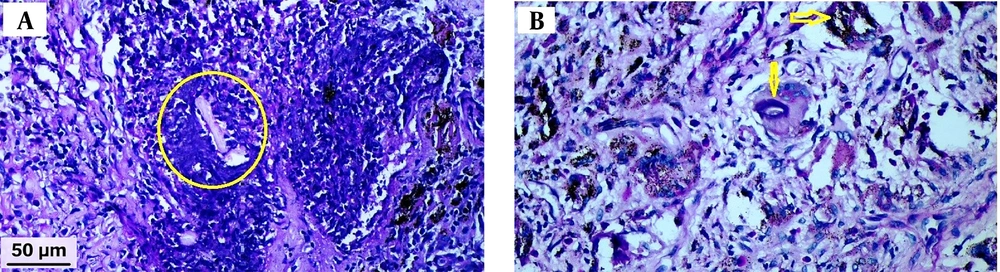
To investigate further, special histochemical stains were performed. Periodic acid-Schiff (PAS) (
Figure 3A and
B), Gomori methenamine silver (GMS), Ziehl-Neelsen (ZN), and Gram stains were all negative. Tissue cultures for bacteria, fungi, and mycobacteria also yielded no growth. These findings significantly reduced the likelihood of an infectious etiology.
Photomicrograph with periodic acid-Schiff (PAS) stain showed absence of magenta staining of the tubular structures (yellow circle) (A). The PAS stain did not show positivity in the curved empty-looking spaces within histiocytes (yellow arrows) (B). These findings are indicative of foreign body granuloma, ruling out fungal aetiology.
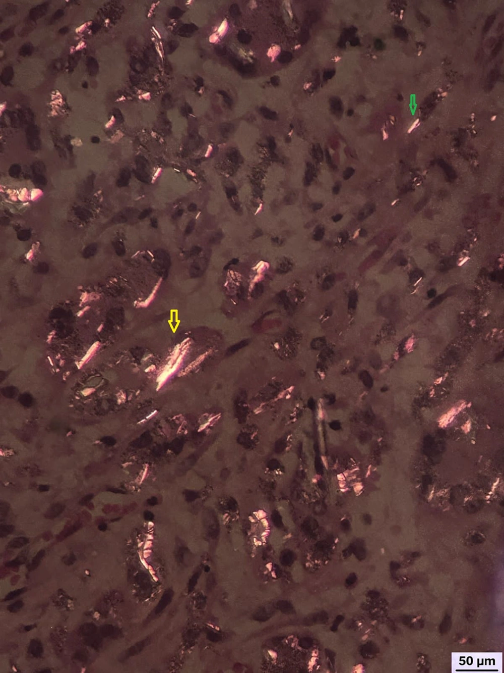
Subsequently, polarized light microscopy was employed to evaluate the refractile structures observed histologically. Under polarized light, numerous birefringent, needle-like, and rod-shaped structures were identified within histiocytes and multinucleated giant cells (
Figure 4). These structures exhibited bright birefringence, characteristic of talc and silica particles, thereby confirming the diagnosis of a foreign body granulomatous reaction induced by adulterants introduced during IV heroin injection.
Polarized microscopy depicting birefringent quadrangular structures (yellow arrow) and needle like spicules (green arrow), confirming the presence of foreign bodies; most likely talc and silica (opioid adulterants) respectively.
The patient was initiated on a combination of anti-inflammatory therapy. Oral doxycycline (100 mg once daily) was prescribed for its anti-inflammatory and antimicrobial properties. Colchicine (0.5 mg twice daily) was added for its efficacy in reducing neutrophil-mediated inflammation. Intralesional corticosteroids were administered to a few selected non-suppurative nodules to further reduce the granulomatous response, whereas the suppurative nodules and ulcers were treated with topical antibiotic (mupirocin 2% ointment) for a short duration of 7 days, to prevent secondary infection. The above treatment was planned for a period of approximately six months, with tailored therapeutic modifications during sequential interim follow-up visits. Concurrently, the patient continued to receive opioid de-addiction therapy, including psychosocial counseling and maintenance pharmacotherapy. Over the next four weeks, significant improvement was noted, with a reduction in tenderness, erythema, and nodule size. The ulcerated lesions began to heal with scarring. Unfortunately, the patient defaulted from follow-up after one month, limiting long-term assessment of therapeutic response.