
1. Introduction
Chromoblastomycosis is a chronic fungal infection of the subcutaneous tissue caused by various melanized dematiaceous fungal species and is typically observed in tropical and subtropical regions (1). The infection usually follows traumatic implantation, most commonly involves the lower extremities, and is more frequently observed among agricultural workers (2). Facial involvement is uncommon and may mimic several chronic granulomatous dermatoses, resulting in a diagnostic delay. We report a rare case of long-standing bilateral facial chromoblastomycosis with a prolonged diagnostic delay and a successful response to antifungal therapy combined with cryotherapy.
2. Case Presentation
A 57-year-old man residing in central Maharashtra and working in an ice cream factory presented with discolored, raised facial lesions of 20 years’ duration. The lesions began as a small papule on the right cheek and gradually enlarged. Subsequently, a similar lesion developed on the left cheek. The right-sided lesion progressively extended to the chin, both angles of the mouth, and the lower lip margin. There was no history of trauma, fever, constitutional symptoms, or travel.
Before presenting to our institute, the patient had received multiple therapies for these lesions, including systemic corticosteroids, methotrexate (7.5 mg weekly), hydroxychloroquine (200 mg daily), dapsone (100 mg daily), and topical tacrolimus, without improvement. Limited details regarding the patient’s prior treatment history were available.
Dermatologic examination revealed multiple well-defined, hyperpigmented to erythematous, indurated plaques with surface crusting and verrucous changes on both cheeks (Figure 1A and B). The right-sided lesion extended inferiorly to the chin and both angles of the mouth, with partial involvement of the lower lip. The plaques were firm on palpation, with underlying induration; no ulceration or discharge was observed. There was no sensory loss or peripheral nerve thickening. The regional submental and submandibular lymph nodes were palpable but nontender. Systemic examination was unremarkable. The differential diagnoses considered included leprosy, cutaneous tuberculosis, sarcoidosis, leishmaniasis, and deep fungal infection.
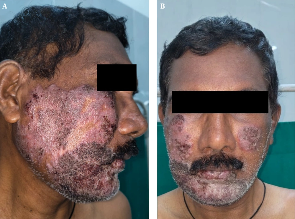
Figure 1.
Clinical presentation of facial chromoblastomycosis (A) Large erythematous to hyperpigmented indurated plaque with surface crusting and verrucous changes involving the right cheek and extending to the chin and lower lip margin. (B) Frontal view showing bilateral facial plaques involving both cheeks.
All routine hematologic and biochemical findings were within acceptable reference ranges. Serologic test results for HIV and hepatitis B and C were nonreactive. A slit-skin smear for Mycobacterium leprae and the Mantoux test were negative, and the chest radiograph showed no abnormality. Plain magnetic resonance imaging of the face demonstrated diffuse cutaneous thickening with subcutaneous fat stranding but no deep tissue extension.
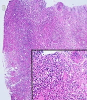
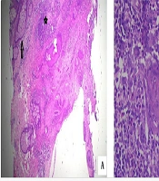
A potassium hydroxide (KOH) mount of scales obtained from the plaque demonstrated numerous muriform (sclerotic/copper-penny) bodies (Figure 2). Histopathologic examination revealed hyperkeratosis and pseudoepitheliomatous hyperplasia of the epidermis. The dermis showed granulomatous inflammation with lymphocytes, epithelioid cells, and multinucleated giant cells containing brown, thick-walled muriform (sclerotic/copper-penny) bodies (Figure 3). Periodic acid-Schiff (PAS) staining confirmed the presence of fungal organisms, whereas Ziehl-Neelsen staining was negative, effectively excluding mycobacterial infection. Fungal culture on Sabouraud dextrose agar yielded black, velvety colonies that were morphologically identified as Exophiala jeanselmei. Species identification was based on colony morphology and microscopic features observed on a lactophenol cotton blue mount, supporting a diagnosis of chromoblastomycosis (Figure 4).
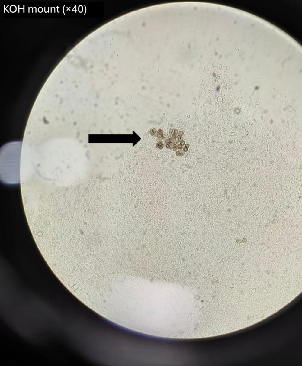
Figure 2.
Direct microscopy findings Potassium hydroxide (KOH) mount (×40) showing characteristic thick-walled, brown muriform (sclerotic/copper-penny) bodies suggestive of chromoblastomycosis.
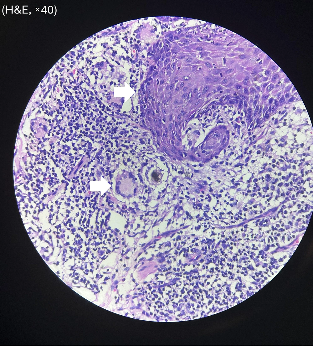
Figure 3.
Histopathologic findings in chromoblastomycosis (hematoxylin and eosin, ×40) Section showing pseudoepitheliomatous hyperplasia of the epidermis with an underlying granulomatous inflammatory infiltrate containing thick-walled, pigmented muriform (sclerotic/copper-penny) bodies.
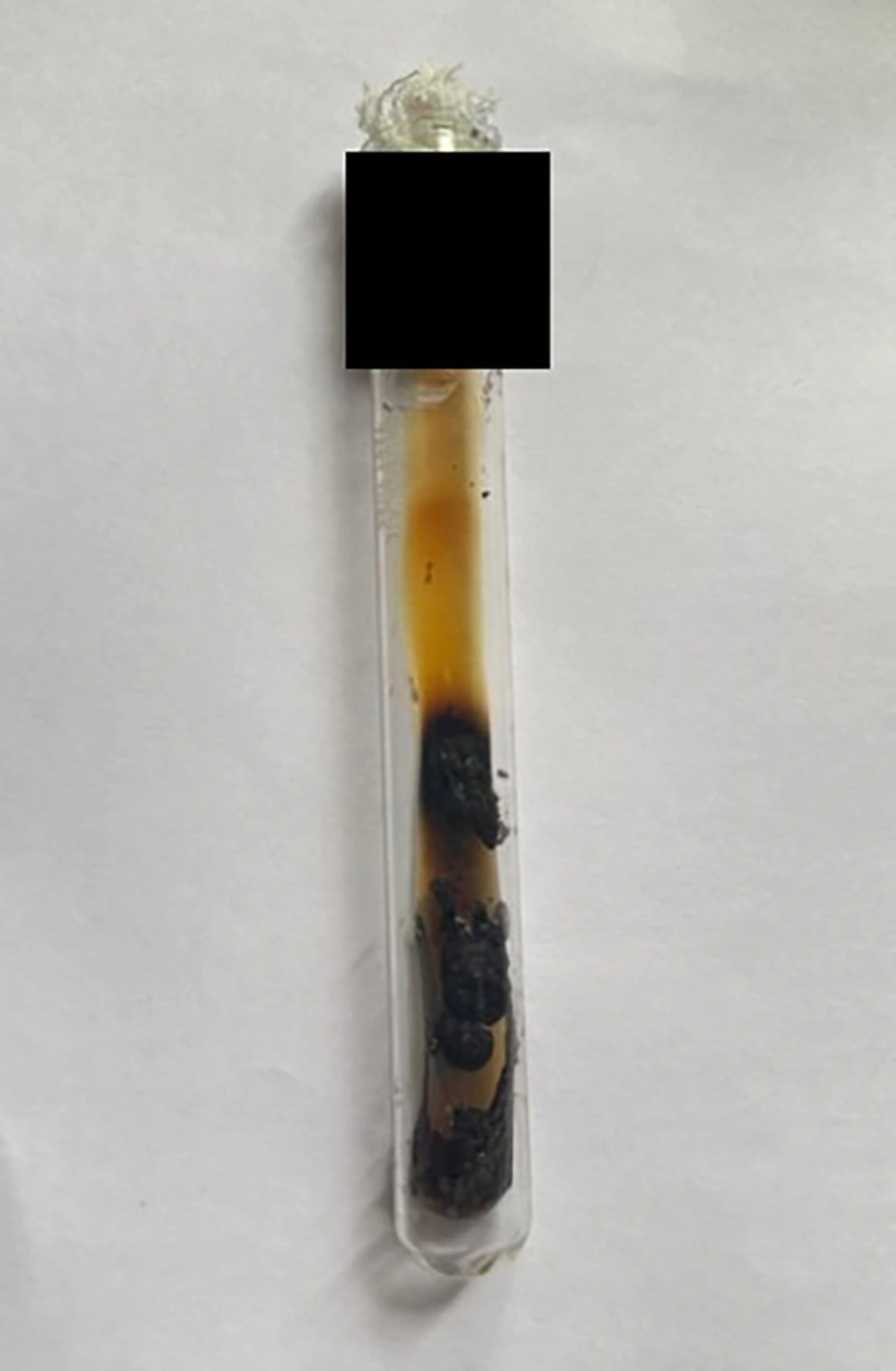
Figure 4.
Fungal culture findings Black, velvety colonies of Exophiala jeanselmei grown on Sabouraud dextrose agar, supporting the diagnosis of chromoblastomycosis.
The patient began oral itraconazole, 200 mg twice daily, which was continued for a total of 1 year. Significant flattening of the plaques was observed after 6 months of therapy. However, a single plaque on the right side of the face remained indurated and verrucous. Therefore, 4 sessions of cryotherapy with liquid nitrogen were administered to the plaque at weekly intervals over 1 month. Near-complete resolution was achieved, with mild residual scarring and postinflammatory pigmentary changes (Figure 5A and B). No relapse was observed during 1.5 years of follow-up. The patient’s liver function was monitored monthly after initiation of oral itraconazole and throughout treatment. The patient remained adherent to the prescribed treatment. The clinical timeline is summarized in Table 1.
Table 1.
Timeline of Clinical Events
| Timeline | Clinical Events |
|---|---|
| 20 years before presentation | Initial papular lesion appeared on the right cheek |
| Following years | Gradual enlargement of the papule into an indurated plaque, with bilateral facial involvement |
| Before diagnosis at our institute | The patient received corticosteroids, methotrexate, hydroxychloroquine, dapsone, and topical tacrolimus without substantial improvement; limited details were available |
| At presentation to our institute | Investigations performed at our institute included a KOH mount, histopathologic examination, PAS staining, fungal culture, and magnetic resonance imaging of the face |
| Final diagnosis | Chromoblastomycosis morphologically consistent with Exophiala jeanselmei based on morphologic and microscopic features |
| Treatment initiation | Oral itraconazole, 200 mg twice daily, was started and continued for 1 year after diagnosis, with monthly liver function monitoring |
| 1 month after treatment initiation | Partial flattening of the plaques was observed |
| 6 months after treatment initiation | One plaque on the right side of the face remained indurated and did not show substantial resolution |
| At 6 months after treatment initiation | Four cryotherapy sessions were administered once weekly to the indurated plaque on the right side of the face; substantial flattening was observed after the fourth session |
| At 1 year after treatment initiation | Near-complete resolution of the lesions was observed, and oral itraconazole was discontinued |
| At 1.5 years after treatment initiation | No relapse was observed after itraconazole was discontinued; only residual scarring and postinflammatory pigmentation remained |
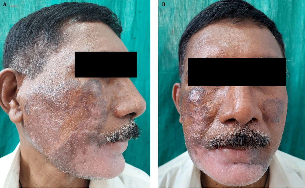
Figure 5.
Post-treatment clinical outcome (A) Marked regression of the plaque on the right cheek, with near-complete resolution following oral itraconazole therapy and adjunctive cryotherapy and mild residual scarring and postinflammatory pigmentation. (B) Frontal view showing near-complete resolution of bilateral facial lesions after treatment.
2.1. Patient Perspective
The patient expressed frustration regarding the prolonged duration of the disease and the multiple ineffective treatments received before diagnosis at our institute. Following antifungal therapy and adjunctive cryotherapy, he reported substantial improvement in his appearance and overall quality of life.
3. Discussion
Chromoblastomycosis is an indolent, granulomatous subcutaneous mycosis caused by infection with melanized dematiaceous fungi belonging to genera such as Fonsecaea, Cladophialophora, Phialophora, and Rhinocladiella and, less commonly, Exophiala (1). The disease shows a strong predilection for tropical and subtropical environments, where climatic and occupational factors favor fungal proliferation and transmission, and it usually follows traumatic implantation of fungal elements into the skin. It is commonly observed among agricultural workers and individuals exposed to soil, wood, and decaying vegetation.
Clinically, chromoblastomycosis typically presents as slowly progressive verrucous plaques, nodules, or cauliflower-like lesions. Lesions often begin as small papules at the site of inoculation and gradually enlarge over months to years. The most commonly affected sites are trauma-prone areas, particularly the lower limbs, especially the feet and legs. Other sites that may be involved include the upper limbs and trunk, whereas facial involvement, as in our case, is distinctly uncommon.
A global literature review identified approximately 7,740 reported cases of chromoblastomycosis worldwide, whereas an Indian systematic review documented 169 cases between 1957 and 2016 (3, 4). Among these, only 12 cases involved the face, highlighting the rarity of facial localization (4). Previously reported facial cases in India presented as verrucous plaques, nodular lesions, or infiltrated crusted plaques and were often initially misdiagnosed as cutaneous tuberculosis, lupus vulgaris, or sarcoidosis (5).
Regarding etiologic distribution, Fonsecaea pedrosoi accounts for most cases, whereas Cladophialophora carrionii and Phialophora verrucosa are the next most frequent causative agents (2). In contrast, infections caused by Exophiala species are rare. Only 2 cases of facial chromoblastomycosis caused by Exophiala species have been reported in the Indian subcontinent, one of which was caused by Exophiala spinifera. Therefore, the present case caused by Exophiala jeanselmei represents an uncommon etiologic agent associated with facial chromoblastomycosis.
In resource-limited settings, the diagnosis can often be established using simple bedside investigations. Direct microscopy with a potassium hydroxide mount can reveal characteristic muriform (sclerotic/copper-penny) bodies that are pathognomonic of chromoblastomycosis (2). Histopathologic examination typically shows pseudoepitheliomatous hyperplasia and granulomatous inflammation containing these pigmented fungal elements. Culture on Sabouraud dextrose agar permits species identification based on colony morphology (1). Contemporary diagnostic platforms, such as matrix-assisted laser desorption/ionization time-of-flight (MALDI-TOF) mass spectrometry and polymerase chain reaction techniques, can provide rapid and precise identification of fungal species in specialized centers. In our case, molecular identification of the species grown on fungal culture was not performed because the necessary resources were unavailable; we acknowledge this as a limitation.
The prolonged diagnostic delay in our patient is a clinically important teaching point. The patient received multiple immunosuppressive agents, including systemic corticosteroids, methotrexate, hydroxychloroquine, dapsone, and topical tacrolimus, during his illness, likely for presumed inflammatory or granulomatous dermatoses such as cutaneous lupus, sarcoidosis, or leprosy. Although the precise duration and sequence of these therapies could not be ascertained because prior records were limited, prolonged immunosuppression in the setting of an undiagnosed deep fungal infection is a recognized risk factor for disease persistence and progression. Corticosteroids and immunomodulatory agents may attenuate the granulomatous host response that partially contains chromoblastomycosis, potentially allowing further fungal proliferation and lesion extension. Therefore, this case highlights the importance of including deep fungal infections in the differential diagnosis of chronic verrucous or infiltrated facial plaques, particularly when lesions fail to respond to or worsen with immunosuppressive therapy.
The differential diagnosis of chromoblastomycosis is broad and includes cutaneous tuberculosis, particularly lupus vulgaris; leprosy; sporotrichosis; cutaneous leishmaniasis; sarcoidosis; verrucous carcinoma; and other deep fungal infections. Slowly progressive chronic verrucous plaques that are unresponsive to conventional therapy should raise suspicion of deep fungal infections such as chromoblastomycosis.
Management of chromoblastomycosis is often prolonged and challenging because of fibrosis and poor penetration of antifungal agents into chronic lesions. Systemic antifungal therapy remains the cornerstone of treatment. Itraconazole and terbinafine are the main pharmacologic treatments and are generally prescribed for several months to 1 year; most cases reported from India have been treated similarly (6). In refractory cases, agents such as posaconazole, voriconazole, amphotericin B, or flucytosine may be used. Adjunctive physical therapies, including cryotherapy, surgical excision, and laser therapy, may enhance the treatment response (7). In our patient, long-term itraconazole therapy combined with cryotherapy resulted in substantial clinical improvement, with near-complete resolution and minimal residual scarring.
Long-standing chromoblastomycosis may lead to complications such as secondary bacterial infection, lymphatic obstruction, and, rarely, malignant transformation into squamous cell carcinoma. Therefore, early diagnosis and treatment remain crucial.
3.1. Conclusions
Chronic verrucous or infiltrated plaques, particularly when long-standing and unresponsive to conventional therapy, should prompt consideration of deep fungal infections such as chromoblastomycosis, even at unusual sites such as the face. Simple bedside investigations, such as a potassium hydroxide mount and early biopsy, can facilitate timely diagnosis and prevent prolonged diagnostic delay and complications.