
1. Introduction
Aspergillus species are among the most important opportunistic fungal pathogens causing human infections and represent the second most common cause of invasive fungal disease after Candida species. These molds are ubiquitous in the environment and can be isolated from soil, air, dust, and decaying vegetation (1). Among the species known to cause human disease, Aspergillus fumigatus is the most commonly implicated, followed by A. flavus, A. terreus, and, less frequently, A. niger (1). Although pulmonary aspergillosis remains the most common manifestation, cutaneous aspergillosis is a relatively uncommon clinical entity (2). It may occur as either primary cutaneous aspergillosis following direct inoculation through breaches in the skin or secondary cutaneous aspergillosis due to hematogenous spread from a primary internal focus, usually the lungs (3).
Primary cutaneous aspergillosis is an uncommon clinical condition that can occur in both immunocompetent and immunocompromised individuals, with a higher frequency reported in the latter. Direct traumatic inoculation, occlusive dressings, indwelling catheters, surgical wounds, and burn injuries are recognized risk factors in immunocompetent patients (4). We report an unusual case of PCA caused by Aspergillus fumigatus in an immunocompetent farmer with a history of remote trauma.
2. Case Presentation
A 47-year-old married Hindu male farmer from Central Maharashtra, India, presented with reddish, painful lesions on the right forearm for 8 months. The lesion gradually increased in size, and the pain worsened, with associated intermittent low-grade fever. He reported a history of trauma at the same site approximately 12 years earlier while working in the fields, after which the wound was surgically drained, leaving a scar. The patient had chronic plaque psoriasis at different sites and had been treated intermittently with topical steroids and emollients. There was no history of systemic corticosteroid therapy, methotrexate, cyclosporine, biologic therapy, or any other systemic immunomodulatory treatment. There was no history of tuberculosis, respiratory complaints, diabetes mellitus, organ transplantation, immunosuppressive therapy, or high-risk behavior.
On dermatological examination, a single large erythematous to dusky-colored, firm, tender plaque measuring approximately 5 × 8.2 cm with a bosselated surface was present on the volar and medial aspects of the right forearm (Figure 1A). A smaller satellite plaque was noted proximally (Figure 1B). No regional lymphadenopathy was observed. Systemic examination findings were within normal limits. Based on the history and examination, the differential diagnoses included sarcoidosis, atypical mycobacterial infection, cutaneous leishmaniasis, bacillary angiomatosis, and deep fungal infection.
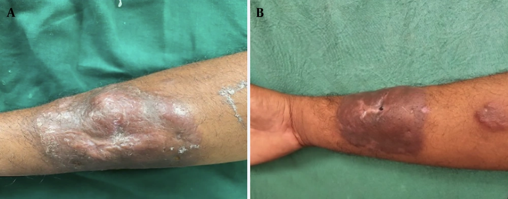
Figure 1.
A, Single large erythematous to dusky-colored plaque measuring approximately 5 × 8.2 cm with a bosselated surface over the volar and medial aspects of the right forearm; B, smaller satellite plaque proximal to the main lesion.
Routine hematological parameters, including the absolute neutrophil count, and biochemical parameters, including fasting blood glucose/HbA1c levels, were within normal limits. HIV testing by enzyme-linked immunosorbent assay was nonreactive. The CD4 count was 498 cells/mm3. Chest radiography and ultrasonography of the abdomen and pelvis did not reveal any systemic focus of infection.
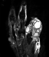
Local ultrasonography with Doppler revealed a fairly well-defined hypoechoic lesion in the subcutaneous plane with significant internal vascularity. Magnetic resonance imaging of the lesion demonstrated a well-defined altered-signal-intensity lesion involving the skin and superficial subcutaneous fat plane, with sparing of the underlying muscle (Figure 2).
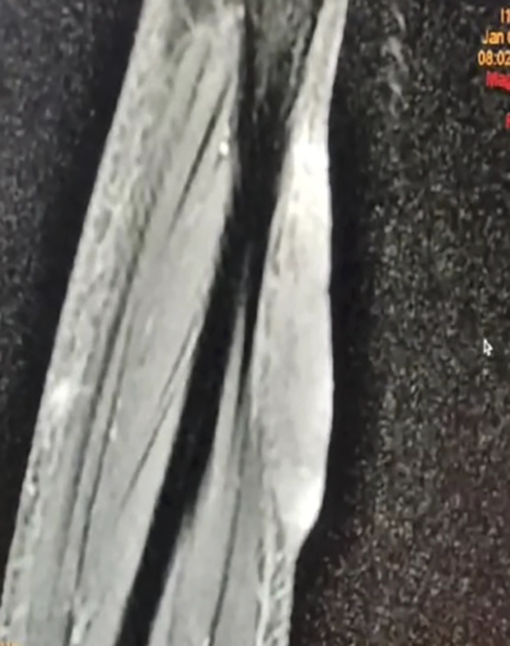
Figure 2.
Magnetic resonance imaging showing a well-defined altered signal intensity lesion involving the skin and superficial subcutaneous tissue, with sparing of the underlying muscle.
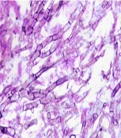
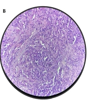
Histopathological examination of the lesion revealed diffuse, ill-defined dermal granulomas composed predominantly of lymphocytes and histiocytes, with multinucleated giant cells in a background of dense lymphomononuclear inflammatory infiltrate admixed with neutrophils (Figure 3A and B). A 10% potassium hydroxide mount showed septate hyphae. Periodic acid-Schiff and Gomori methenamine silver stains highlighted the fungal elements.
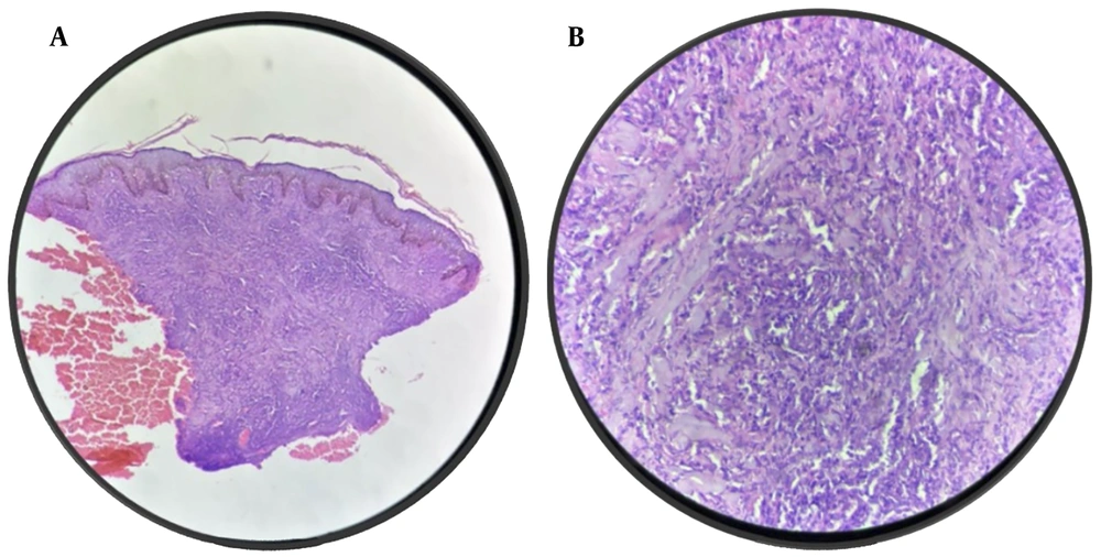
Figure 3.
A, Photomicrograph (hematoxylin and eosin stain, 10x) showing diffuse ill-defined dermal granulomatous inflammation; B, photomicrograph (hematoxylin and eosin stain, 40x) showing granulomas composed predominantly of lymphocytes and histiocytes with multinucleated giant cells and admixed neutrophils.
Fungal culture on Sabouraud dextrose agar showed velvety smoky blue-green colonies (Figure 4). On microscopic morphologic examination, a lactophenol cotton blue mount demonstrated septate hyphae with short, smooth conidiophores terminating in flask-shaped vesicles with uniseriate phialides covering the upper two-thirds of the vesicle, suggestive of Aspergillus fumigatus. Molecular confirmation and antifungal susceptibility testing could not be performed because of resource limitations.
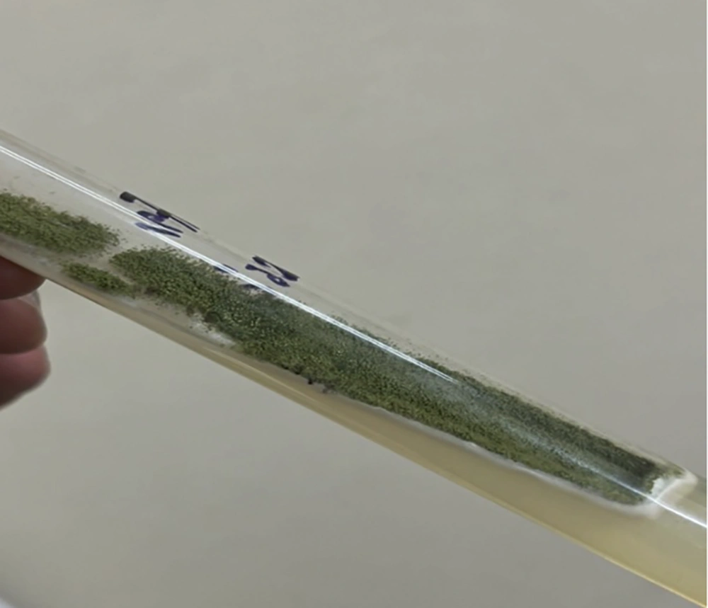
Figure 4.
Fungal culture on Sabouraud dextrose agar showing velvety smoky green colonies morphologically suggestive of Aspergillus fumigatus.
A final diagnosis of primary cutaneous aspergillosis was established based on the clinical presentation, histopathological findings, positive stains, fungal culture, and the absence of any identifiable systemic focus of infection.
After confirmation by fungal culture, the patient was started immediately on intravenous liposomal amphotericin B at a dose of 3 mg/kg; however, the drug was discontinued after 3 days of infusion because of fever, chills, and worsening renal function, with the serum creatinine level rising from a baseline value of 1.2 mg/dL to 3.4 mg/dL during therapy. The cumulative dose received was approximately 540 mg over 3 days.
Five days after diagnostic confirmation, oral itraconazole 200 mg twice daily was initiated. The patient had good adherence to therapy, and no significant drug interactions were identified. Therapeutic drug monitoring for itraconazole could not be performed because of resource limitations.
After 2 months of oral itraconazole therapy, there was no appreciable clinical improvement in lesion size, pain, induration, or plaque thickness. Therefore, oral voriconazole 200 mg twice daily was initiated. Baseline and periodic liver function tests were monitored during therapy and remained within normal limits. No major adverse effects, such as visual disturbances, hepatotoxicity, or phototoxicity, were observed during treatment.
After 5 months of oral voriconazole therapy, marked regression was observed, with complete flattening of the plaque and residual hyperpigmentation compared with baseline (Figure 5). The patient remains under regular follow-up and has been receiving maintenance therapy with oral voriconazole 100 mg daily for the past 8 months, with no evidence of recurrence or systemic involvement.
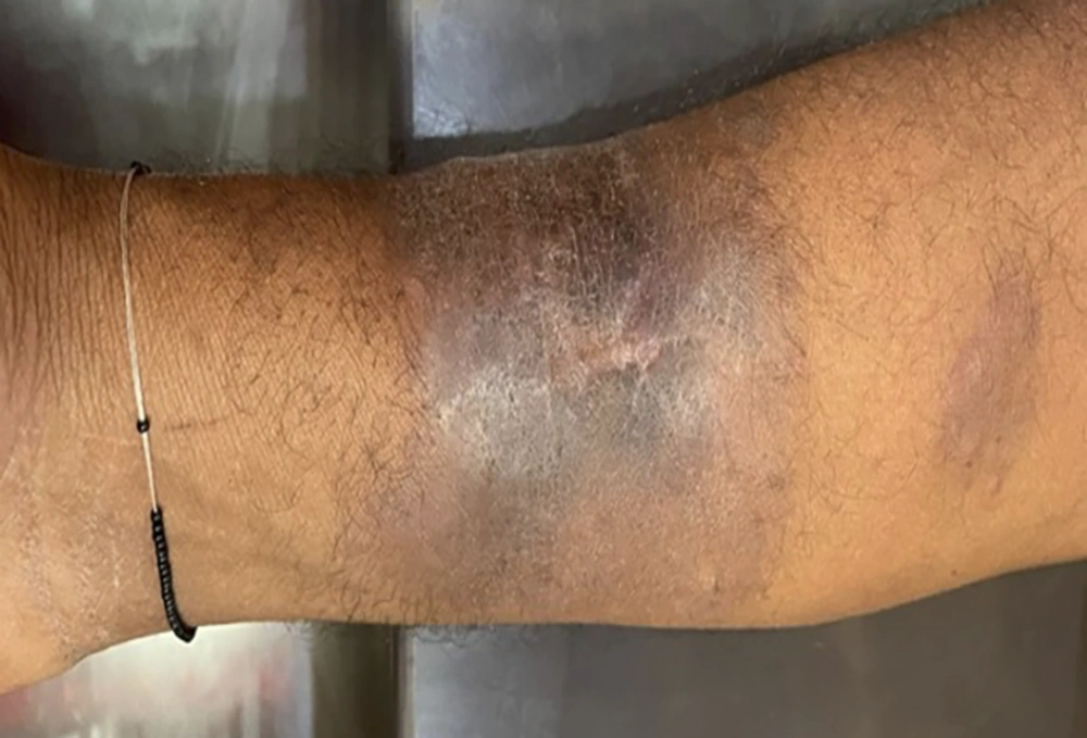
Figure 5.
Marked clinical improvement after 5 months of oral voriconazole therapy showing complete flattening of the plaque with residual hyperpigmentation.
3. Discussion
Cutaneous aspergillosis is a rare manifestation caused by Aspergillus species and presents as either a primary localized disease or secondary involvement due to systemic dissemination. Pulmonary involvement is most common and typically presents as invasive aspergillosis. It predominantly affects immunocompromised individuals, including patients with hematologic malignancies undergoing chemotherapy, those receiving prolonged corticosteroid therapy, recipients of broad-spectrum antibiotics, individuals with HIV infection, and patients with inherited immunodeficiency disorders such as chronic granulomatous disease (4). Prolonged neutropenia and sustained immunosuppression significantly increase the risk of invasive disease. Our patient did not have any of the above-mentioned predisposing factors. Although he had a history of chronic plaque psoriasis, it had been managed only with intermittent topical corticosteroids and emollients. He had not received any treatment for 4 years and was in complete remission. Furthermore, there was no history of the current lesion developing at the site of a pre-existing psoriatic plaque. His CD4 count (498 cells/mm3) was at the lower end of the normal range; however, there was no neutropenia, evidence of systemic infection, or other clinical features suggestive of clinically significant immunodeficiency. Therefore, although the patient was considered immunocompetent in the clinical context, the borderline CD4 count should be interpreted with caution.
Cutaneous aspergillosis occurs either as primary cutaneous aspergillosis following direct inoculation through breaches in the skin or as secondary cutaneous aspergillosis due to hematogenous spread from a primary internal focus, usually the lungs (4).
The global literature includes a limited number of case reports of primary cutaneous aspergillosis. This condition occurs mostly in immunocompromised individuals but can rarely develop in immunocompetent hosts after direct traumatic inoculation of spores into the skin, particularly in settings involving occlusive dressings, indwelling catheters, surgical wounds, or burn injuries. Our patient had a similar history of trauma followed by surgical intervention.
Clinical manifestations are heterogeneous and may range from violaceous macules, papules, plaques, and subcutaneous nodules to florid presentations characterized by hemorrhagic bullae, black eschar, pustular lesions, and ulceration with central necrosis (5). Our case presented with bosselated swelling with a central atrophic scar.
A comprehensive literature review by Alp et al. highlighted the occurrence of PCA in 3 immunocompetent individuals treated with oral voriconazole along with surgical debridement (6). Zhang et al. described an immunocompetent adult with PCA due to A. fumigatus after tattooing, treated successfully with oral itraconazole 200 mg/day for 3 months, with complete resolution and no recurrence at 6 months (7). Tahir et al. reported primary cutaneous aspergillosis in an immunocompetent woman after presumed traumatic inoculation from shaving, successfully managed with combined itraconazole therapy and surgical excision (8). Camus et al. reported primary cutaneous aspergillosis in an immunocompetent adult with a kerion-like facial lesion due to A. fumigatus, which resolved completely with systemic voriconazole (9). Elbayomi et al. reported PCA in an immunocompetent patient from India caused by Aspergillus flavus who responded to itraconazole antifungal therapy and surgical debridement (10). According to current clinical practice guidelines, voriconazole is regarded as first-line therapy for primary cutaneous aspergillosis because of its proven efficacy and improved survival outcomes (5).
Given its rarity and variable presentation, timely diagnosis of primary cutaneous aspergillosis remains challenging. Histopathological examination with special stains and fungal culture remain the cornerstone of diagnosis. Our case underscores the need to consider primary cutaneous aspergillosis in chronic cutaneous lesions, even in apparently immunocompetent individuals, and highlights the effectiveness of voriconazole in refractory cases.
3.1. Patient Perspective
At initial presentation, the patient reported significant discomfort and limitations in daily activities due to pain and the gradual enlargement of the lesion. He expressed satisfaction with the clinical improvement after voriconazole therapy, particularly the complete resolution of pain and flattening of the lesion.