The Association of Brain-Derived Neurotrophic Factor with Long-Term Potentiation

authors:

avatar Zahra Salimi 1 , 2 , avatar Farshad Moradpour 1 , 2 , avatar Zahra Rashidi 1 , avatar Fatemeh Zarei 1 , 2 , avatar Mohammad Rasool Khazaei 1 , avatar Seyed Mojtaba Ahmadi 1 , *

Fertility and Infertility Research Center, Health Technology Institute, Kermanshah University of Medical Sciences, Kermanshah, Iran
Department of Physiology, Kermanshah University of Medical Sciences, Kermanshah, Iran

how to cite: Salimi Z, Moradpour F, Rashidi Z, Zarei F, Khazaei M R, et al. The Association of Brain-Derived Neurotrophic Factor with Long-Term Potentiation. Shiraz E-Med J. 2022;23(2):e112278. https://doi.org/10.5812/semj.112278.

Abstract

Long-term potentiation (LTP) is one of the most important topics in neuroscience. It refers to a long-lasting increase in synaptic efficacy and is considered as a molecular and cellular mechanism of learning and memory. Neurotrophins play essential roles in different processes in the central nervous system (CNS), such as synaptogenesis, survival of specific populations of neurons, and neuroplasticity. Some evidence suggests that neurotrophins also participate in the synaptic plasticity related to learning and memory formation. Brain-derived neurotrophic factor (BDNF) is an important neurotrophic factor that is extensively expressed in the hippocampus and cerebral cortex, where it promotes neuroprotection, increases synaptogenesis and neurotransmission, and mediates synapse formation and synaptic plasticity. In this review, we first focused on the research investigating the effects of BDNF on synaptic plasticity and LTP induction and then reviewed the neuronal signaling molecules employed by BDNF to promote its effects on these processes.

1. Context

Learning and memory are the most important concepts in neuroscience. Learning can be defined as the mechanism by which new knowledge about the world is attained, and memory as the mechanism by which that information is retained. Learning and memory are dependent on synaptic plasticity that includes major changes in neurons and synapses (1). In 1984, Konorski presented the term “synaptic plasticity” for the first time to explain persistent and activity-dependent modifications in synaptic strength (2). Long-term potentiation (LTP) is described as the long-lasting activity-dependent enhancement of synaptic strength, which is commonly regarded as the closest neural model for explaining the molecular and cellular mechanisms involved in learning and memory (3). Long-term potentiation comprises three phases, including initial-LTP, early-LTP, and late-LTP, which occur consecutively over time to create what we call LTP. Initial-LTP (i.e., short-term potentiation) lasts for about 30 - 60 min and is a constant type of synaptic plasticity and dependent on the N-methyl-D-aspartate receptor (NMDAR). This phase of LTP does not require the activity of protein kinases. Early-LTP lasts for about 2 - 3 h and is not dependent on protein kinases’ activity. Finally, late-LTP persists for 5 - 6 h and requires gene expression and protein synthesis. Each phase is performed by different mediators and regulators (4). The high-frequency stimulation of presynaptic neurons leads to the secretion of neurotransmitters, chiefly glutamate. The binding of this neurotransmitter to the 2-amino-3-(3-hydroxy-5 methylisoxazol-4-yl) propanoic acid receptor (AMPAR) of postsynaptic neurons causes an influx of sodium into these neurons, leading to neuron depolarization. When the extent of depolarization is enough to eliminate the Mg2+-induced block of NMDAR, the influx of calcium into the postsynaptic neuron starts through this receptor. It has been well established that an optimal level of intracellular calcium-mediated NMDAR activation is essential for LTP induction. An increase in intracellular calcium concentration activates the signaling pathways that modify gene transcription and induce the synthesis of new proteins (5).

The induction of LTP needs the activation of many molecules and signaling cascades. Brain-derived neurotrophic factor (BDNF) is a key mediator that plays critical roles in the CNS, including promoting the survival and growth of neurons and inducing long-term synaptic plasticity (6-8). The mechanisms by which BDNF induces LTP are not fully understood. Substantial number of studies have suggested various roles for BDNF in the induction of hippocampal LTP. On the other hand, there is a relationship between BDNF and the neuronal mediators and pathways involved in LTP induction, including neurotransmitter systems, cellular receptors, and presynaptic and postsynaptic proteins. Thus, the present review primarily aimed to explain the impacts of BDNF on synaptic plasticity and LTP induction and then to elaborate the possible mechanisms responsible for these effects.

2. BDNF and LTP

Neurotrophins are a main group of signaling molecules widely investigated for their roles in promoting the survival, growth, and differentiation of neurons during development. Numerous studies have revealed that neurotrophins can also act as the regulators of synaptic plasticity. As one of the most important neurotrophic factors, BDNF is a 119-amino-acid basic peptide and has long been recognized as an anti-apoptotic factor extensively expressed in the brain. This factor shares important roles in the CNS, including neuronal maturation, synaptogenesis, synaptic plasticity, and hippocampal LTP induction (6, 7). Because of its essential role in neuronal development and function, changes in BDNF level have been described in a variety of psychiatric and neurological disorders (8, 9). Likewise, BDNF levels have been detected to decline in the CNS of patients with neurodegenerative disorders, including Huntington, Parkinson’s, and Alzheimer’s disease, neuropsychiatric disorders such as schizophrenia, and individuals with depressive-like behaviors (10-14).

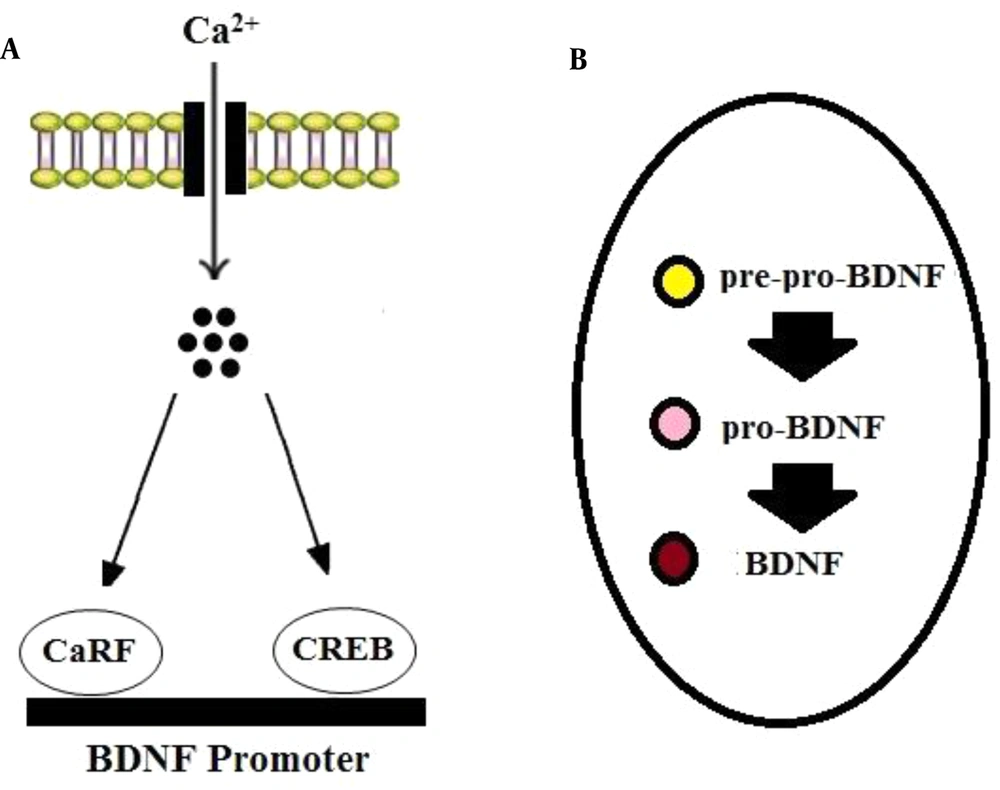
Brain-derived neurotrophic factor is produced in the soma of neurons and neuroglia and then is translocated to the terminals of pre-synapses (15, 16). The transcription of the gene encoding BDNF can be modulated by calcium influx. It has been established that calcium induces the binding of transcription factors, including cyclic AMP response element-binding protein (CREB) and calcium response factor (CaRF), to the bdnf gene’s promoters (17) (Figure 1A). In neurons, BDNF is synthesized as a precursor peptide known as pre-pro-BDNF that is cleaved into pro-BDNF (35 kDa). Then pro-BDNF can be cleaved by proteases into mature BDNF (mBDNF or BDNF: 14 kDa) (15, 16) (Figure 1B).

BDNF synthesis. BDNF mRNA transcription can be modulated via calcium influx through calcium channels. A, Ca2+ induces the binding of transcription factors, including CREB and CaRF, to BDNF promoters. B, In neurons, BDNF is first synthesized as pre-pro-BDNF in the endoplasmic reticulum and converted to pro-BDNF, which is then cleaved to mature BDNF.

Many studies propose that BDNF induces the complex neuronal signaling cascades responsible for the promotion of the cellular events involved in neuroplasticity and LTP induction (18, 19). For the first time, in the 1990s, it was reported that the treatment of hippocampus slices with BDNF enhanced early-LTP induced by theta-burst stimulation (20). Later, it was revealed that BDNF was involved in the early and late phases of LTP (21, 22). Consistent with these findings, De Vincent et al. reported that BDNF regulated the extent of LTP in the presynaptic neuron whereas in the postsynaptic neuron, it was involved in the maintenance of LTP (23). Furthermore, Messaoudi et al. assessed the impact of the intrahippocampal infusion of BDNF on synaptic plasticity and reported that the intrahippocampal microinjection of BDNF enhanced synaptic plasticity in the dentate gyrus of the hippocampus (24). Other studies also described a key role for BDNF in LTP induction, confirming that the knockout mice lacking the bdnf gene exhibited hippocampal LTP impairment (25-27).

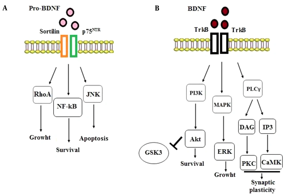
neurotrophic factors in response to physiological changes bind to to the tyrosine kinase (Trk) and p75 neurotrophin (p75NTR) receptors. Research findings propose that pro-BDNF and mature BDNF activate distinct cellular signaling pathways. Non-cleaved pro-BDNF leads to generally undesirable cellular events such as apoptosis and LTD induction through its binding to p75NTR; on the other hand, BDNF (i.e., mature BDNF) promotes positive events, including survival, growth, synaptic plasticity, and LTP induction through binding to TrkB receptors (28, 29). These findings suggest that pro-neurotrophins can elicit impacts opposite to mature-neurotrophins via binding to a different receptor (i.e., p75NTR) (30). In other words, the activation of TrkB receptors promotes cell survival, synaptogenesis, and synaptic plasticity whereas the engagement of p75 receptors leads to cell death and synaptic pruning (31). Consistent with these findings, LTD induction in the hippocampus has been reported to be dependent on the activation of p75 receptors (32). Overall, the binding of BDNF to the TrkB receptor mediates anti-apoptotic functions while pro-BDNF binding to p75NTR triggers apoptosis, a phenomenon that needs interaction with its co-receptor, sortilin. The pro-BDNF/p75NTR/sortilin complex begins the signaling pathways activating Ras-homolog gene family member A (RhoA), c-Jun amino terminal kinase (JNK), and nuclear factor kappa B (NF-ҡB) (33, 34). The activation of the RhoA-dependent signaling cascade regulates neuronal growth cone, development, and motility (34). The JNK-dependent signaling pathway stimulated by the proBDNF/p75NTR/sortilin complex induces neuronal apoptosis (33). Ultimately, the p75NTR-dependent activation of NF-ҡB triggers the processes stimulating neurons’ survival and maintaining their quantity during brain development (34) (Figure 2A).

The intracellular signaling pathways of BDNF-p75NTR and BDNF-TrkB. A, Binding of pro-BDNF to the p75NTR and sortilin receptors activates the signaling cascades related to RhoA, NF-ҡB, and JNK, stimulating the processes involved in neuronal development, survival, and programmed cell death; B, Binding of BDNF to the TrkB receptor initiates the MAPK, PI3K, and PLC γ pathways that promotes neuronal survival and growth and synaptic plasticity.

In addition, BDNF (mature BDNF) has a high affinity for binding to TrkB receptors, which subsequently induces the dimerization and autophosphorylation of this receptor, activating the downstream mitogen-activated protein kinase [MAPK, or Extracellular Signal Related Kinase (ERK)], phosphatidylinositol 3-kinase (PI3K), and phospholipase C-γ (PLC-γ) pathways (35, 36). The MAPK/ERK signaling pathway causes cell growth and differentiation, and the PLC-γ signaling route stimulates the inositol trisphosphate (IP3) receptor to release calcium, boosting calmodulin kinase (CamK) function and thus synaptic plasticity. Also, the PI3K signaling pathway activates protein kinase B (AKT), extending cell survival. Evidence from different studies suggests that the PI3K/Akt pathway is essential for axonal elongation. Furthermore, when the PI3K signaling pathway is activated, it leads to the inhibition of the downstream glycogen synthase kinase-3 (GSK-3), a serine/threonine kinase originally recognized as a regulator of glycogen metabolism (35, 37) and known to be extensively expressed throughout the CNS (38). On the other hand, GSK-3 has been shown to play an important role in NMDAR-dependent LTD at CA3–CA1 synapses of the hippocampus (39, 40). Previous studies reported that the local inactivation of GSK-3 was essential for axonal growth (41, 42). Moreover, other studies showed that various GSK-3 blockers could prevent the induction of LTD, when loaded into neuronal cells by a patch pipette (40, 43). Interestingly, it has been reported that BDNF inhibits GSK-3 by activating a PI3K-dependent cascade (44) (Figure 2B). Finally, these signaling pathways affect the cAMP response element-binding protein (CREB) transcription factor that can up-regulate the expression of relevant survival genes. It is notable that rapid effects on synapses and ion channels are dependent on PLCγ-mediated calcium release, and more long-lasting impacts, such as gene transcription, are downstream to the PI3K and MAPK signaling pathways. The activation of these pathways promotes neuronal survival and growth and neuroplasticity (8, 35, 36).

Dendrites are known to integrate synaptic inputs into neuronal cells, and their branching is associated with their representational capacity. Interestingly, the branching forms of dendritic trees are associated with the extent of the compartmentalization of inputs to neurons, and a stronger compartmentalization potential has been suggested to increase the representational capacity of the neuron, leading to a greater neuroplasticity capacity (45). The administration of diverse types of BDNF molecules mediates different morphological alterations. For example, the acute application of BDNF stimulated neurite elongation and spine head enlargement while its gradual application enhanced the branching of dendritic and filopodia-like spines (46). The effects of BDNF on cellular responses are also complex. For instance, the rapid perfusion of BDNF (i.e., acute exposure) increases synaptic transmission at neuromuscular synapses (47) while long-term (chronic) treatment with BDNF stimulates synapse maturation (48). A previous study found that the perfusion of BDNF in hippocampal slices induced a long-lasting improvement in basal-synaptic transmission (49). However, some studies have demonstrated that LTP, but not basal synaptic transmission, is facilitated in the hippocampal slices perfused with BDNF (50, 51). In support of these findings, Ji et al. reported that cellular responses to BDNF differed dramatically depending on how BDNF was delivered. In the neonatal slices of the hippocampus, slow BDNF perfusion (i.e., chronic exposure) facilitated the LTP induced by theta burst stimulation, without altering the baseline synaptic strength. In contrast, in adult hippocampus slices, the rapid perfusion of BDNF (acute) stimulated the synaptic growth necessary for the establishment of neuronal networks during development and induced structural and functional modifications in synapses (46). Kang et al. reported that the chronic or acute application of BDNF induced the long-lasting enhancement of basal synaptic transmission in the CA1 area of the hippocampus (49). However, some studies reported that slow or chronic BDNF perfusion did not induce such an enhancement (26, 50). On the other hand, in hippocampus slices, the rapid and slow increase of BDNF enhanced basal synaptic transmission and LTP induction, respectively (46). Overall, BDNF regulates an extensive range of cellular functions in the CNS, such as dendritic and axonal growth, survival of neurons, signal transmission in synapses, and LTP induction in the hippocampus.

3. BDNF and Neurotransmission

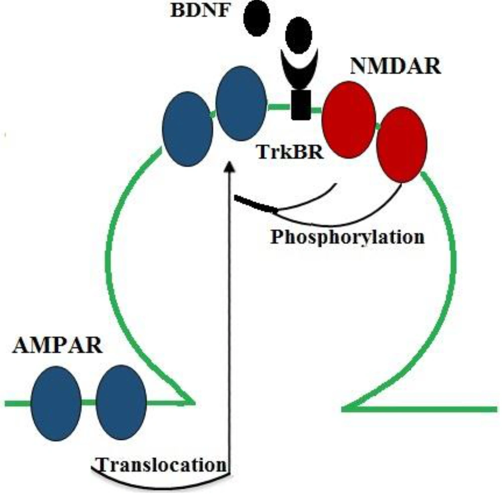
A great number of studies suggest that optimal brain function requires an intermediate range of neurotransmitters, and both high and low levels of neurotransmitters are involved in the pathophysiology of many mental diseases. Synaptic plasticity is commonly regulated by the release of various neurotransmitters from presynaptic neurons. Actually, dysregulated neurotransmitter systems can impair synaptic plasticity (52, 53). Some studies have reported that BDNF can regulate neurotransmission and LTP induction through both presynaptic and postsynaptic pathways. Consistent with these findings, the application of BDNF to cortical and hippocampal neuronal cultures and slice preparations induced excitatory neurotransmission and increased glutamate release (36, 49, 54, 55). Glutamate is one of the most important excitatory neurotransmitters in the CNS, and glutamate ionotropic receptors mediate most of excitatory neurotransmission. In fact, NMDAR and AMPAR are two types of ionotropic glutamate receptors (ligand-gated ion channels) that have critical roles in neuroplasticity and LTP induction. Glutamatergic synapses act mainly through AMPAR to generate rapid synaptic excitation, and NMDAR can also contribute to neurotransmission and plasticity (56). The activation of NMDAR increases calcium levels in the postsynaptic neuron and modulates the downstream signaling cascades involved in LTP induction (57). Considerable evidence suggests that BDNF promotes glutamatergic synaptic transmission in postsynaptic neurons by modulating NMDAR phosphorylation (58, 59). In line with these findings, it was reported that BDNF increased the phosphorylation of the NR1 and NR2B subunits of NMDAR (58) and enhanced the probability of the opening of NMDAR channels (54) to increase calcium influx (60). Moreover, AMPAR mediates most of the fast excitatory synaptic transmission in the brain of mammals. In the hippocampus, early-LTP maintenance is accompanied by an increase in AMPAR. According to some studies, changes in the number and type of the subunits of this receptor can impair synaptic plasticity. A considerable number of studies have also reported that BDNF modulates the surface expression of AMPAR through regulating its quick surface translocation to enhance excitatory transmission (61, 62) (Figure 3).

BDNF/TrkB actions on NMDAR and AMPAR, which mediate synaptic transmission in neurons. BDNF increases glutamatergic neurotransmission through increasing the probability of the opening of NMDAR (by promoting its phosphorylation) and through inducing the surface expression of AMPAR (by inducing the rapid translocation of its subunits to the surface) to enhance excitatory transmission.

Evidence suggests that a decrease in the level of monoamine neurotransmitters, including serotonin, dopamine and norepinephrine, impairs brain functions. Interestingly, BDNF regulates the activity of the dopaminergic, serotonergic, and noradrenergic systems (63, 64) and changes the secretion of neurotransmitters by altering protein expression in the presynaptic neurons responsible for releasing these mediators (65). Glutamate production in glutamatergic neurons is induced via a rise in the number of docked vesicles in the active zones of synapses (66). A previous study reported that the mice lacking BDNF showed a decline in the release of neurotransmitters in neuronal synapses, which was associated with a drop in the number of docked synaptic vesicles (67) and a reduction in the content of different presynaptic proteins, including synapsin, synaptophysin, and synaptobrevin, which are essential for vesicle docking and exocytosis in synaptic active zones (27). In line with these findings, Jovanovic et al. have reported that BDNF binds to the TrkB receptor and activates MAPK in synaptosome, inducing synapsin I phosphorylation at MAPK-dependent P-sites and the release of neurotransmitters (55).

Further evidence suggests that BDNF, in addition to influencing presynaptic proteins, alters the levels of postsynaptic proteins. As the most abundant postsynaptic protein, PSD-95 regulates different types of synaptic transmission, synapse structure and stability, and the formation and long-term stabilization of memory. The expression of PSD-95 enhances synapse maturation while the knockdown of this protein weakens synaptic strength and reduces spine density (68). Also, PSD-95-dependent protein complexes interact with NMDAR- and AMPAR-type glutamate receptors (69), which in the postsynaptic space can be vital for neuroplasticity and synaptic plasticity (70). Furthermore, the BDNF-TrkB pathway has an essential role in the development of synapses through the synaptic delivery of PSD-95 by vesicular transport (71) and its initial association with the membrane (71, 72).

Moreover, it appears that BDNF can affect synaptic plasticity and LTP induction via interaction with the sigma-1 receptor (Sig-1Rs) that is extensively distributed in different areas of the CNS, such as the prefrontal and parietal cortex, hypothalamus, and hippocampus (73). This receptor plays an important role in regulating different types of ion channels (74, 75), including voltage-gated ion channels that are critical for initiating and shaping action potentials (76) and NMDA-induced neuronal firing in the hippocampal CA3 region (77). The agonists of Sig-1Rs enhance synaptic plasticity, neuroplasticity, and memory functions (78, 79). It also has been reported that Sig-1Rs ligands affect memory functions via modulating NMDA-dependent LTP (75) and increasing BDNF level (80, 81). In a previous study, Malik et al. reported that the administration of a Sig-1Rs agonist stimulated the release of BDNF in rat primary cortical cultures (82), proposing that Sig-1Rs may facilitate BDNF secretion. Furthermore, Lever et al. reported that a Sig-1Rs agonist, cutamesine (SA4503), contributed to the trafficking, processing, and also release of BDNF from the neuronal B104 cell line (83). In agreement, the release of BDNF was shown to be inhibited by a Sig-1Rs antagonist (i.e., NE-100) (84). Overall, these findings confirmed a relationship between Sig-1Rs and BDNF, which can mediate synaptic plasticity and LTP induction.

Androgens have been suggested to exert profound effects on hippocampal structure and LTP induction (85, 86). According to some reports, it seems that the binding of androgens to androgenic receptors (ARs) induces a series of molecular mechanisms that activate BDNF via a TrkB receptor-dependent manner, finally resulting in enhanced synaptic plasticity and LTP induction. In a previous study, Salimi et al. reported a link between ARs and BDNF, mediated by calcium influx through Ca2+ channels (87). Therefore, the interaction between ARs and BDNF may affect neuroplasticity and LTP induction. In conclusion, BDNF is a neurotrophic factor that plays a fundamental role in the activation of cellular receptors and neurotransmitter systems and the induction of the presynaptic and postsynaptic proteins involved in synaptic responses. These properties make BDNF an essential modulator of the LTP process and synaptic plasticity.

4. BDNF and Free Radicals

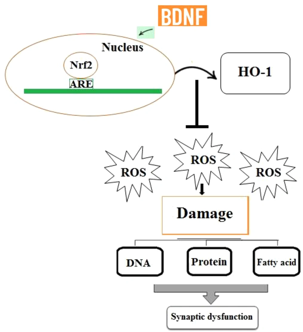
Reactive oxygen species (ROS) encompass the initial species formed by oxygen reduction (i.e., hydrogen peroxide (H2O2) or superoxide (O2-), as well as their secondary reactive products. These radicals are commonly generated in cells during metabolism, and the disruption of the balance between the formation of these radicals and the antioxidant capacity of cells can lead to oxidative stress. The increased production of ROS can pose a threat to cells by oxidizing proteins, inducing fatty acid peroxidation, and causing damage to nucleic acids. The brain is especially vulnerable to oxidative stress since it uses a large amount of oxygen and has a plentiful supply of lipids while it has a low antioxidant capacity (88, 89). Therefore, the accumulation of free radicals is very dangerous to the brain and can lead to memory impairment, synaptic dysfunction, neuronal death, and cognitive decay, and this is while antioxidants can abolish these effects (87). One of the most important cellular antioxidant defense mechanisms is the activation of the nuclear factor erythroid 2-related factor 2 (Nrf2) signaling pathway that regulates the expression of the genes whose protein products are involved in the detoxification and removal of reactive oxidants via conjugative reactions and increasing cells’ antioxidant capacity. Furthermore, Nrf2 is itself an important transcription factor regulating the expression of endogenous antioxidants, including heme oxygenase-1 (HO-1) (90). Indeed, Nrf2 deletion suppresses neurogenesis and leads to LTP impairment (91). Together, these findings reflect that the Nrf2 signaling pathway contributes to brain health and synaptic plasticity preservation in the hippocampus. According to the results of a previous study, there is a potential relationship between BDNF and Nrf2 activity. It has been reported that BDNF, as a homeostatic regulator of Nrf2 activity, induces its translocation into the nucleus in basic conditions, persistently activating antioxidant mechanisms (92) (Figure 4). In line with these findings, Bouvier et al. reported that low BDNF levels inhibited the activation of Nrf2-dependent endogenous antioxidant defense mechanisms, finally leading to persistent oxidative stress (92). Therefore, BDNF can enhance the antioxidant capacity of cells through inducing the translocation of Nrf2 into the nucleus. Overall, synaptic dysfunction and LTP impairment are associated with the accumulation of oxidative damage to macromolecules, and these negative effects can be reversed by the BDNF/Nrf2 pathway. In conclusion, BDNF shares a critical role in promoting Nrf2 nuclear translocation to preserve synaptic responses and LTP induction.

Increased production of ROS can cause damage to biomolecules including nucleic acids, fatty acids, and proteins. Therefore, ROS accumulation may be involved in synaptic dysfunction and LTP impairment. BDNF plays a critical role in promoting the nuclear translocation of Nrf2, which contributes to the expression of antioxidant genes and ROS removal.

4. Conclusions

As a member of the family of neurotrophins, BDNF has a fundamental role in the survival and differentiation of neurons during development and is involved in the plastic changes related to synaptic plasticity, as well as in learning and memory formation. This review aimed to discuss the effects of BDNF on LTP induction and also to describe the neuronal pathways involved in these effects of BDNF in the CNS. As one of the most important neurotrophins, BDNF induces complex neuronal signaling pathways that are essential for the cellular modifications underlying synaptic plasticity and LTP induction. Indeed, BDNF regulates a variety of functions in the CNS, such as dendritic and axonal growth, neuronal survival, synaptic signal transmission, activation of presynaptic and postsynaptic proteins, and LTP induction in the hippocampus. The brain is highly vulnerable to oxidative stress due to its high oxygen turnover that results in the formation of oxygen-related free radicals. In addition, the brain contains a high level of polyunsaturated fatty acids and a low antioxidant capacity compared to other organs and tissues. On the other hand, the Nrf2 transcriptional signaling pathway plays an essential role in regulating the genes encoding the proteins sharing critical roles in the detoxification and removal of free radicals. As described in this review, BDNF induces Nrf2 translocation into the nucleus in basic conditions. Thus, BDNF seems to promote its effects on neuroplasticity and LTP induction at least partly through activating antioxidant mechanisms and reducing free radical production. However, further research is needed to understand the interactions of BDNF-derived signaling with other mediators of synaptic
References

  • 1.

    Lynch MA. Long-term potentiation and memory. Physiol Rev. 2004;84(1):87-136. [PubMed ID: 14715912]. https://doi.org/10.1152/physrev.00014.2003.

  • 2.

    Konorski J. Conditioned reflexes and neuron organization. Cambridge, England: Cambridge University Press; 1948.

  • 3.

    Malenka RC, Bear MF. LTP and LTD: An embarrassment of riches. Neuron. 2004;44(1):5-21. [PubMed ID: 15450156]. https://doi.org/10.1016/j.neuron.2004.09.012.

  • 4.

    Kumar A. Long-term potentiation at CA3-CA1 hippocampal synapses with special emphasis on aging, disease, and stress. Front Aging Neurosci. 2011;3:7. [PubMed ID: 21647396]. [PubMed Central ID: PMC3102214]. https://doi.org/10.3389/fnagi.2011.00007.

  • 5.

    Sweatt JD. Toward a molecular explanation for long-term potentiation. Learn Mem. 1999;6(5):399-416. [PubMed ID: 10541462]. https://doi.org/10.1101/lm.6.5.399.

  • 6.

    Pillai A, Buckley PF. Reliable biomarkers and predictors of schizophrenia and its treatment. Psychiatr Clin North Am. 2012;35(3):645-59. [PubMed ID: 22929871]. https://doi.org/10.1016/j.psc.2012.06.006.

  • 7.

    Nieto R, Kukuljan M, Silva H. BDNF and schizophrenia: From neurodevelopment to neuronal plasticity, learning, and memory. Front Psychiatry. 2013;4:45. [PubMed ID: 23785335]. [PubMed Central ID: PMC3683823]. https://doi.org/10.3389/fpsyt.2013.00045.

  • 8.

    Park H, Poo MM. Neurotrophin regulation of neural circuit development and function. Nat Rev Neurosci. 2013;14(1):7-23. [PubMed ID: 23254191]. https://doi.org/10.1038/nrn3379.

  • 9.

    Angoa-Perez M, H. Anneken J, M. Kuhn D. The role of brain-derived neurotrophic factor in the pathophysiology of psychiatric and neurological disorders. Journal of Psychiatry and Psychiatric Disorders. 2017;1(5):252-69. https://doi.org/10.26502/jppd.2572-519X0025.

  • 10.

    Yang Y, Liu Y, Wang G, Hei G, Wang X, Li R, et al. Brain-derived neurotrophic factor is associated with cognitive impairments in first-episode and chronic schizophrenia. Psychiatry Res. 2019;273:528-36. [PubMed ID: 30710808]. https://doi.org/10.1016/j.psychres.2019.01.051.

  • 11.

    Fukuchi M, Okuno Y, Nakayama H, Nakano A, Mori H, Mitazaki S, et al. Screening inducers of neuronal BDNF gene transcription using primary cortical cell cultures from BDNF-luciferase transgenic mice. Sci Rep. 2019;9(1):11833. [PubMed ID: 31413298]. [PubMed Central ID: PMC6694194]. https://doi.org/10.1038/s41598-019-48361-4.

  • 12.

    Dwivedi Y, Rizavi HS, Conley RR, Roberts RC, Tamminga CA, Pandey GN. Altered gene expression of brain-derived neurotrophic factor and receptor tyrosine kinase B in postmortem brain of suicide subjects. Arch Gen Psychiatry. 2003;60(8):804-15. [PubMed ID: 12912764]. https://doi.org/10.1001/archpsyc.60.8.804.

  • 13.

    Weickert CS, Hyde TM, Lipska BK, Herman MM, Weinberger DR, Kleinman JE. Reduced brain-derived neurotrophic factor in prefrontal cortex of patients with schizophrenia. Mol Psychiatry. 2003;8(6):592-610. [PubMed ID: 12851636]. https://doi.org/10.1038/sj.mp.4001308.

  • 14.

    Zuccato C, Ciammola A, Rigamonti D, Leavitt BR, Goffredo D, Conti L, et al. Loss of huntingtin-mediated BDNF gene transcription in Huntington's disease. Science. 2001;293(5529):493-8. [PubMed ID: 11408619]. https://doi.org/10.1126/science.1059581.

  • 15.

    Lessmann V, Brigadski T. Mechanisms, locations, and kinetics of synaptic BDNF secretion: An update. Neurosci Res. 2009;65(1):11-22. [PubMed ID: 19523993]. https://doi.org/10.1016/j.neures.2009.06.004.

  • 16.

    Lau AG, Irier HA, Gu J, Tian D, Ku L, Liu G, et al. Distinct 3'UTRs differentially regulate activity-dependent translation of brain-derived neurotrophic factor (BDNF). Proc Natl Acad Sci U S A. 2010;107(36):15945-50. [PubMed ID: 20733072]. [PubMed Central ID: PMC2936648]. https://doi.org/10.1073/pnas.1002929107.

  • 17.

    Tao X, West AE, Chen WG, Corfas G, Greenberg ME. A calcium-responsive transcription factor, CaRF, that regulates neuronal activity-dependent expression of BDNF. Neuron. 2002;33(3):383-95. [PubMed ID: 11832226]. https://doi.org/10.1016/s0896-6273(01)00561-x.

  • 18.

    Matsumoto T, Rauskolb S, Polack M, Klose J, Kolbeck R, Korte M, et al. Biosynthesis and processing of endogenous BDNF: CNS neurons store and secrete BDNF, not pro-BDNF. Nat Neurosci. 2008;11(2):131-3. [PubMed ID: 18204444]. https://doi.org/10.1038/nn2038.

  • 19.

    Yang J, Siao CJ, Nagappan G, Marinic T, Jing D, McGrath K, et al. Neuronal release of proBDNF. Nat Neurosci. 2009;12(2):113-5. [PubMed ID: 19136973]. [PubMed Central ID: PMC2737352]. https://doi.org/10.1038/nn.2244.

  • 20.

    Figurov A, Pozzo-Miller LD, Olafsson P, Wang T, Lu B. Regulation of synaptic responses to high-frequency stimulation and LTP by neurotrophins in the hippocampus. Nature. 1996;381(6584):706-9. [PubMed ID: 8649517]. https://doi.org/10.1038/381706a0.

  • 21.

    Lu Y, Christian K, Lu B. BDNF: a key regulator for protein synthesis-dependent LTP and long-term memory? Neurobiol Learn Mem. 2008;89(3):312-23. [PubMed ID: 17942328]. [PubMed Central ID: PMC2387254]. https://doi.org/10.1016/j.nlm.2007.08.018.

  • 22.

    Bramham CR, Messaoudi E. BDNF function in adult synaptic plasticity: The synaptic consolidation hypothesis. Prog Neurobiol. 2005;76(2):99-125. [PubMed ID: 16099088]. https://doi.org/10.1016/j.pneurobio.2005.06.003.

  • 23.

    De Vincenti AP, Rios AS, Paratcha G, Ledda F. Mechanisms that modulate and diversify BDNF functions: Implications for hippocampal synaptic plasticity. Front Cell Neurosci. 2019;13:135. [PubMed ID: 31024262]. [PubMed Central ID: PMC6465932]. https://doi.org/10.3389/fncel.2019.00135.

  • 24.

    Messaoudi E, Bardsen K, Srebro B, Bramham CR. Acute intrahippocampal infusion of BDNF induces lasting potentiation of synaptic transmission in the rat dentate gyrus. J Neurophysiol. 1998;79(1):496-9. [PubMed ID: 9425220]. https://doi.org/10.1152/jn.1998.79.1.496.

  • 25.

    Korte M, Carroll P, Wolf E, Brem G, Thoenen H, Bonhoeffer T. Hippocampal long-term potentiation is impaired in mice lacking brain-derived neurotrophic factor. Proc Natl Acad Sci U S A. 1995;92(19):8856-60. [PubMed ID: 7568031]. [PubMed Central ID: PMC41066]. https://doi.org/10.1073/pnas.92.19.8856.

  • 26.

    Patterson SL, Abel T, Deuel TA, Martin KC, Rose JC, Kandel ER. Recombinant BDNF rescues deficits in basal synaptic transmission and hippocampal LTP in BDNF knockout mice. Neuron. 1996;16(6):1137-45. [PubMed ID: 8663990]. https://doi.org/10.1016/s0896-6273(00)80140-3.

  • 27.

    Pozzo-Miller LD, Gottschalk W, Zhang L, McDermott K, Du J, Gopalakrishnan R, et al. Impairments in high-frequency transmission, synaptic vesicle docking, and synaptic protein distribution in the hippocampus of BDNF knockout mice. J Neurosci. 1999;19(12):4972-83. [PubMed ID: 10366630]. [PubMed Central ID: PMC6782660].

  • 28.

    Lessmann V, Gottmann K, Malcangio M. Neurotrophin secretion: Current facts and future prospects. Prog Neurobiol. 2003;69(5):341-74. [PubMed ID: 12787574]. https://doi.org/10.1016/s0301-0082(03)00019-4.

  • 29.

    Roux PP, Barker PA. Neurotrophin signaling through the p75 neurotrophin receptor. Prog Neurobiol. 2002;67(3):203-33. [PubMed ID: 12169297]. https://doi.org/10.1016/s0301-0082(02)00016-3.

  • 30.

    Meeker RB, Williams KS. The p75 neurotrophin receptor: At the crossroad of neural repair and death. Neural Regen Res. 2015;10(5):721-5. [PubMed ID: 26109945]. [PubMed Central ID: PMC4468762]. https://doi.org/10.4103/1673-5374.156967.

  • 31.

    Deinhardt K, Chao MV. Trk receptors. Handb Exp Pharmacol. 2014;220:103-19. [PubMed ID: 24668471]. https://doi.org/10.1007/978-3-642-45106-5_5.

  • 32.

    Woo NH, Teng HK, Siao CJ, Chiaruttini C, Pang PT, Milner TA, et al. Activation of p75NTR by proBDNF facilitates hippocampal long-term depression. Nat Neurosci. 2005;8(8):1069-77. [PubMed ID: 16025106]. https://doi.org/10.1038/nn1510.

  • 33.

    Anastasia A, Deinhardt K, Chao MV, Will NE, Irmady K, Lee FS, et al. Val66Met polymorphism of BDNF alters prodomain structure to induce neuronal growth cone retraction. Nat Commun. 2013;4:2490. [PubMed ID: 24048383]. [PubMed Central ID: PMC3820160]. https://doi.org/10.1038/ncomms3490.

  • 34.

    Reichardt LF. Neurotrophin-regulated signalling pathways. Philos Trans R Soc Lond B Biol Sci. 2006;361(1473):1545-64. [PubMed ID: 16939974]. [PubMed Central ID: PMC1664664]. https://doi.org/10.1098/rstb.2006.1894.

  • 35.

    Autry AE, Monteggia LM. Brain-derived neurotrophic factor and neuropsychiatric disorders. Pharmacol Rev. 2012;64(2):238-58. [PubMed ID: 22407616]. [PubMed Central ID: PMC3310485]. https://doi.org/10.1124/pr.111.005108.

  • 36.

    Levine ES, Crozier RA, Black IB, Plummer MR. Brain-derived neurotrophic factor modulates hippocampal synaptic transmission by increasing N-methyl-D-aspartic acid receptor activity. Proc Natl Acad Sci U S A. 1998;95(17):10235-9. [PubMed ID: 9707630]. [PubMed Central ID: PMC21491]. https://doi.org/10.1073/pnas.95.17.10235.

  • 37.

    Hermida MA, Dinesh Kumar J, Leslie NR. GSK3 and its interactions with the PI3K/AKT/mTOR signalling network. Adv Biol Regul. 2017;65:5-15. [PubMed ID: 28712664]. https://doi.org/10.1016/j.jbior.2017.06.003.

  • 38.

    Leroy K, Brion JP. Developmental expression and localization of glycogen synthase kinase-3beta in rat brain. J Chem Neuroanat. 1999;16(4):279-93. [PubMed ID: 10450875]. https://doi.org/10.1016/s0891-0618(99)00012-5.

  • 39.

    Bradley CA, Peineau S, Taghibiglou C, Nicolas CS, Whitcomb DJ, Bortolotto ZA, et al. A pivotal role of GSK-3 in synaptic plasticity. Front Mol Neurosci. 2012;5:13. [PubMed ID: 22363262]. [PubMed Central ID: PMC3279748]. https://doi.org/10.3389/fnmol.2012.00013.

  • 40.

    Peineau S, Taghibiglou C, Bradley C, Wong TP, Liu L, Lu J, et al. LTP inhibits LTD in the hippocampus via regulation of GSK3beta. Neuron. 2007;53(5):703-17. [PubMed ID: 17329210]. https://doi.org/10.1016/j.neuron.2007.01.029.

  • 41.

    Jiang H, Guo W, Liang X, Rao Y. Both the establishment and the maintenance of neuronal polarity require active mechanisms: Critical roles of GSK-3beta and its upstream regulators. Cell. 2005;120(1):123-35. [PubMed ID: 15652487]. https://doi.org/10.1016/j.cell.2004.12.033.

  • 42.

    Eickholt BJ, Walsh FS, Doherty P. An inactive pool of GSK-3 at the leading edge of growth cones is implicated in Semaphorin 3A signaling. J Cell Biol. 2002;157(2):211-7. [PubMed ID: 11956225]. [PubMed Central ID: PMC2199247]. https://doi.org/10.1083/jcb.200201098.

  • 43.

    Peineau S, Bradley C, Taghibiglou C, Doherty A, Bortolotto ZA, Wang YT, et al. The role of GSK-3 in synaptic plasticity. Br J Pharmacol. 2008;153 Suppl 1:S428-37. [PubMed ID: 18311157]. [PubMed Central ID: PMC2268071]. https://doi.org/10.1038/bjp.2008.2.

  • 44.

    Smillie KJ, Pawson J, Perkins EM, Jackson M, Cousin MA. Control of synaptic vesicle endocytosis by an extracellular signalling molecule. Nat Commun. 2013;4:2394. [PubMed ID: 23999152]. [PubMed Central ID: PMC3778765]. https://doi.org/10.1038/ncomms3394.

  • 45.

    Poirazi P, Mel BW. Impact of active dendrites and structural plasticity on the memory capacity of neural tissue. Neuron. 2001;29(3):779-96. [PubMed ID: 11301036]. https://doi.org/10.1016/s0896-6273(01)00252-5.

  • 46.

    Ji Y, Lu Y, Yang F, Shen W, Tang TT, Feng L, et al. Acute and gradual increases in BDNF concentration elicit distinct signaling and functions in neurons. Nat Neurosci. 2010;13(3):302-9. [PubMed ID: 20173744]. [PubMed Central ID: PMC4780419]. https://doi.org/10.1038/nn.2505.

  • 47.

    Lohof AM, Ip NY, Poo MM. Potentiation of developing neuromuscular synapses by the neurotrophins NT-3 and BDNF. Nature. 1993;363(6427):350-3. [PubMed ID: 8497318]. https://doi.org/10.1038/363350a0.

  • 48.

    Wang T, Xie K, Lu B. Neurotrophins promote maturation of developing neuromuscular synapses. J Neurosci. 1995;15(7 Pt 1):4796-805. [PubMed ID: 7623111]. [PubMed Central ID: PMC6577890].

  • 49.

    Kang H, Schuman EM. Long-lasting neurotrophin-induced enhancement of synaptic transmission in the adult hippocampus. Science. 1995;267(5204):1658-62. [PubMed ID: 7886457]. https://doi.org/10.1126/science.7886457.

  • 50.

    Tanaka T, Saito H, Matsuki N. Inhibition of GABAA synaptic responses by brain-derived neurotrophic factor (BDNF) in rat hippocampus. J Neurosci. 1997;17(9):2959-66. [PubMed ID: 9096132]. [PubMed Central ID: PMC6573653].

  • 51.

    Kang H, Jia LZ, Suh KY, Tang L, Schuman EM. Determinants of BDNF-induced hippocampal synaptic plasticity: Role of the Trk B receptor and the kinetics of neurotrophin delivery. Learn Mem. 1996;3(2-3):188-96. [PubMed ID: 10456089]. https://doi.org/10.1101/lm.3.2-3.188.

  • 52.

    Janowsky JS, Oviatt SK, Orwoll ES. Testosterone influences spatial cognition in older men. Behav Neurosci. 1994;108(2):325-32. [PubMed ID: 8037876]. https://doi.org/10.1037//0735-7044.108.2.325.

  • 53.

    Babanejad S, Naghdi N, Haeri Rohani SA. Microinjection of dihydrotestosterone as a 5alpha-reduced metabolite of testosterone into CA1 region of hippocampus could improve spatial learning in the adult male rats. Iran J Pharm Res. 2012;11(2):661-9. [PubMed ID: 24250491]. [PubMed Central ID: PMC3832172].

  • 54.

    Levine ES, Kolb JE. Brain-derived neurotrophic factor increases activity of NR2B-containing N-methyl-D-aspartate receptors in excised patches from hippocampal neurons. J Neurosci Res. 2000;62(3):357-62. [PubMed ID: 11054804]. https://doi.org/10.1002/1097-4547(20001101)62:3<357::AID-JNR5>3.0.CO;2-6.

  • 55.

    Jovanovic JN, Czernik AJ, Fienberg AA, Greengard P, Sihra TS. Synapsins as mediators of BDNF-enhanced neurotransmitter release. Nat Neurosci. 2000;3(4):323-9. [PubMed ID: 10725920]. https://doi.org/10.1038/73888.

  • 56.

    Calabresi P, Centonze D, Gubellini P, Marfia GA, Pisani A, Sancesario G, et al. Synaptic transmission in the striatum: From plasticity to neurodegeneration. Prog Neurobiol. 2000;61(3):231-65. [PubMed ID: 10727775]. https://doi.org/10.1016/s0301-0082(99)00030-1.

  • 57.

    Rosen LB, Ginty DD, Weber MJ, Greenberg ME. Membrane depolarization and calcium influx stimulate MEK and MAP kinase via activation of Ras. Neuron. 1994;12(6):1207-21. [PubMed ID: 8011335]. https://doi.org/10.1016/0896-6273(94)90438-3.

  • 58.

    Lin SY, Wu K, Levine ES, Mount HT, Suen PC, Black IB. BDNF acutely increases tyrosine phosphorylation of the NMDA receptor subunit 2B in cortical and hippocampal postsynaptic densities. Brain Res Mol Brain Res. 1998;55(1):20-7. [PubMed ID: 9645956]. https://doi.org/10.1016/s0169-328x(97)00349-5.

  • 59.

    Alder J, Thakker-Varia S, Crozier RA, Shaheen A, Plummer MR, Black IB. Early presynaptic and late postsynaptic components contribute independently to brain-derived neurotrophic factor-induced synaptic plasticity. J Neurosci. 2005;25(12):3080-5. [PubMed ID: 15788764]. [PubMed Central ID: PMC6725079]. https://doi.org/10.1523/JNEUROSCI.2970-04.2005.

  • 60.

    Sakai N, Yamada M, Numakawa T, Ogura A, Hatanaka H. BDNF potentiates spontaneous Ca2+ oscillations in cultured hippocampal neurons. Brain Res. 1997;778(2):318-28. [PubMed ID: 9459549]. https://doi.org/10.1016/s0006-8993(97)01052-4.

  • 61.

    Narisawa-Saito M, Iwakura Y, Kawamura M, Araki K, Kozaki S, Takei N, et al. Brain-derived neurotrophic factor regulates surface expression of alpha-amino-3-hydroxy-5-methyl-4-isoxazoleproprionic acid receptors by enhancing the N-ethylmaleimide-sensitive factor/GluR2 interaction in developing neocortical neurons. J Biol Chem. 2002;277(43):40901-10. [PubMed ID: 12130635]. https://doi.org/10.1074/jbc.M202158200.

  • 62.

    Li W, Keifer J. BDNF-induced synaptic delivery of AMPAR subunits is differentially dependent on NMDA receptors and requires ERK. Neurobiol Learn Mem. 2009;91(3):243-9. [PubMed ID: 18977306]. [PubMed Central ID: PMC2649981]. https://doi.org/10.1016/j.nlm.2008.10.002.

  • 63.

    Tronci E, Napolitano F, Munoz A, Fidalgo C, Rossi F, Bjorklund A, et al. BDNF over-expression induces striatal serotonin fiber sprouting and increases the susceptibility to l-DOPA-induced dyskinesia in 6-OHDA-lesioned rats. Exp Neurol. 2017;297:73-81. [PubMed ID: 28757258]. https://doi.org/10.1016/j.expneurol.2017.07.017.

  • 64.

    Palasz E, Wysocka A, Gasiorowska A, Chalimoniuk M, Niewiadomski W, Niewiadomska G. BDNF as a promising therapeutic agent in Parkinson's disease. Int J Mol Sci. 2020;21(3). [PubMed ID: 32050617]. [PubMed Central ID: PMC7037114]. https://doi.org/10.3390/ijms21031170.

  • 65.

    Andreae LC, Burrone J. The role of spontaneous neurotransmission in synapse and circuit development. J Neurosci Res. 2018;96(3):354-9. [PubMed ID: 29034487]. [PubMed Central ID: PMC5813191]. https://doi.org/10.1002/jnr.24154.

  • 66.

    Tyler WJ, Pozzo-Miller LD. BDNF enhances quantal neurotransmitter release and increases the number of docked vesicles at the active zones of hippocampal excitatory synapses. J Neurosci. 2001;21(12):4249-58. [PubMed ID: 11404410]. [PubMed Central ID: PMC2806848].

  • 67.

    Carter AR, Chen C, Schwartz PM, Segal RA. Brain-derived neurotrophic factor modulates cerebellar plasticity and synaptic ultrastructure. J Neurosci. 2002;22(4):1316-27. [PubMed ID: 11850459]. [PubMed Central ID: PMC6757568].

  • 68.

    Ehrlich I, Klein M, Rumpel S, Malinow R. PSD-95 is required for activity-driven synapse stabilization. Proc Natl Acad Sci U S A. 2007;104(10):4176-81. [PubMed ID: 17360496]. [PubMed Central ID: PMC1820728]. https://doi.org/10.1073/pnas.0609307104.

  • 69.

    Opazo P, Sainlos M, Choquet D. Regulation of AMPA receptor surface diffusion by PSD-95 slots. Curr Opin Neurobiol. 2012;22(3):453-60. [PubMed ID: 22051694]. https://doi.org/10.1016/j.conb.2011.10.010.

  • 70.

    Hata Y, Takai Y. Roles of postsynaptic density-95/synapse-associated protein 90 and its interacting proteins in the organization of synapses. Cell Mol Life Sci. 1999;56(5-6):461-72. [PubMed ID: 11212298]. https://doi.org/10.1007/s000180050445.

  • 71.

    Yoshii A, Constantine-Paton M. BDNF induces transport of PSD-95 to dendrites through PI3K-AKT signaling after NMDA receptor activation. Nat Neurosci. 2007;10(6):702-11. [PubMed ID: 17515902]. https://doi.org/10.1038/nn1903.

  • 72.

    Yoshii A, Murata Y, Kim J, Zhang C, Shokat KM, Constantine-Paton M. TrkB and protein kinase Mzeta regulate synaptic localization of PSD-95 in developing cortex. J Neurosci. 2011;31(33):11894-904. [PubMed ID: 21849550]. [PubMed Central ID: PMC3158490]. https://doi.org/10.1523/JNEUROSCI.2190-11.2011.

  • 73.

    Alonso G, Phan V, Guillemain I, Saunier M, Legrand A, Anoal M, et al. Immunocytochemical localization of the sigma(1) receptor in the adult rat central nervous system. Neuroscience. 2000;97(1):155-70. [PubMed ID: 10771347]. https://doi.org/10.1016/s0306-4522(00)00014-2.

  • 74.

    Kim HW, Roh DH, Yoon SY, Seo HS, Kwon YB, Han HJ, et al. Activation of the spinal sigma-1 receptor enhances NMDA-induced pain via PKC- and PKA-dependent phosphorylation of the NR1 subunit in mice. Br J Pharmacol. 2008;154(5):1125-34. [PubMed ID: 18493253]. [PubMed Central ID: PMC2465573]. https://doi.org/10.1038/bjp.2008.159.

  • 75.

    Maurice T, Su TP. The pharmacology of sigma-1 receptors. Pharmacol Ther. 2009;124(2):195-206. [PubMed ID: 19619582]. [PubMed Central ID: PMC2785038]. https://doi.org/10.1016/j.pharmthera.2009.07.001.

  • 76.

    Zhang H, Cuevas J. Sigma receptors inhibit high-voltage-activated calcium channels in rat sympathetic and parasympathetic neurons. J Neurophysiol. 2002;87(6):2867-79. [PubMed ID: 12037190]. https://doi.org/10.1152/jn.2002.87.6.2867.

  • 77.

    Martina M, Turcotte ME, Halman S, Bergeron R. The sigma-1 receptor modulates NMDA receptor synaptic transmission and plasticity via SK channels in rat hippocampus. J Physiol. 2007;578(Pt 1):143-57. [PubMed ID: 17068104]. [PubMed Central ID: PMC2075134]. https://doi.org/10.1113/jphysiol.2006.116178.

  • 78.

    Sabeti J, Nelson TE, Purdy RH, Gruol DL. Steroid pregnenolone sulfate enhances NMDA-receptor-independent long-term potentiation at hippocampal CA1 synapses: role for L-type calcium channels and sigma-receptors. Hippocampus. 2007;17(5):349-69. [PubMed ID: 17330865]. https://doi.org/10.1002/hipo.20273.

  • 79.

    Sabeti J, Gruol DL. Emergence of NMDAR-independent long-term potentiation at hippocampal CA1 synapses following early adolescent exposure to chronic intermittent ethanol: Role for sigma-receptors. Hippocampus. 2008;18(2):148-68. [PubMed ID: 17960647]. https://doi.org/10.1002/hipo.20379.

  • 80.

    Peviani M, Salvaneschi E, Bontempi L, Petese A, Manzo A, Rossi D, et al. Neuroprotective effects of the sigma-1 receptor (S1R) agonist PRE-084, in a mouse model of motor neuron disease not linked to SOD1 mutation. Neurobiol Dis. 2014;62:218-32. [PubMed ID: 24141020]. https://doi.org/10.1016/j.nbd.2013.10.010.

  • 81.

    Ring RM, Regan CM. Captodiamine, a putative antidepressant, enhances hypothalamic BDNF expression in vivo by synergistic 5-HT2c receptor antagonism and sigma-1 receptor agonism. J Psychopharmacol. 2013;27(10):930-9. [PubMed ID: 23863923]. https://doi.org/10.1177/0269881113497614.

  • 82.

    Malik M, Rangel-Barajas C, Sumien N, Su C, Singh M, Chen Z, et al. The effects of sigma (sigma1) receptor-selective ligands on muscarinic receptor antagonist-induced cognitive deficits in mice. Br J Pharmacol. 2015;172(10):2519-31. [PubMed ID: 25573298]. [PubMed Central ID: PMC4409904]. https://doi.org/10.1111/bph.13076.

  • 83.

    Lever JR, Gustafson JL, Xu R, Allmon RL, Lever SZ. Sigma1 and sigma2 receptor binding affinity and selectivity of SA4503 and fluoroethyl SA4503. Synapse. 2006;59(6):350-8. [PubMed ID: 16463398]. https://doi.org/10.1002/syn.20253.

  • 84.

    Fujimoto M, Hayashi T, Urfer R, Mita S, Su TP. Sigma-1 receptor chaperones regulate the secretion of brain-derived neurotrophic factor. Synapse. 2012;66(7):630-9. [PubMed ID: 22337473]. [PubMed Central ID: PMC3824965]. https://doi.org/10.1002/syn.21549.

  • 85.

    MacLusky NJ, Hajszan T, Prange-Kiel J, Leranth C. Androgen modulation of hippocampal synaptic plasticity. Neuroscience. 2006;138(3):957-65. [PubMed Central ID: PMC16488544]. https://doi.org/10.1016/j.neuroscience.2005.12.054.

  • 86.

    Salimi Z, Khajehpour L, Moradpour F, Moazedi AA, Pourmotabbed A, Zarei F. Nandrolone improve synaptic plasticity at the hippocampus CA1 area and spatial localization in the Morris water maze of male adolescent rats. Neurosci Res. 2020;158:21-9. [PubMed ID: 31499080]. https://doi.org/10.1016/j.neures.2019.09.001.

  • 87.

    Salimi Z, Moradpour F, Zarei F, Rashidi Z, Khazaei MR, Ahmadi SM. The effect of blockade of androgen receptors by flutamide on learning and memory, synaptic plasticity and behavioral performances: A review study. Cell Tissue Biol. 2021;15:337-46. https://doi.org/10.1134/S1990519X21040088.

  • 88.

    Riley PA. Free radicals in biology: Oxidative stress and the effects of ionizing radiation. Int J Radiat Biol. 1994;65(1):27-33. [PubMed ID: 7905906]. https://doi.org/10.1080/09553009414550041.

  • 89.

    Maurya PK, Noto C, Rizzo LB, Rios AC, Nunes SO, Barbosa DS, et al. The role of oxidative and nitrosative stress in accelerated aging and major depressive disorder. Prog Neuropsychopharmacol Biol Psychiatry. 2016;65:134-44. [PubMed ID: 26348786]. https://doi.org/10.1016/j.pnpbp.2015.08.016.

  • 90.

    Gergues MM, Moiseyenko A, Saad SZ, Kong AN, Wagner GC. Nrf2 deletion results in impaired performance in memory tasks and hyperactivity in mature and aged mice. Brain Res. 2018;1701:103-11. [PubMed ID: 30194014]. [PubMed Central ID: PMC8111504]. https://doi.org/10.1016/j.brainres.2018.08.033.

  • 91.

    Robledinos-Anton N, Rojo AI, Ferreiro E, Nunez A, Krause KH, Jaquet V, et al. Transcription factor NRF2 controls the fate of neural stem cells in the subgranular zone of the hippocampus. Redox Biol. 2017;13:393-401. [PubMed ID: 28667908]. [PubMed Central ID: PMC5493838]. https://doi.org/10.1016/j.redox.2017.06.010.

  • 92.

    Bouvier E, Brouillard F, Molet J, Claverie D, Cabungcal JH, Cresto N, et al. Nrf2-dependent persistent oxidative stress results in stress-induced vulnerability to depression. Mol Psychiatry. 2017;22(12):1701-13. [PubMed ID: 27646262]. https://doi.org/10.1038/mp.2016.144.