
1. Introduction
Hepatic multifocal nodular fatty infiltration (HMNFI) is an uncommon benign entity that can mimic malignant hepatic lesions on imaging (1, 2). It typically presents as multiple hypodense nodules on unenhanced computed tomography (CT) without enhancement (1, 6). Magnetic resonance imaging (MRI) demonstrates characteristic signal dropout on fat-suppressed and opposed-phase sequences, confirming intracellular fat deposition (2, 6).
Patients who undergo hepatocellular carcinoma (HCC) resection often have metabolic disturbances. Prolonged parenteral nutrition (PN) is a well-recognized risk factor for hepatic steatosis, particularly when lipid supply is excessive (3-5). This case report describes PN-induced HMNFI after HCC resection and highlights imaging features that may help avoid misdiagnosis as recurrence or metastasis (7).
2. Case Presentation
A 50-year-old woman with a 16-year history of chronic hepatitis B was admitted with dull pain in the right upper quadrant. Her body mass index was 23.1 kg/m2. She had no diabetes, dyslipidemia, alcohol intake, or history of hepatotoxic medication use. Liver function was Child-Pugh class A. Imaging confirmed HCC in the left hepatic lobe (4, 8).
She underwent laparoscopic left hepatectomy in April 2024. Postoperatively, she developed gastrointestinal dysfunction and was unable to tolerate oral intake. Therefore, PN was administered from May 11 to May 30, 2024 (20 days). The PN regimen included C6 - 24 mixed lipid emulsion (medium- and long-chain triglycerides), 100 mL/d; lipid-amino acid 17-glucose (11%) injection, 900 mL/d; and compound amino acids, glucose, electrolytes, and vitamins.
The clinical timeline was as follows: laparoscopic left hepatectomy was performed in April 2024; PN was initiated on May 11, 2024; multiple nodules were detected on unenhanced CT on May 15, 2024 (postoperative day 25); MRI confirmed HMNFI on June 4, 2024; PN was discontinued on May 30, 2024; and follow-up MRI on July 15, 2024, showed partial regression of the lesions (Figure 1).
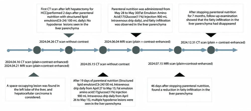
Figure 1.
Patient treatment and imaging follow-up timeline
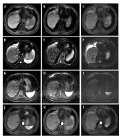
Unenhanced CT showed multiple well-circumscribed hypodense nodules. MRI revealed mild hyperintensity on T1-weighted imaging and T2-weighted imaging, signal loss on fat-suppressed T2-weighted imaging, signal reduction on opposed-phase imaging compared with in-phase imaging, no restricted diffusion, and no enhancement on dynamic contrast-enhanced sequences (Figure 2).
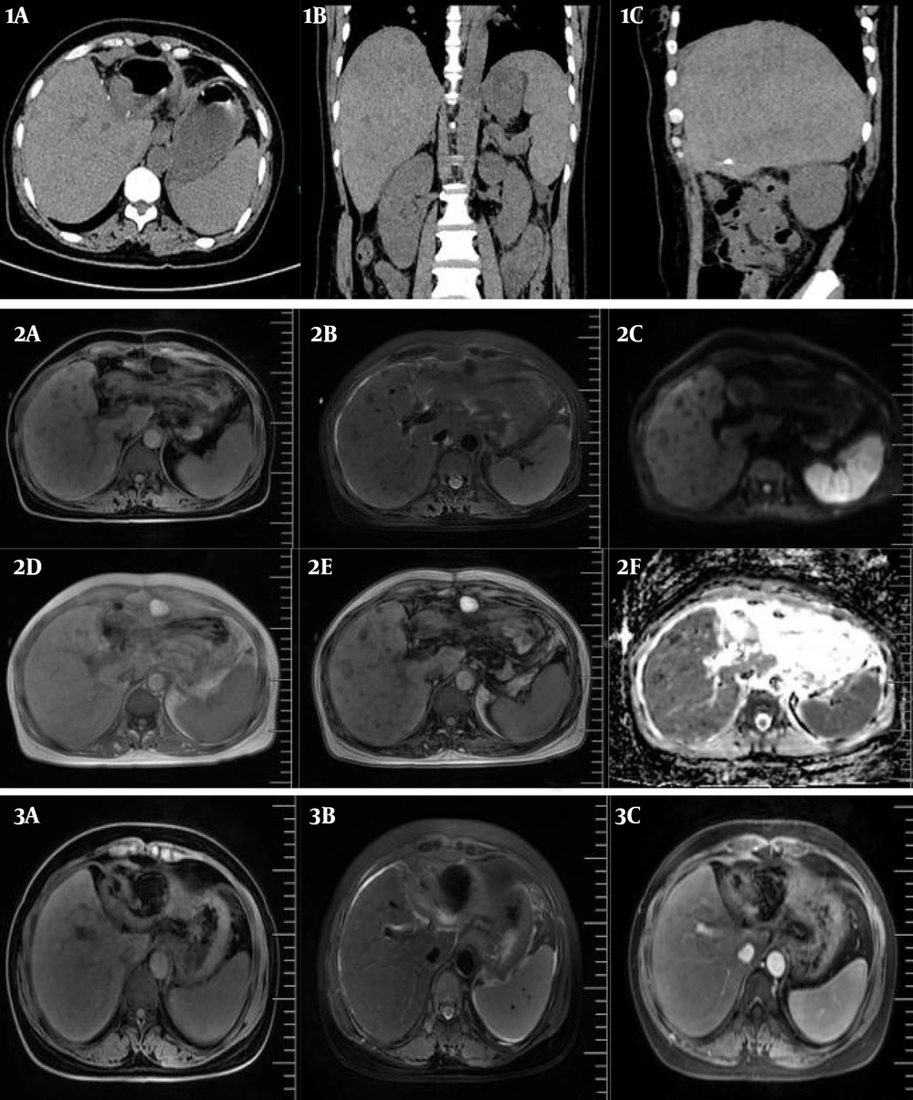
Figure 2.
Imaging findings. (1A-1C) Unenhanced computed tomography shows multiple hypodense nodules; (2A) T1-weighted imaging shows hyperintensity; (2B) T2-weighted imaging shows hyperintensity; (2C) diffusion-weighted imaging shows hypointensity; (2D-2E) opposed-phase imaging shows signal reduction consistent with fat; (2F) apparent diffusion coefficient mapping shows no restricted diffusion; and (3A-3C) follow-up magnetic resonance imaging after parenteral nutrition discontinuation shows partial lesion regression.
These findings were diagnostic of HMNFI (1, 2). Alternative diagnoses, including HCC recurrence, metastasis, hemangioma, cyst, and perfusion disorder, were systematically excluded. Biopsy was not performed because the imaging features were pathognomonic and the lesions regressed after PN withdrawal.
Serial liver function tests are shown in Table 1. Transient elevations in alanine aminotransferase (ALT), aspartate aminotransferase (AST), alkaline phosphatase (ALP), and gamma-glutamyl transferase (GGT) were observed. Total bilirubin, direct bilirubin, and albumin remained stable. After PN cessation, all markers improved, consistent with imaging regression.
Table 1.
Liver Function Parameters During the Treatment Course a
| Date | ALT (U/L) | AST (U/L) | ALP (U/L) | GGT (U/L) | TBIL (μmol/L) | DBIL (μmol/L) | ALB (g/L) |
|---|---|---|---|---|---|---|---|
| April 24, 2024 | 204 | 196 | 76 | 36 | 22.6 | 7.8 | 33.6 |
| April 29, 2024 | 91 | 46 | 65 | 72 | 37.1 | 29.1 | 31.2 |
| April 30, 2024 | 61 | 43 | 53 | 45 | 38.7 | 30.1 | 32 |
| May 3, 2024 | 51 | 51 | 66 | 57 | 33.9 | 19.4 | 30.7 |
| May 7, 2024 | 46 | 49 | 73 | 40 | 60.2 | 36.6 | 39.7 |
| May 9, 2024 | 50 | 54 | 80 | 41 | 78.1 | 41 | 41.3 |
| May 11, 2024 | 55 | 63 | 108 | 54 | 80.1 | 53.1 | 39.6 |
| May 14, 2024 | 106 | 140 | 251 | 277 | 50.5 | 34.3 | 37.9 |
| May 17, 2024 | 74 | 57 | 153 | 180 | 44.7 | 29.8 | 34.4 |
| May 19, 2024 | 51 | 36 | 157 | 136 | 34.2 | 23.1 | 34.5 |
| May 21, 2024 | 53 | 45 | 135 | 134 | 34.0 | 23.5 | 34.5 |
| May 23, 2024 | 54 | 50 | 145 | 131 | 36.5 | 23.3 | 35.4 |
| May 27, 2024 | 72 | 63 | 145 | 126 | 40.3 | 26.2 | 37.0 |
| May 30, 2024 | 108 | 80 | 136 | 191 | 45.8 | 26.0 | 32.4 |
| June 2, 2024 | 107 | 111 | 176 | 250 | 25.2 | 17.4 | 34.5 |
| June 8, 2024 | 176 | 100 | 336 | 434 | 17.2 | 11.5 | 33.1 |
| June 12, 2024 | 82 | 59 | 227 | 266 | 15.1 | 9.5 | 32.8 |
| June 16, 2024 | 53 | 50 | 188 | 213 | 13.6 | 8.0 | 34.5 |
| June 20, 2024 | 43 | 44 | 192 | 191 | 15.2 | 8.2 | 34.2 |
| July 14, 2024 | 31 | 44 | 127 | 160 | 10.7 | 6.5 | 42.4 |
| July 22, 2024 | 39 | 52 | 125 | 143 | 11.7 | 4.7 | 44.7 |
| December 30, 2024 | 45 | 46 | 75 | 79 | 14.7 | 10.8 | 39.3 |
a Abbreviations: ALB, albumin; ALP, alkaline phosphatase; ALT, alanine aminotransferase; AST, aspartate aminotransferase; DBIL, direct bilirubin; GGT, gamma-glutamyl transferase; TBIL, total bilirubin.
2.1. Differential Diagnosis
HCC recurrence was excluded because recurrent HCC typically shows wash-in and washout enhancement, which was not observed in this case (4). Liver metastases were excluded because peripheral enhancement and the bull's-eye sign were absent (4). Hepatic hemangioma was excluded because peripheral nodular enhancement was not present (4). Liver cyst was excluded because the lesions did not show water attenuation and demonstrated fat-signal characteristics (4). Focal nodular hyperplasia was excluded because a central scar and characteristic flow features were absent (2).
3. Discussion
HCC resection disrupts hepatic metabolism, and prolonged PN may trigger fatty infiltration (3). Exogenous lipid overload may exceed the liver’s oxidative capacity, leading to focal fat accumulation (3).
HMNFI exhibits typical MRI features that allow for a confident diagnosis (1, 2). Unlike metabolic dysfunction-associated fatty liver disease, PN-related HMNFI is reversible after lipid withdrawal (3, 4).
We recommend monitoring hepatobiliary function 2 weeks after PN initiation to enable early detection of steatosis (1).
Prolonged PN with high lipid content can induce HMNFI after HCC resection. Its characteristic imaging features allow for reliable differentiation from malignant lesions. Early discontinuation of PN and nutritional adjustment can lead to lesion regression.