Research by other workers has demonstrated that glutamate is a key neurotransmitter in the ischemia induced excitotoxicity process. An excess of glutamate is thought to play an important role in the progress of cerebral infarction via NMDA receptor activation that leads to intracellular calcium overload which causes in turn toxic reaction and ultimately leads to cell damage and death (
29–
31). Although blocking these receptors could help to attenuate neuronal damage, inhibition of other positive effects of NMDA receptors put a limit on usefulness of this treatment. Therefore, other strategies had to be used to treat hypoxic-ischemic brain damage for moderating NMDA receptor function or interaction in the cascade of toxic reactions that led to excitotoxicity.
According to the previous investigations, sigma receptors could act as putative therapeutic targets for reduction of excitotoxic and neurodegenerative damage (
29,
32). Sigma receptors are important because they can greatly affect function of glutamate receptors during excitotoxicity (
33). Ligands of both types of sigma receptor subtypes possibly synergistic influenced by several aspects of neurodegeneration and made it possible to modulator effect on neuronal degeneration process far more than traditional drugs (
29). More recently a greater role has been attributed to the sigma-1 receptor subtype in some pathological and physiological processes (
2). Sigma-1 receptors are located mostly on the endoplasmic reticulum (ER), but when cells are exposed to prolonged stress or their sigma- one agonists, these receptors could translocate to the plasma membrane and modulate different ion channels activities such as voltage-gated and ligand-gated Ca
2+, K
+, Na
+ and Cl
- (
34). Therefore, based on these observations it has been suggested that sigma-1 receptor ligands hold promise as protective agents for the treatment of cerebral ischemia (
34–
36).
Many studies have shown that sigma receptor ligands have a wide variety of actions including protecting of neuronal cells and treatment of cerebral ischemia in several cells such as retinal ganglion cells, cerebral cortex, primary neuronal cultures, and cells used in ischemic stroke models (
29,
33–
35 and
37). For example, it has been revealed that the potent sigma-1 receptor ligand 4-phenyl-1-(4-phenylbutyl) piperidine (PPBP) attenuated neuronal injury of primary cortical neuronal cultures when exposed to 120 min OGD or 100 µM glutamate (
37). Yang
et al. (
35) have demonstrated that therapeutic use of sigma-1 receptor ligand PPBP protected striatal neurons in newborn piglets model of neonatal global hypoxia-ischemia devoid of showing adverse effects associated with completely blocking NMDA receptors in the developing brain. Similarly, PPBP used in an
in-vitro model of hypoxia/hypoglycemia in rat primary neuronal cultures and could decrease neurotoxicity caused by cerebral ischemia (
32). However, the exact mechanism of this effect is unclear.
In previous studies it has been shown that noscapine could induce the protective effects in variety of ischemic conditions. Khanmoradi
et al. (
23) showed that noscapine exerted renoprotective effects via down regulation of the inflammatory mediators such as tumor necrosis factor-α (TNF-α) and monocyte chemoattractant protein1 (MCP-1) in renal ischemia–recovery injury in rat models. Mahmoudian
et al. (
22) demonstrated that noscapine as a bradykinin antagonist reduced inflammation in a rat model of prenatal hypoxic-ischemic brain edema. Vahabzadeh
et al. (
24) showed that noscapine decreased NO levels and modulation intracellular calcium levels in primary murine fetal cortical. Kamei
et al. (
38) showed that the antitussive effect of noscapine was dose dependently reduced by pre-treatment with rimcazole, a specific antagonist of sigma sites in mice. This is appearing that noscapine has a protective role in ischemic conditions. Therefore, it could be postulated that this protective effect afforded by noscapine were to some extent dependent upon its action on sigma-one receptors.
As such, the aim of this research is to investigate the effects of noscapine in the presence of 5 µM BD1047, a sigma-1 receptor antagonist in primary cortical culture on 60 min oxygen-glucose deprivation/24 h recovery and show that if different concentrations of noscapine could protect neuronal cells against sigma-1 antagonist in this condition and which mechanisms were involved.
Lack of oxygen and/or glucose is the main reason for ischemic brain injury (
39–
41). Thus, models have been developed to simulate
, in-vitro conditions of low or no oxygen/glucose to mimic the pathologic process of ischemic stroke (
42,
43).
Goldberg and Choi (
44) subjected primary neuronal cultures to no oxygen/ no glucose conditions followed by normal oxygen and glucose exposure. He provided evidence that this model
in-vitro produced cell damage similar to neurodegeneration that occurs in cerebral ischemic events in man. Therefore, the Goldberg OGD/R model was adopted in the present work.
MTT assay is widely used to measure cell viability and is a good indicator of cellular metabolic activity (
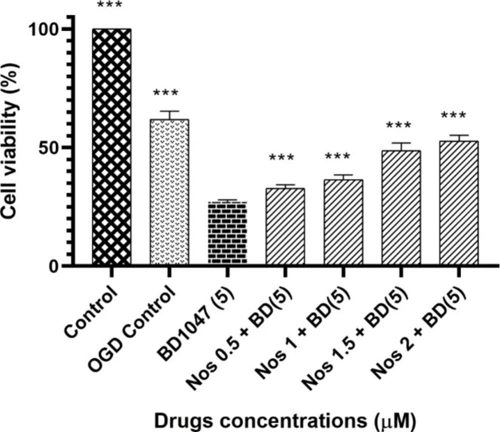
45). In this experiment we used MTT assay to measure the number of living cells and were able to show that 5 µM BD1047 did not have neuroprotective effects on primary cortical culture after 60 min OGD, when used alone. However, when cells incubated in 0.5-2 µM noscapine together with 5 µM BD1047 after 60 min OGD/R, the neuroprotective effects of noscapine were increased in a dose-dependent manner compared to BD1047alone but this effect was not complete.
It can be concluded that the neuroprotective effect of noscapine was at least, partially reversed by pretreating cells with BD1047, a selective sigma-1 antagonist. This suggests that activation of sigma-1 receptors is probably involved in the neuroprotective effects of noscapine against OGD-induced cell injury.
During the period of oxygen and/or glucose deprivation, or in the process of nerve damage, amounts of cellular and neuronal energy is diminished; this change results in increase in glutamate and aspartate concentration. Thus, NMDA receptor stimulation, followed by intracellular Ca
2+ change and Ca
2+-CaM pathway activation ensue. It is known that excessive calcium in neuron and endothelial cells can activate the Ca
2+-dependent enzyme cascades such as neuronal and endothelial isoforms of NOS (nNOS and eNOS). Therefore, increased intracellular NO levels after OGD/R, is expected. (
30,
46 and
47). In the target cells, NO is converted to the cytotoxic molecule peroxynitrite which causes damage to DNA and therefore leads to cell injury or death (
30,
46). In several
in-vitro and
in-vivo animal ischemic models, neurotoxicity of NO has been demonstrated. It is also known that under certain pathological conditions, NO production increases
e.g. during ischemia. This can lead to cell injury and apoptotic cells death by induction of glutamate (
11,
35,
46 and
47).
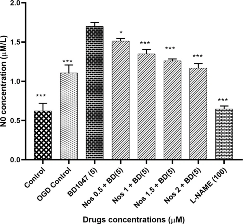
In this work, we were able to show that OGD/R caused an increase in the amount of intracellular NO. Also, our data show that BD1047 enhanced the NO generation due to OGD/R treat. In addition, noscapine was able to partially restore NO levels in the presence of BD1047.
Many studies have demonstrated that in the presence of ischemia, the generation of NO increases (
11,
30,
35,
41 and
48). For example, Goyagi
et al. (
11) showed that PPBP, the potent sigma-1 agonist, had neuroprotective effects and decreased NO production after
in-vivo striatal tissue damage due to 90 min middle cerebral artery occlusion (MCAO) in Wistar rats. Also, these effects of PPBP disappeared when nNOS was absent or was inhibited. In another research, Yang
et al. (
35) demonstrated that in striatal neurons of newborn piglets exposed to global hypoxia-ischemia, PPBP protected neurons by inhibiting NOS activity that was linked to the decreased coupling of nNOS to postsynaptic density-95 (PSD-95).
Therefore, the data obtained in this work is consistent with other investigations in so far as it shows the importance of sigma-one receptors in lowering the NO induced by OGD/R treatment. In addition, we were able to show that NO-attenuating effects of noscapine were also dependent, at least partially, on sigma-one receptor function.
Change in intracellular calcium ion concentration induced by ischemic condition, result to cell death. Increasing intracellular calcium via stimulation of calcium-sensitive ion channels led to disestablishment of plasma membrane function and finally boosted processes such as proteolysis, lipolysis, and the production of reactive oxygen species that caused cell injury and death (
3). In this regard, one important mechanism by which sigma-1 receptor ligands protected the neuronal cells in glutamate receptor-mediated excitotoxicity, was modulating intracellular calcium homeostasis and inhibiting ion channel function (
2,
3 and
9). For example; it has been shown that in neurocortical culture which was exposed to ischemia, activation of sigma-1 receptors protects the neuronal cells by decreasing in intracellular calcium concentrations (
3).
Sodium azide/glucose deprivation model (chemical OGD) has been demonstrated to provoke electrophysiological and neurochemical changes similar to what is observed in the OGD model and could simulate ischemic conditions. This model is preferable to OGD because of producing neurochemical changes more rapidly and reproducibly, therefore making it easier to record the events that are related to changes in [Ca
2+]
i (
49). Thus, in this study, sodium azide/glucose deprivation model was used. Our results showed that at the time of chemical OGD, the [Ca
2+]i was significantly increased compared to the external control condition. This result is consistent with previous reports, which showed that sodium azide could increase neuronal [Ca
2+]i, to reduced energy production (
3,
49). So, we examined the [Ca
2+]i level in neuronal cells exposed to 2 µM noscapine after 30 min of sodium azide and glucose deprivation treatment by using the Ca+
2-indicator Fura-2. The results suggested that exposure to 2 µM noscapine was able to reverse the effects of chemical OGD on intracellular Ca
2+ levels in primary cortical culture. This study also showed that increase in intracellular calcium by 5 µM BD1047, sigma-1 receptor antagonist, was diminished by noscapine, suggesting that noscapine acted as a sigma-1 receptor agonist. Our results support previous observations in other tissues that sigma-1 receptor ligands could modify intracellular calcium ion concentration. Katnik
et al. (
3) demonstrated that 1,3-di-o-tolyl- guanidine (DTG), a sigma receptor agonist, was able to decrease [Ca
2+]i elevations in cultured cortical neurons from the embryonic rats exposed to chemical OGD. Also when the cells were treated with BD1047, DTG was not able to prevent ischemia induced increases in [Ca
2+]i. Mueller
et al. (
34) showed that sigma-1 receptors exert neuroprotective effects on retinal ganglionic cells by suppression of calcium signaling through L-type VGCCs. Hayashi
et al. (50) also showed that in a neuroblastoma-glioma cell line (NG108), sigma-1 receptors modulated calcium signaling. Our data in the line with these works, demonstrate that noscapine has a protective effect on primary cortical culture exposed to sodium azide and glucose deprivation via reduction of intracellular Ca
2+ levels thorough sigma-1 receptors. Thus, the role of sigma-1 receptor activation by noscapine was confirmed from the antagonism observed by BD1047.
The effect of 5 µM BD1047 in the presence of different concentrations of noscapine on primary cultured murine cortical neurons subjected to 60 min oxygen-glucose deprivation/24 h recovery-induced cell injury. Cell viability was determined using MTT assay. The values are presented as the mean ± SEM. ***P < 0.001 vs. BD1047 group
The effect of 5 µM BD1047 on NO production in the presence of different concentrations of noscapine on murine primary cultured cortical neurons during 30 min oxygen-glucose deprivation/24 h recovery-induced cell injury. The values are presented as the mean ± SEM. *P < 0.05, ***P < 0.001 vs. BD1047 group
Changes in [Ca2+]i levels induced by chemical OGD (4 mM NaN3) in the presence of 2 µM noscapine in primary cortical neurons. (A) Time course of NaN3-induced [Ca2+]i rise in presence of 2 µM noscapine. NaN3 (4 mM) stimulated [Ca2+]i levels increase. After 30 min ischemic insult, the cells were treated with 2 µM noscapine during chemical ischemia. Two micromolar noscapine could decrease [Ca2+]i levels during chemical ischemia. (B) Bar graph of mean change in [Ca2+]i levels acquired in response to chemical ischemia in presence of 2 µM noscapine. ***P < 0.001 vs. NaN3 group
Changes in [Ca2+]i levels induced by chemical OGD (4 mM NaN3) in the presence of BD1047 with or without 2 µM noscapine in primary cortical neurons. (A) Time course of NaN3-induced [Ca2+]i rise in presence and absence of 2 µM noscapine and 5 µM BD1047. NaN3 (4 mM) stimulated [Ca2+]i levels increase. After 30 min ischemic insult, the cells treated with 5 µM BD1047. BD1047 could rise [Ca2+]i levels during chemical ischemia. Two micromolar noscapine added to 5 µM BD1047 during ischemia and reduced the [Ca2+]i levels compare to BD1047 alone. (B) Bar graph of mean change in [Ca2+]i levels acquired in response to chemical ischemia in presence and absence of 2 µM noscapine and 5 µM BD1047. ***P < 0.001 vs. NaN3 group


![Changes in [Ca<sup>2+</sup>]i levels induced by chemical OGD (4 mM NaN<sub>3</sub>) in the presence of 2 µM noscapine in primary cortical neurons. (A) Time course of NaN<sub>3</sub>-induced [Ca<sup>2+</sup>]i rise in presence of 2 µM noscapine. NaN<sub>3</sub> (4 mM) stimulated [Ca<sup>2+</sup>]i levels increase. After 30 min ischemic insult, the cells were treated with 2 µM noscapine during chemical ischemia. Two micromolar noscapine could decrease [Ca<sup>2+</sup>]i levels during chemical ischemia. (B) Bar graph of mean change in [Ca<sup>2+</sup>]<sub>i</sub> levels acquired in response to chemical ischemia in presence of 2 µM noscapine. <sup>***</sup><i>P </i>< 0.001<i> vs.</i> NaN3 group](https://brieflands.com/journals/ijpr/articles/124433/figures/ijpr-19-331-g003-preview.webp)
![Changes in [Ca<sup>2+</sup>]i levels induced by chemical OGD (4 mM NaN<sub>3</sub>) in the presence of BD1047 with or without 2 µM noscapine in primary cortical neurons. (A) Time course of NaN<sub>3</sub>-induced [Ca<sup>2+</sup>]i rise in presence and absence of 2 µM noscapine and 5 µM BD1047. NaN<sub>3</sub> (4 mM) stimulated [Ca<sup>2+</sup>]i levels increase. After 30 min ischemic insult, the cells treated with 5 µM BD1047. BD1047 could rise [Ca<sup>2+</sup>]i levels during chemical ischemia. Two micromolar noscapine added to 5 µM BD1047 during ischemia and reduced the [Ca<sup>2+</sup>]i levels compare to BD1047 alone. (B) Bar graph of mean change in [Ca<sup>2+</sup>]<sub>i</sub> levels acquired in response to chemical ischemia in presence and absence of 2 µM noscapine and 5 µM BD1047. <sup>***</sup><i>P </i>< 0.001<i> vs.</i> NaN3 group](https://brieflands.com/journals/ijpr/articles/124433/figures/ijpr-19-331-g004-preview.webp)
