

Introduction
Experimental
Results
| IC10 (µM) | IC50 (µM) | |
|---|---|---|
| LNCaP (24 h) | 44 | - |
| LNCaP (48 h) | 20 | 116.5 |
| DU-145 (24 h) | 39 | - |
| DU-145 (48 h) | 20 | 235 |
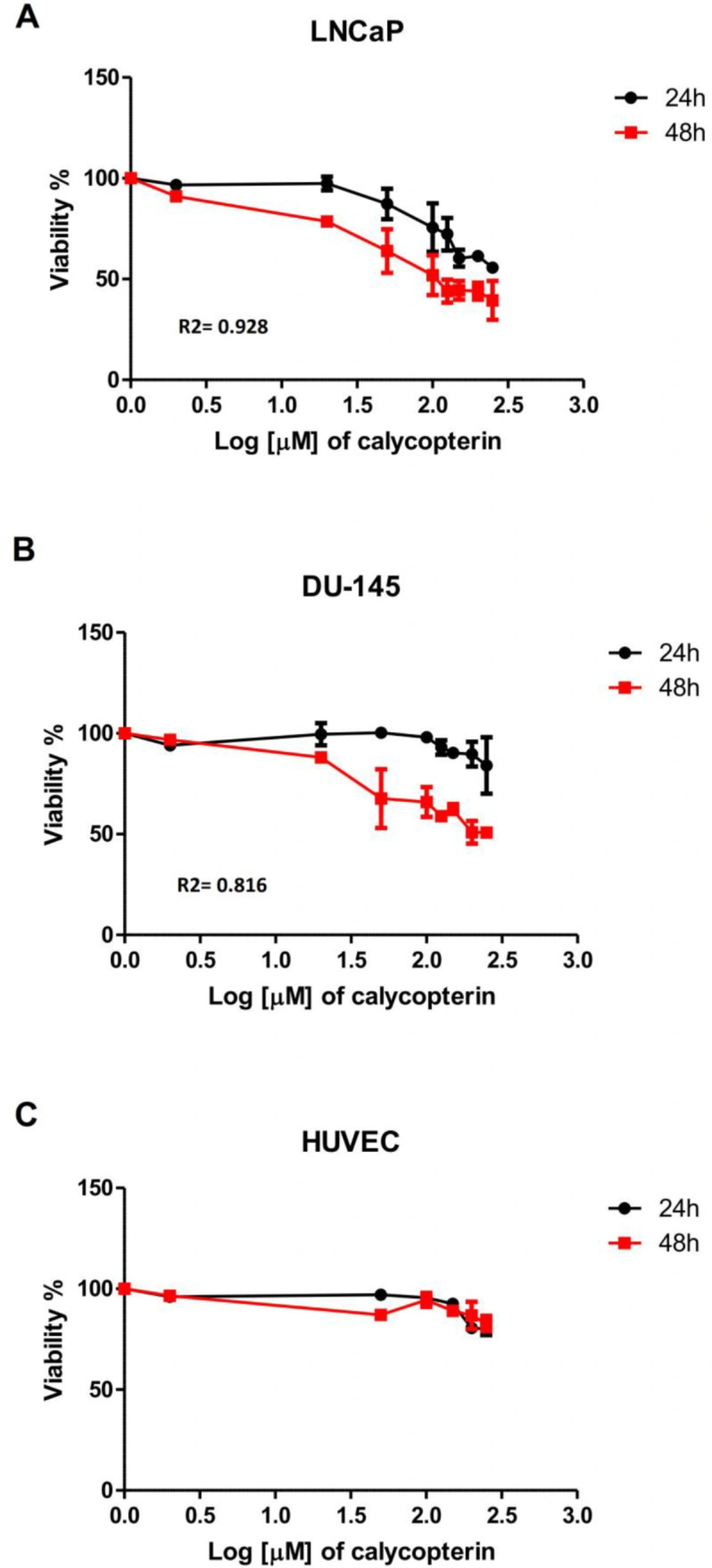
Effect of different concentrations of calycopterin (0 to 250 μM in Log scale) on cell viability after 24 and 48 h assessed by MTT assay. (A) Viability of human prostate cancer LNCaP following calycopterin treatment. (B) Viability of human prostate cancer DU-145 cells following calycopterin treatment. (C) Viability of HUVEC cells following calycopterin treatment. The results are illustrated from four experimental and three biological replicates for each sample (mean ± SD).
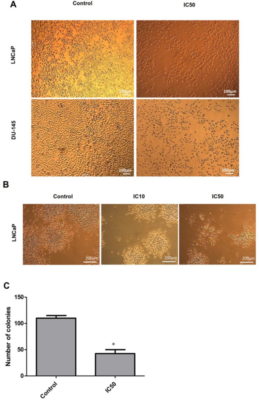
Phase-contrast microscopy images of LNCaP and DU-145 cells. (A) Microscopic images of LNCaP and DU-145 cells treated with IC50 concentration of calycopterin. The apparent reduction in cell numbers in the IC50 treated cells is observed. (B) Morphology of LNCaP colonies in control, IC10, and IC50 concentrations of calycopterin after 14 days. (C) Quantification of colony numbers in control and IC50-treated LNCaP cells after 14 days. * P < 0.05
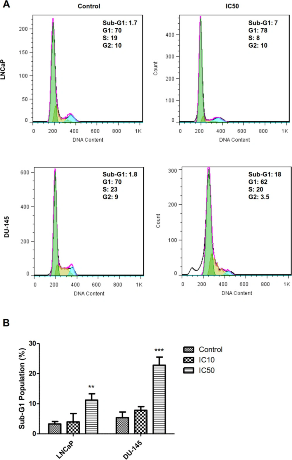
Cell cycle analysis of LNCaP and DU-145 cells after 48 h treatments with IC50 concentration of calycopterin, which was analyzed by PI staining flowcytometric assay. The sub-G1 phase is observed in the IC50 concentration of both treated cells. (B) Quantification of sub-G1 population in control, IC10, and IC50 treated cells (Three biological replicates). ** P < 0.01 and *** P < 0.001
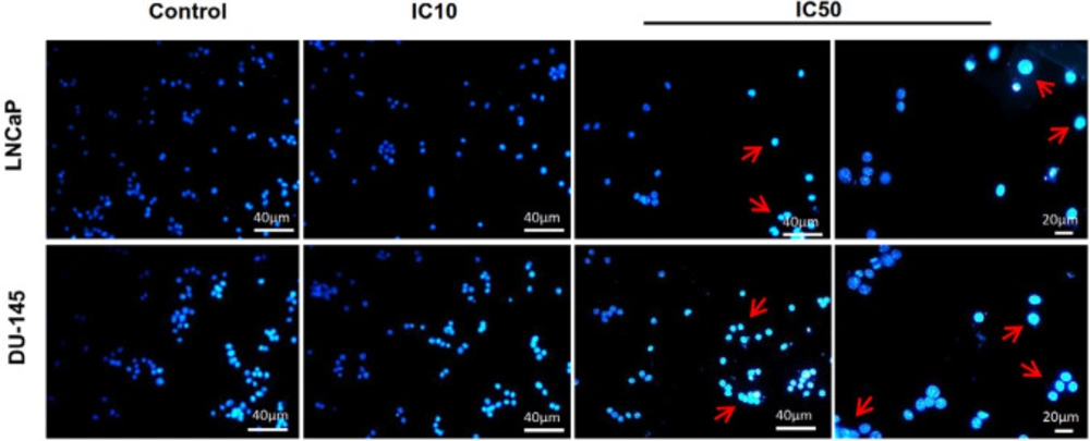
Fluorescent staining of nuclei of calycopterin-treated LNCaP and DU-145 cells by Hoechst dye. Cells were treated with IC10 and IC50 concentrations of calycopterin. Apoptotic nuclei are indicated by red arrows
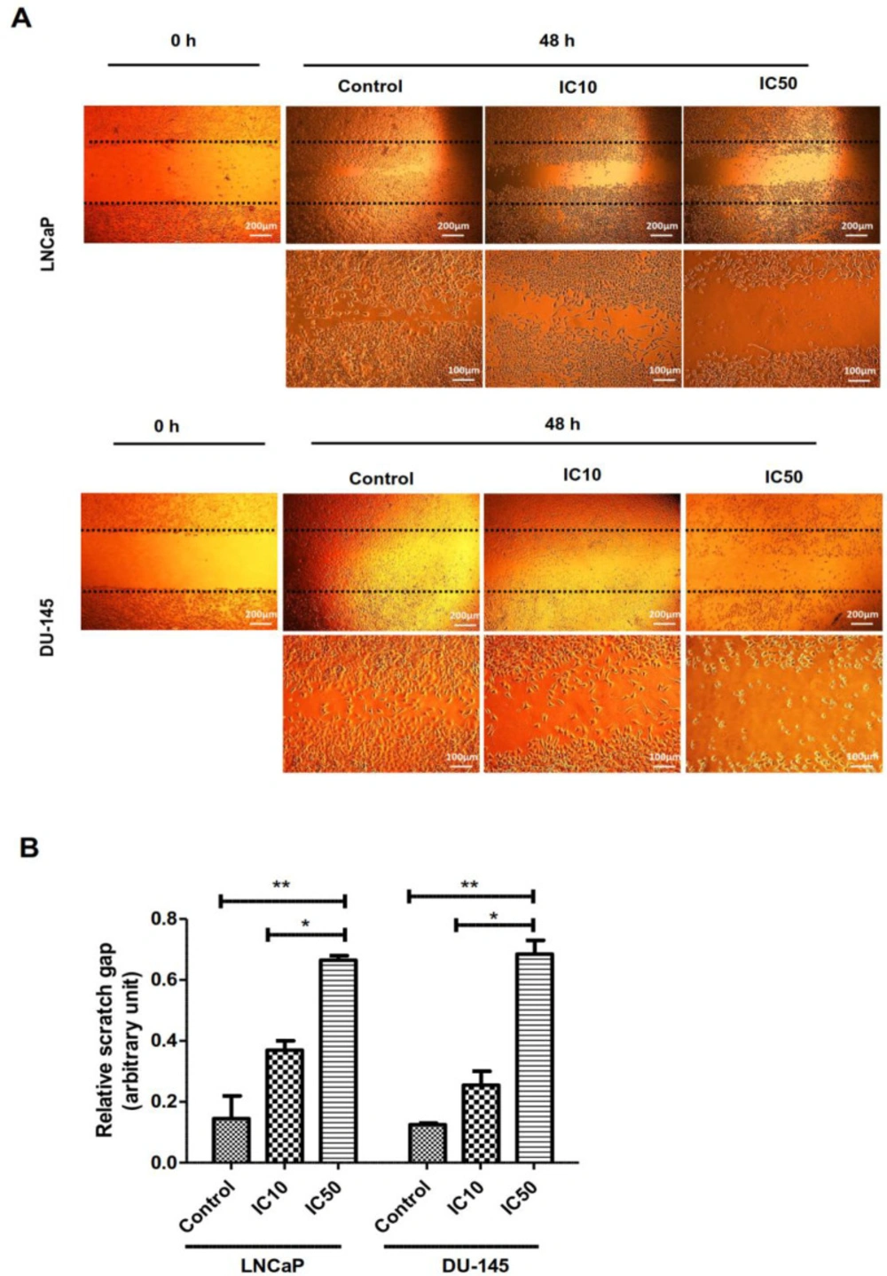
Effect of calycopterin on the potential of LNCaP and DU-145 cell migration assessed by wound healing assay. The scratched monolayer was treated with IC10 and IC50 concentrations of calycopterin. Black dotted lines indicate the wound borders at the beginning of the assay and were recorded at 0 and 48 h post-scratching. (B) Relative scratch gap was calculated as the ratio of the remaining scratch gap after 48 h and the original gap at 0 h. * P < 0.05 and ** P < 0.01
