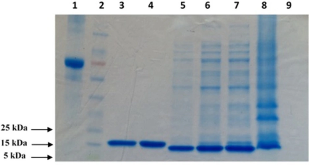
Large scale expression and purification of TNF-α protein
DNA sequence corresponding to the soluble form of human TNF-α protein was codon optimized and synthesized by Bioneer Inc. (Daejeon, South Korea) for
E. coli cell expression, and cloned into a pET28a vector (Invitrogen, Carlsbad, California, United States) containing an expression cassette with an N-terminal polyhistidine tag. Protein expression was conducted under native conditions (
15) and purification was accomplished by the Ni-NTA agarose column using ion-exchange metal affinity chromatography (IMAC) method according to the manufacturer’s instructions (Qiagen, Hilden, Germany). The purity and integrity of the produced recombinant protein was analyzed by sodium dodecyl sulfate polyacrylamide gel electrophoresis (SDS-PAGE) as previously described (
16).
Panning of phage display library
Tomlinson I naïve scFv phage library was used for the selection of specific binders against recombinant TNF-α protein. The panning procedure was carried out by immobilizing recombinant TNF-α protein (100 µg/mL) overnight onto immunotubes (Nunc, Roskilde, Denmark), which had been washed with 1X PBS buffer, blocked with 2% skim milk in 1X PBS (w/v), and incubated with phage suspension (~1013 cfu) at room temperature. Phage particles with affinity for the recombinant TNF-α were eluted using 100 mM triethylamine, and amplified in exponentially growing E. coli TG1 cells (Source BioScience, Nottingham, United Kingdom). The panning procedure was repeated for three rounds and the total eluted phage titer was determined after each round.
Mini-induction of scFvs
After three rounds of panning, E. coli non-suppressor HB2151 cells and also TG1 cells were infected with 1 µL of eluted phage and plated on TYE agar plates containing 1% (w/v) glucose and 100 µg/mL ampicillin. In order to induce scFv production, 96 colonies of TG1 and HB2151 containing the scFv phagemids were randomly picked up and inoculated in 2xTY medium supplemented with 100 µg/mL ampicillin and 0.1% (w/v) glucose in a microtiter plate. The cultures were grown while shaking (250 rpm) at 37 °C until the OD600 reached about 0.9. The transcription of scFv cassette was then induced with isopropyl β-D-thiogalactopyranoside (IPTG), which was added to the culture at a final concentration of 1 mM. Shaking was continued at 200 rpm in 30 °C environment overnight. The cells were isolated by centrifugation and the supernatants were used for subsequent assays.
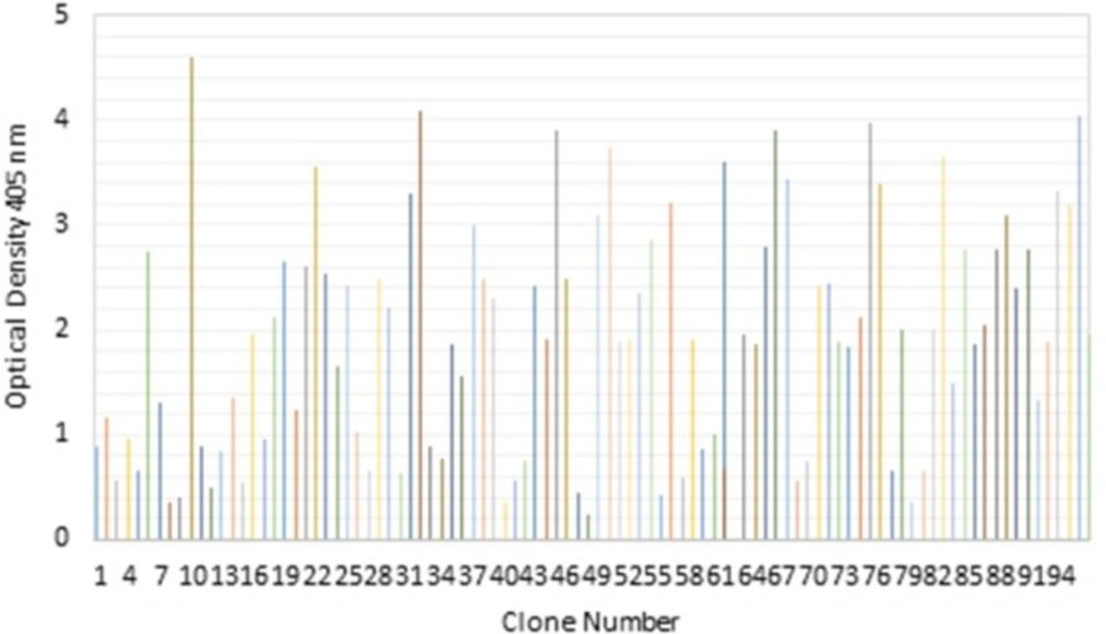
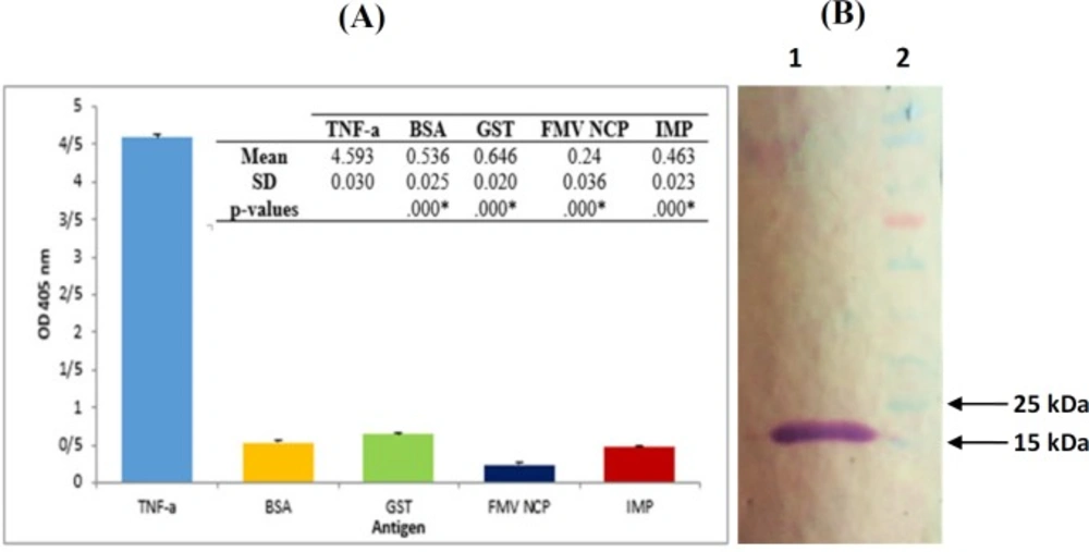
Binding activity of scFv against TNF-α protein
The binding activities of selected clones were investigated by Enzyme-Linked Immunosorbent Assay (ELISA). Briefly, About 100 µg/mL recombinant TNF-α in 1X PBS buffer was directly coated on high-binding Maxisorp microtiter plates (Nunc, Roskilde, Denmark). The wells were subsequently blocked with 2% BSA, in 1X PBS. 100 µL of scFv solutions (as described above) were then applied to the plates and incubated at 37 °C for 2 h. Bound scFvs were detected using anti c-Myc monoclonal antibody 9E10 followed by horseradish peroxidase conjugated to goat anti-mouse polyclonal antibodies. The plates were then evaluated using Biotek ELISA reader (Winooski, Vermont, United States). Positive clones were further characterized by western blotting.
Blotting analysis
In Western blotting, electrophoretically separated TNF-α protein was transferred from SDS-PAGE gel to nitrocellulose membrane (0.45 µm). The membrane was blocked with PBS buffer containing 2% (w/v) skim milk.
The scFv proteins were detected by the anti c-Myc monoclonal antibody 9E10, followed by Goat anti-mouse IgG, coupled to alkaline phosphatase (Sigma-Aldrich, St. Louis, Missouri, United States). Blots were developed using 5-bromo-4-chloro-3-indolyl phosphate (BCIP) and nitro blue tetrazolium chloride (NBT) as substrate.
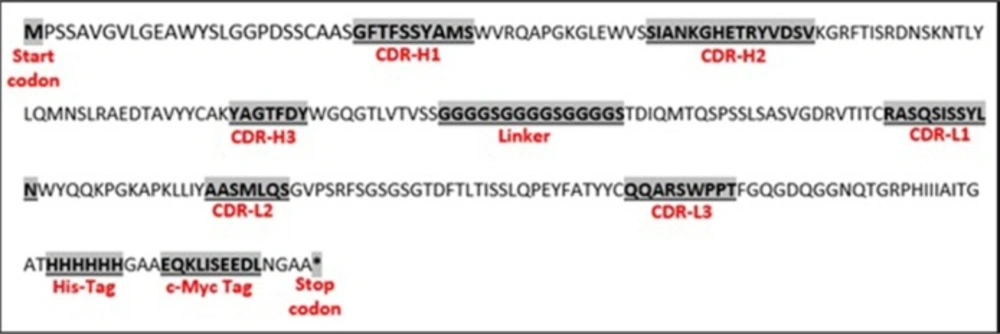
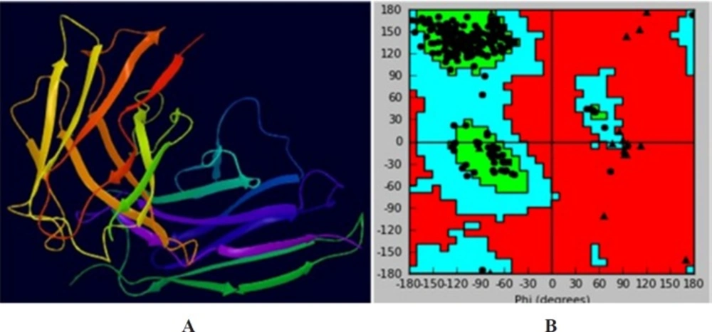
Homology modeling
The 3D structure of the anti-TNF-α scFv was built by homology modeling based on the template crystal structure. To do so, the Anti-TNF-α scFv complex models were predicted using the structures determined by X-Ray crystallography of the different partners. The calculations were performed using the ModWeb version r182 web server (https://modbase.compbio.ucsf.edu/modweb). Briefly, the heavy and light chain sequences were searched against antibody structure database followed by the identification of the template for framework (FRH/FRL) region.
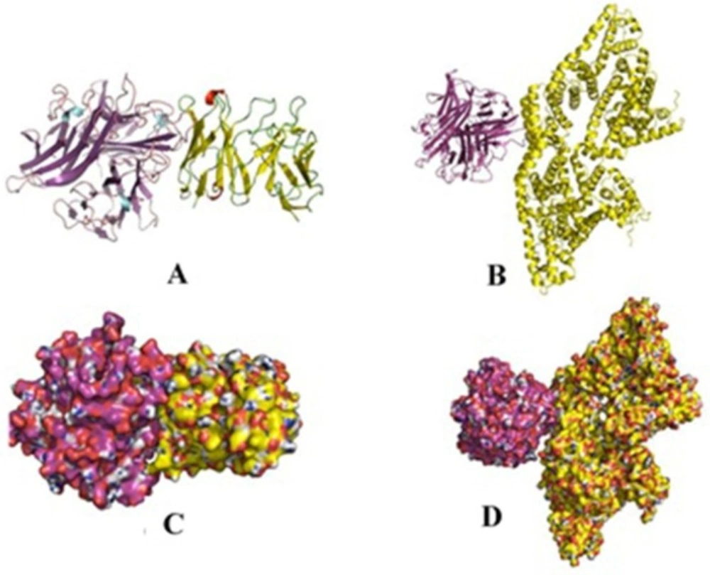
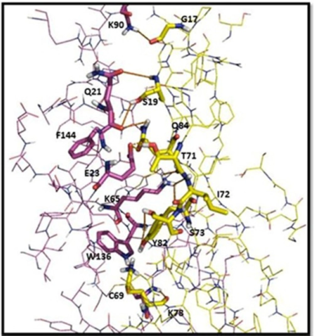
Molecular docking
Hex program (version 6.0, http://hex.loria.fr/) was used to investigate the mode of interaction between the TNF-α and BSA (as negative control) with scFv. All water molecules were removed from the scFv model structure, and hydrogen atoms were added followed by minimal minimization in the presence of bound ligand using swis-pdb Viewer. The active site for docking was defined for all atoms within the 10 Å radius of the co-crystallized ligand. The correlation type was set to shape with electrostatic and subset parameters set at 0. The possible interactions were analyzed using Pymol software version 1.5.0.1 (http://pymol.findmysoft.com). The final model was selected based on the cluster size as described in the results session.