
Introduction
Experimental
Results
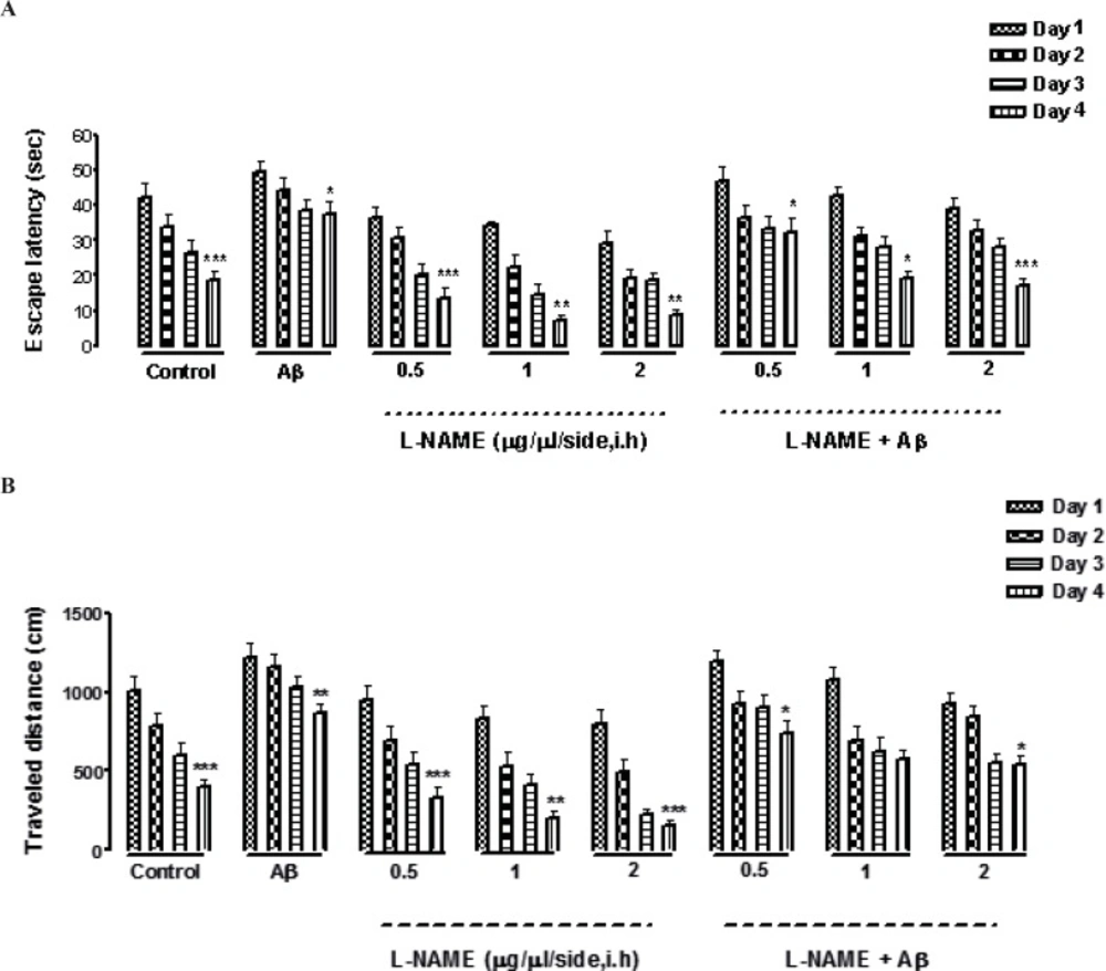
Effects of four-training trials on escape latency (A) and traveled distance (B) in L-NAME and Aβ pretreated animals. There was a significant reduction in escape latency and traveled distance in all the groups through four days of training (Figures 1 A and B). There was a significant difference between the first and the fourth day of training, measured by two factors of escape latency and traveled distance except in (Aβ + L-NAME 1 μg/side) treatment group which the traveled distance didn’t decrease significantly from first day to the fourth day of training. The swimming speed was consistent during the four training days in all of the groups (data not shown). Each value explains Mean ± S.E.M for 6-8 rats. (*P < 0.05; **P < 0.01; ***P < 0.001 shows difference between the first and the fourth days of training).
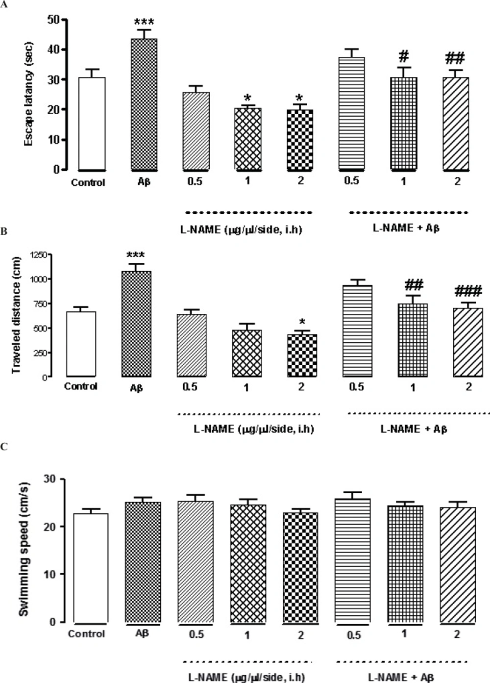
Effects of different concentrations of L-NAME infusion on escape latency (A), traveled distance (B), and swimming speed (C) during the testing trials. Hippocampus infusion of L-NAME (1, 2 μg/side, i.h) showed a significant decrease in escape latency compared to the control group. Treatment by L-NAME (2 μg/side, i.h) decreased traveled distance significantly comparing to the control group (Figures 2 A and B).There was no significant difference in swimming speed between the control and the treatment groups (Figure 2C). Intra-hippocampal infusion of Aβ significantly increased the time and distance for finding the hidden platform comparing with the control group. Intra-hippocampal infusion of 1, 2 (μg/side) of L-NAME with Aβ decreased the time and distance for finding the hidden platform significantly in comparison with Aβ treated animals. Each point explains Mean ± S.E.M for 6-8 rats. (*P < 0.05; ***P < 0.001 different from control group, #P < 0.05; ##P < 0.01; ###P < 0.001 different from Aβ-treated group
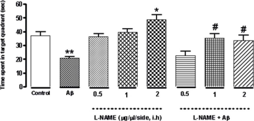
Interactive effect of administration of bilateral intra-hippocampal infusion of Aβ and different doses of L-NAME on time spent in target quadrant in 90 sec probe test. Treatment with L-NAME (2 μg/side, i.h) significantly increased the time animals spent in the target quadrant comparing to control. Furthermore administration of Aβ (30 ng/side) into the CA1 region of the hippocampus decreased this time significantly compared to the control. The time increased significantly by administration of 1or 2 (μg/side) of L-NAME after infusion of amyloid beta comparing to Aβ-treatment group. Each point shows the mean ± SEM for 6–8 rats. (*P < 0.05; **P < 0.01 different from control group, #P < 0.05 different from Aβ-treatment group).
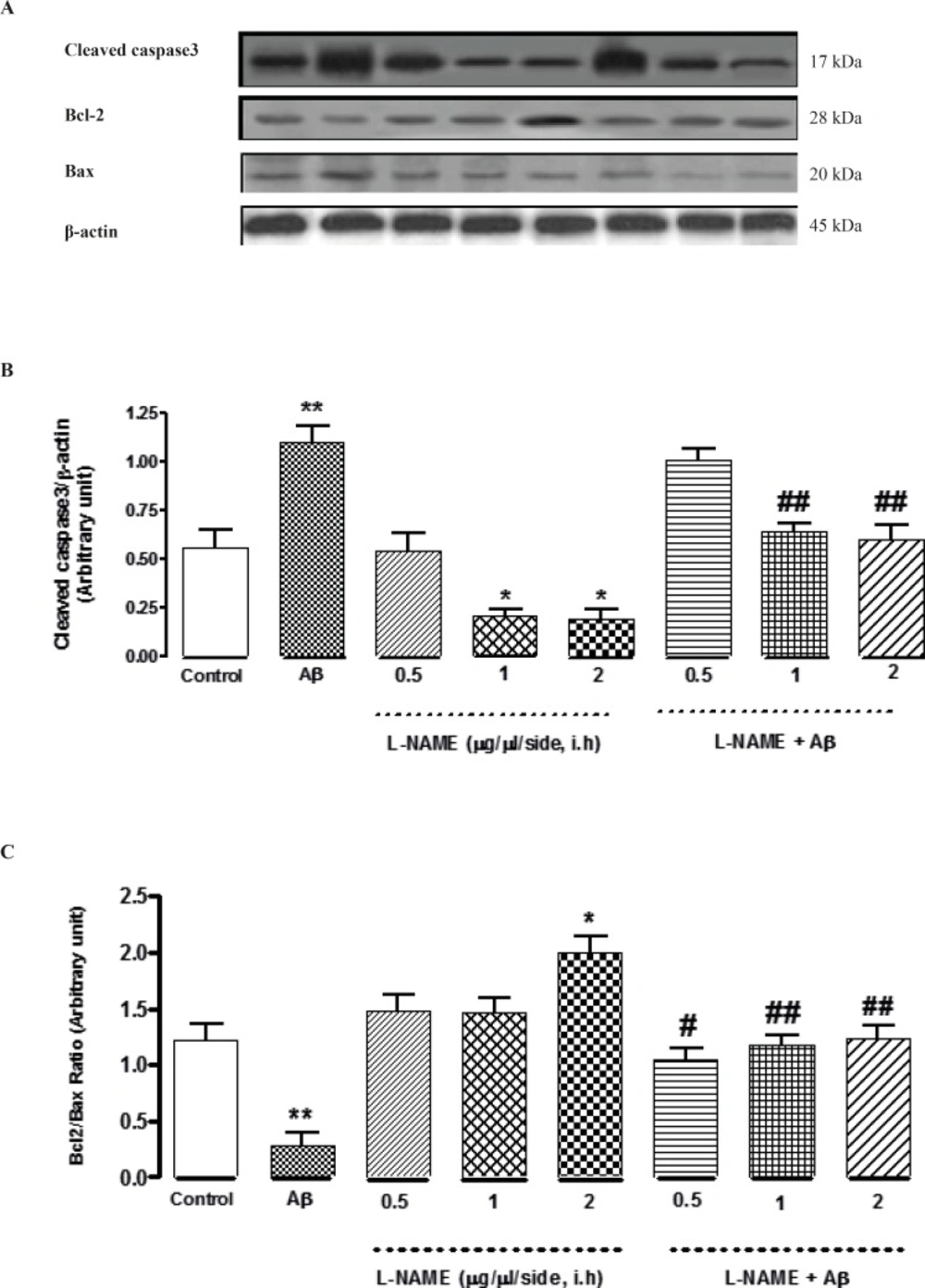
Western blot analysis to measure the effect of L-NAME treatment on the expression of Caspase-3, Bcl-2 and Bax in the hippocampus of rats. (A) 60 μg proteins were alienated on SDS-PAGE, western blotted, probed with specific primary antibodies, and reprobed with control loading antibody (One typical western blot of each antibody is shown: n = 6). (B) The densities of cleaved caspase-3 bands were evaluated and their ratios to β-actin were measured. (C) The densities of Bcl-2 and Bax bands were measured and the ratio of Bcl-2 to Bax was evaluated. Each point shows the mean ± SEM. (*P < 0.05; **P < 0.01 different from the control group, #P < 0.05; ##P < 0.01 different from the Aβ-injected group
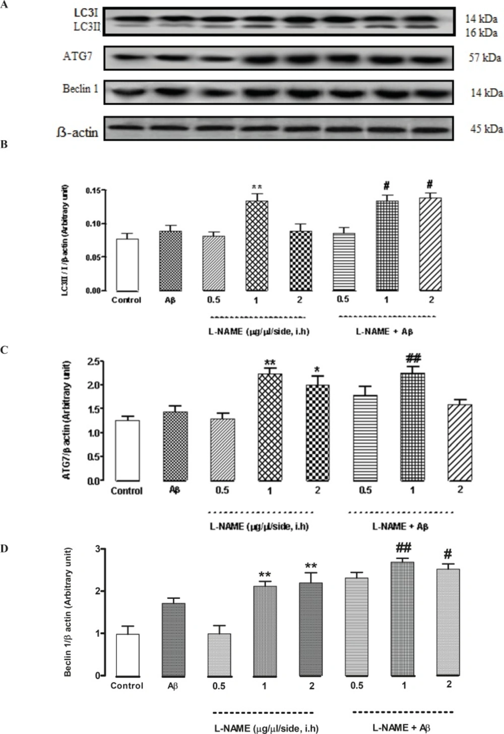
Western blot analysis to measure the effect of L-NAME treatment on the LC3, ATG7 and Beclin 1 expression in the hippocampus of rats. (A) 60 μg proteins were alienated on SDS-PAGE, western blotted, probed with specific primary antibodies, and reprobed with control loading antibody (One typical western blot of each antibody is shown: n = 6). (B) The densities of LC3І and LC3ІІ bands were evaluated and the LC3ІІ/LC3І ratio was measured. (C) The densities of ATG7 bands were evaluated and their ratios to β-actin were measured. (D) The densities of Beclin 1 bands were evaluated and their ratios to β-actin were measured. Each point shows the mean ± SEM. (*P < 0.05; **P < 0.01 different from the control group, #P < 0.05; ##P < 0.01 different from the Aβ-injected group


