
Introduction
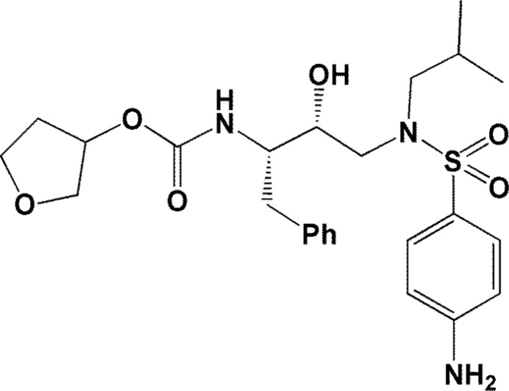
Chemical structure of Amprenavir
Experimental
Results
| PDB code | GA runs | Maximum No. of energy evaluations | RMSD from reference structure (Å) |
|---|---|---|---|
| 4DJO | 100 | 2.5×10⁷ | 3.53 |
| 4DJP | 100 | 2.5×10⁷ | 3.17 |
| 4DJQ | 100 | 2.5×10⁷ | 3.49 |
| 4DJR | 100 | 2.5×10⁷ | 2.20 |
| 4DQB | 100 | 2.5×10⁷ | 2.73 |
| 3SA7 | 100 | 2.5×10⁷ | 3.17 |
| 3SA8 | 100 | 2.5×10⁷ | 2.61 |
| 3SA9 | 100 | 2.5×10⁷ | 3.37 |
| 3NLS | 100 | 2.5×10⁷ | 2.36 |
| 3O9G | 100 | 2.5×10⁷ | 2.63 |
| 3O9F | 100 | 2.5×10⁷ | 3.76 |
| 3O9H | 100 | 2.5×10⁷ | 2.99 |
| 3O9I | 100 | 2.5×10⁷ | 3.05 |
| 3SA3 | 100 | 2.5×10⁷ | 3.03 |
| 3SA4 | 100 | 2.5×10⁷ | 2.26 |
| 3SA5 | 100 | 2.5×10⁷ | 1.60 |
| 3SA6 | 100 | 2.5×10⁷ | 2.34 |
| 3SAC | 100 | 2.5×10⁷ | 1.14 |
| 2Q5K | 100 | 2.5×10⁷ | 3.13 |
| 2Q54 | 100 | 2.5×10⁷ | 4.16 |
| 2Q55 | 100 | 2.5×10⁷ | 3.33 |
| 3EM3 | 100 | 2.5×10⁷ | 2.39 |
| 3EKV | 100 | 2.5×10⁷ | 1.53 |
| 3EKX | 100 | 2.5×10⁷ | 2.30 |
| 3MXD | 100 | 2.5×10⁷ | 3.00 |
| 3MXE | 100 | 2.5×10⁷ | 2.89 |
| 2PSV | 100 | 2.5×10⁷ | 2.49 |
| 2PQZ | 100 | 2.5×10⁷ | 0.34 |
| 2PSU | 100 | 2.5×10⁷ | 2.94 |
| 1T3R | 100 | 2.5×10⁷ | 2.56 |
| 1MUI | 100 | 2.5×10⁷ | 2.97 |
| 2I0A | 100 | 2.5×10⁷ | 2.85 |
| 1IDB | 100 | 2.5×10⁷ | 2.02 |
| 1T7J | 100 | 2.5×10⁷ | 0.47 |
| 1XL2 | 100 | 2.5×10⁷ | 2.91 |
| 1XL5 | 100 | 2.5×10⁷ | 2.28 |
| 1GNO | 100 | 2.5×10⁷ | 3.85 |
| 1A9M | 100 | 2.5×10⁷ | 3.45 |
| 1DIF | 100 | 2.5×10⁷ | 4.45 |
| 1AJV | 100 | 2.5×10⁷ | 1.18 |
| 1AJX | 100 | 2.5×10⁷ | 0.64 |
| 1CPI | 100 | 2.5×10⁷ | 1.72 |
| 1BWA | 100 | 2.5×10⁷ | 2.06 |
| 1BWB | 100 | 2.5×10⁷ | 1.56 |
| 1BV9 | 100 | 2.5×10⁷ | 2.13 |
| 3SAB | 100 | 2.5×10⁷ | 2.28 |
| 3SAA | 100 | 2.5×10⁷ | 1.00 |
| 2I0D | 100 | 2.5×10⁷ | 2.15 |
| 1HPV | 100 | 2.5×10⁷ | 1.80 |
| 3IXO a | - | - | - |
3IXO is an apo file.
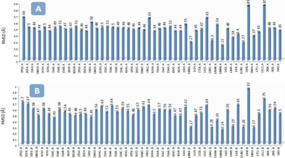
The RMSD (Ǻ) of the backbone carbon atoms (Cα) in the A) chain A of HIV-1 PR and B) chain B of HIV-1 PR structures with regard to the Amprenavir/HIV PR complex (PDB code: IHPV) To run the project, Amprenavir was docked into the active site of multiple HIV-1 PR conformations. AutoDock binding affinities and Amprenavir binding conformations (ligand binding ensembles) are represented in Table 2 and Figure 3, respectively.
| PDB code of the receptor | AutoDock binding energy (kcal/mol) | PDB code of the receptor | AutoDock binding energy (kcal/mol) |
|---|---|---|---|
| 1A8G | -7.28 | 3EKX | -7.95 |
| 1A9M | -8.30 | 3EM3 | -8.90 |
| 1AJV | -7.77 | 3MXD | -7.86 |
| 1AJX | -7.24 | 3MXE | -7.66 |
| 1BV9 | -7.38 | 3NLS | -8.24 |
| 1BWA | -9.25 | 3O9F | -8.42 |
| 1BWB | -8.67 | 3O9H | -7.81 |
| 1CPI | -7.41 | 3O9I | -7.59 |
| 1DIF | -6.64 | 3O9G | -7.37 |
| 1GNO | -8.60 | 3SA3 | -7.90 |
| 1HPV | -7.83 | 3SA4 | -7.92 |
| 1IDB | -6.90 | 3SA5 | -7.97 |
| 1MUI | -7.09 | 3SA6 | -7.72 |
| 1T3R | -8.11 | 3SA7 | -7.51 |
| 1T7J | -7.45 | 3SA8 | -8.11 |
| 1XL2 | -6.73 | 3SA9 | -7.87 |
| 1XL5 | -7.60 | 3SAA | -9.15 |
| 2I0A | -7.88 | 3SAB | -8.16 |
| 2I0D | -7.37 | 3SAC | -8.06 |
| 2PQZ | -7.32 | 4DJO | -7.85 |
| 2PSU | -7.66 | 4DJP | -7.46 |
| 2PSV | -8.12 | 4DJQ | -7.79 |
| 2Q5K | -8.00 | 4DJR | -7.67 |
| 2Q54 | -8.19 | 4DQB | -7.85 |
| 2Q55 | -7.33 | 3IXO | -4.94 |
| 3EKV | -8.10 |
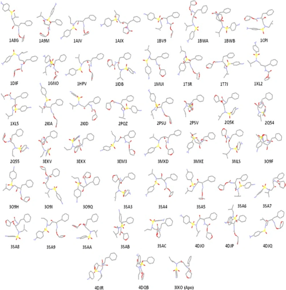
Amprenavir binding ensembles as the result of docking into the different conformations of the HIV-1 PR; each conformation of the target is designated by its relevant PDB code
| Ile50(B) | Gly48(B) | His48(B) | Asp29(B) | Asp30(B) | Asp25(B) | Asp25(A) | Il50(A) | Asp29(A) | Gly27(A) | Gly27(B) | Asp30(A) | Thr80(A) | Arg8(A) | Leu50(B) | Leu50(A) | Val82(A) | Arg8(B) | Gly48(A) | |
|---|---|---|---|---|---|---|---|---|---|---|---|---|---|---|---|---|---|---|---|
| 2PQZ | 2.12 | 2.14 | 1.91 | ||||||||||||||||
| 2Q5K | 2.07 | 2.48 | 2.21 | 1.69 | 2.37 | ||||||||||||||
| 3EKV | 1.83 | 1.81 | 2.08 | ||||||||||||||||
| 3MXD | 2.4 | 2.24 | 2.36 | ||||||||||||||||
| 3O9F | 1.98 | 1.87 | 3.12 | ||||||||||||||||
| 3O9I | 2.24 | 1.9 | 1.99 | ||||||||||||||||
| 3SA5 | 2.67 | 2.05 | 3.01 | 2.19 | |||||||||||||||
| 3SA8 | 1.96 | 1.85 | 2.01 | 1.88 | 2.18 | ||||||||||||||
| 3SAB | 2.34 | 2.04 | 1.74 | 2.08 | 2.04 | 2.28 | |||||||||||||
| 4DJP | 2.26 | 1.78 | 1.98 | 2.12 | |||||||||||||||
| 4DQB | 2.01 | 2.26 | 2.42 | 2.06 | |||||||||||||||
| 2PSU | 2.31 | 1.81 | 1.99 | 2.37 | |||||||||||||||
| 2Q54 | 2.4 | 1.9 | 2.61 | 1.86 | 2.12 | 2.13 | |||||||||||||
| 3EKX | 2.28 | 2.34 | 2.39, 2.4 | ||||||||||||||||
| 3MXE | 2.19 | 2.25 | 1.93 | 1.98 | |||||||||||||||
| 3O9G | 1.96 | 1.88 | 2.02 | 2.20 | |||||||||||||||
| 3SA3 | 2.05 | 1.84 | 2.09 | 4.9 | |||||||||||||||
| 3SA6 | 2.35 | 1.99 | 2.04 | 2.32 | |||||||||||||||
| 3SA9 | 2.22 | 1.86 | 2.16 | 2.36 | 2.41 | 2.04 | |||||||||||||
| 3SAC | 2.42 | 2.81 | 2.12 | ||||||||||||||||
| 4DJQ | 2.31 | 2.04 | 1.86 | 1.86 | 2.18 | ||||||||||||||
| 2PSV | 2.34 | 2.13 | |||||||||||||||||
| 2Q55 | 2.46 | ||||||||||||||||||
| 3EM3 | 1.94 | 1.75 | 2.33 | 2.15 | 2.05 | 2.06 | |||||||||||||
| 3NLS | 1.9 | 1.99 | 2.32 | 2.02 | |||||||||||||||
| 3O9H | 2.43 | 1.71 | 2.09 | 2.71 | 2.77 | 2.43 | |||||||||||||
| 3SA4 | 1.94 | 2.4 | 2.24 | 2.03 | 2.12 | ||||||||||||||
| 3SA7 | 2.49 | 1.91 | 2.67 | 2.14 | 2.18 | ||||||||||||||
| 3SAA | 2.33 | 1.94 | |||||||||||||||||
| 4DJO | 2.15 | 1.96 | 2.21 | 2.35 | 2.58 | ||||||||||||||
| 4DJR | 1.92 | 1.89 | 2.14 | 1.75 | |||||||||||||||
| 1AJV | 2.32 | 1.94 | |||||||||||||||||
| 1BWA | 2.79 | 2.7 | |||||||||||||||||
| 1CPI | 1.84 | 2.17 | 1.85 | 1.81 | |||||||||||||||
| 1HPV | 2.11 | 2.34 | 2.31 | 2.28 | |||||||||||||||
| 1T3R | 1.83 | 2.27 | 2.38 | 2.23 | |||||||||||||||
| 1XL5 | 1.97 | 2.29, 2.21 | 2.08 | ||||||||||||||||
| 1A8G | 2.19 | 2.10, 1.91 | |||||||||||||||||
| 1A9M | 2.32 | 2.22 | 2.22 | 1.89 | 2.07 | 2.41 | |||||||||||||
| 1BWB | 2.11 | ||||||||||||||||||
| 1AJX | 2.26 | 2.32 | 2.2 | 2.2 | 2.13 | ||||||||||||||
| 1BV9 | 1.98 | 2.39 | 2.11 | 2.07 | |||||||||||||||
| 1DIF | 2.15 | 3.25 | 2.88 | ||||||||||||||||
| 1GNO | 1.81 | 1.96 | 1.94 | 1.99 | |||||||||||||||
| 1IDB | 2.04 | 1.78 | |||||||||||||||||
| 1MUI | 2.04 | 1.77 | 2.38 | ||||||||||||||||
| 1T7J | 1.82 | 2.12 | 1.92 | 2.17 | |||||||||||||||
| 1XL2 | 2.34 | 1.95 | |||||||||||||||||
| 2I0A | 1.91 | 1.95 | 2.14 | 2.06 | |||||||||||||||
| 2I0D | 2.11 | 1.87 | 2.32 | 1.79 | 1.89 | ||||||||||||||
| Apo | 2.53 |
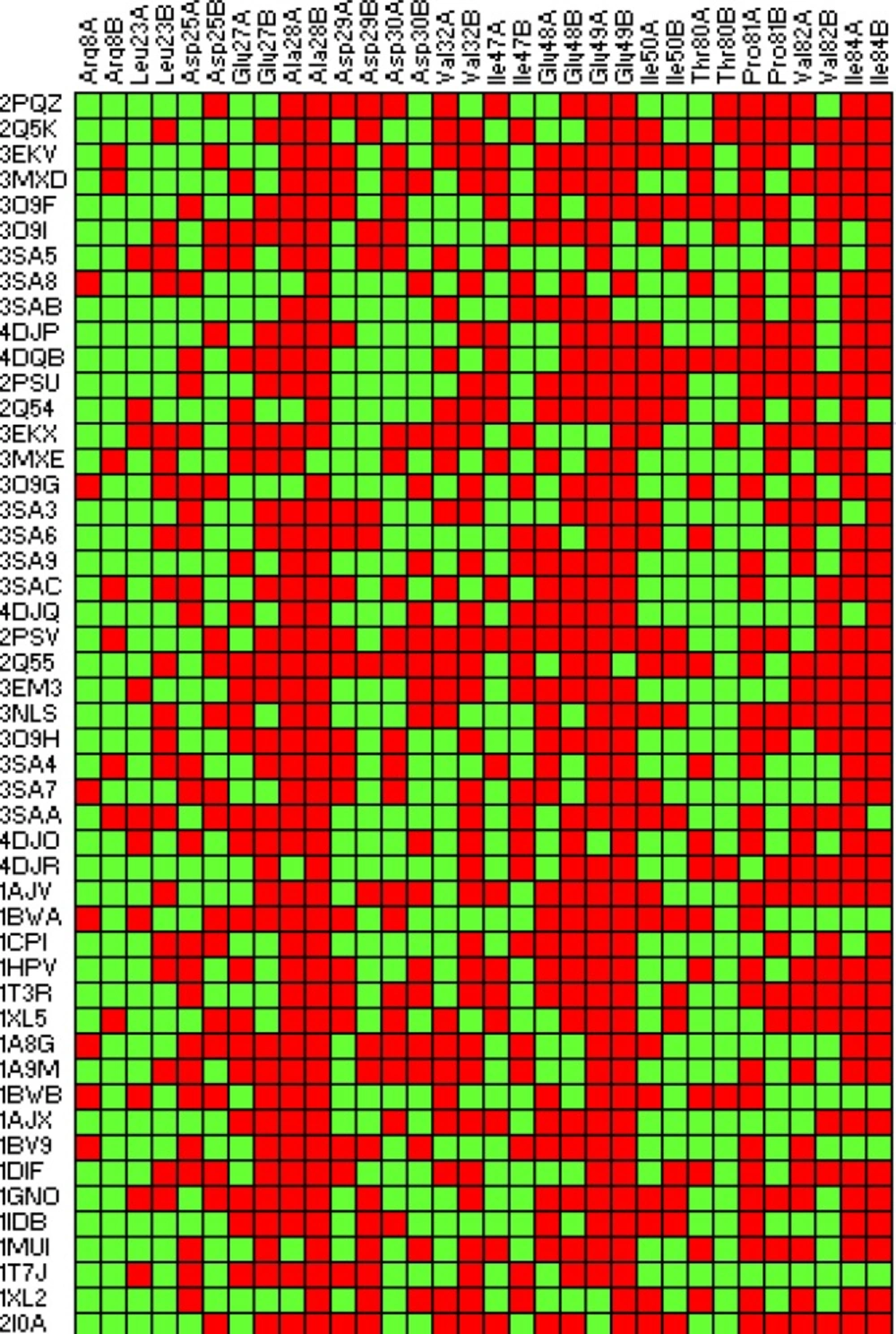
2D fingerprint representation of lipophilic contacts in binding of Amprenavir to the HIV-1 PR ensembles. Red: interacted; Green: Non-interacted
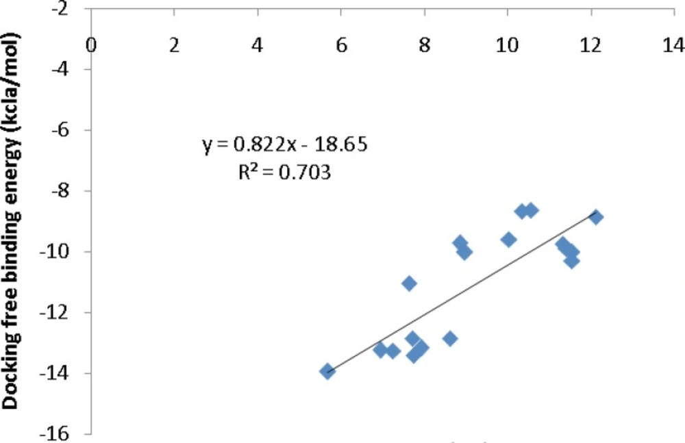
Correlation of AutoDock binding affinities and biological activities (PDB bind database) for a series of co-crystallographic HIV-1 PR inhibitors (PDB codes: 4DJO, 4DJP, 4DJQ, 4DJR, 2Q5K, 3MXD, 3MXE, 2PSV, 2PQZ, 2PSU, 2I0A, 1GNO, 1A9M, 1AJV, 1AJX, 1BWA, 1BV9, 2I0D
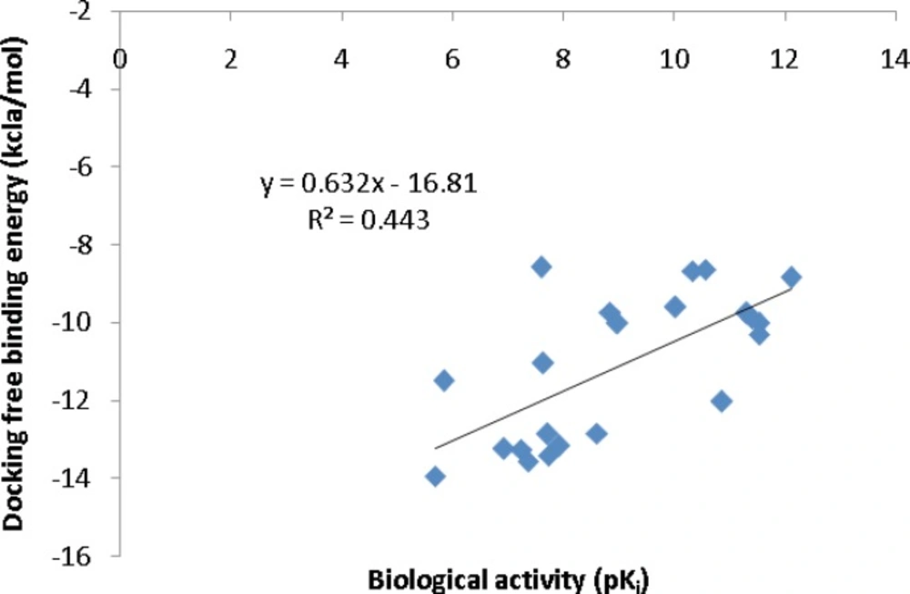
Correlation of AutoDock binding affinities and biological activities (Binding MOAD database) for a series of co-crystallographic HIV-1 PR inhibitors (PDB codes: 4DJO, 4DJP, 4DJQ, 4DJR, 2Q5K, 2Q5S, 3MXD, 3MXE, 2PSV, 2PQZ, 2PSU, 1T3R, 2I0A, 1XL2, 1XL5, 1GNO, 1A9M, 1AJV, 1AJX, 1BWA, 1BV9, 2I0D).
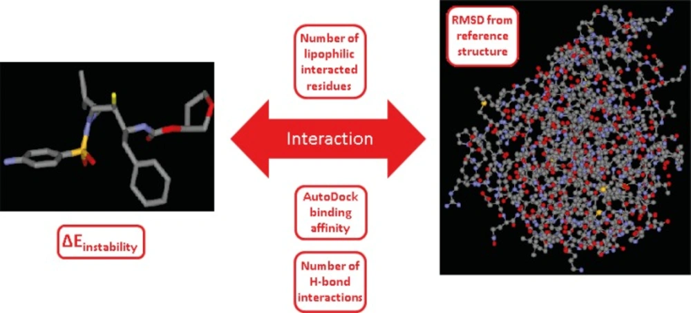
Schematic representation of the ligand-enzyme binding system and its related descriptors, system constituents in our study are ligand (Amprenavir), enzyme (HIV-1 PR) and their interaction. System descriptors are AutoDock score, protein deviation from apo form, number of lipophilic interacted residues, number of H-bond interactions and instability energy of ligand binding pose (∆Einstability). ∆Einstability indicates the instability gain of geometrically optimum conformation as the result of binding to the receptor (19).
| Code of docked HIV-1 PR file | AutoDock binding affinity | Number of lipophilic interacted residues | Number of H-bond interactions | RMSD from reference structure b | ∆Einstability c | |
|---|---|---|---|---|---|---|
| 2PQZ | -7.32 | 17 | 3 | 0.70 | 74.63 | |
| 2Q5K | -8.00 | 18 | 5 | 0.57 | 47.69 | |
| 3EKV | -8.10 | 21 | 3 | 0.53 | 69.81 | |
| 3MXD | -7.86 | 19 | 3 | 0.49 | 83.93 | |
| 3O9F | -8.42 | 19 | 3 | 0.56 | 65.42 | |
| 3O9I | -7.59 | 17 | 3 | 0.49 | 65.56 | |
| 3SA5 | -7.97 | 15 | 4 | 0.52 | 80.89 | |
| 3SA8 | -8.11 | 14 | 5 | 0.55 | 41.07 | |
| 3SAB | -8.16 | 11 | 6 | 0.54 | 73.27 | |
| 4DJP | -7.46 | 16 | 4 | 0.49 | 67.26 | |
| 4DQB | -7.85 | 19 | 4 | 0.57 | 66.53 | |
| 2PSU | -7.66 | 18 | 4 | 0.50 | 71.54 | |
| 2Q54 | -8.19 | 15 | 6 | 0.51 | 64.30 | |
| 3EKX | -7.95 | 21 | 4 | 0.60 | 75.94 | |
| 3MXE | -7.66 | 14 | 4 | 0.55 | 76.66 | |
| 3O9G | -7.37 | 16 | 4 | 0.57 | 81.69 | |
| 3SA3 | -7.90 | 16 | 4 | 0.52 | 69.02 | |
| 3SA6 | -7.72 | 16 | 4 | 0.55 | 71.66 | |
| 3SA9 | -7.87 | 14 | 6 | 0.51 | 96.32 | |
| 3SAC | -8.06 | 18 | 3 | 0.54 | 74.84 | |
| 4DJQ | -7.79 | 13 | 5 | 0.52 | 89.37 | |
| 2PSV | -8.12 | 23 | 2 | 0.48 | 75.17 | |
| 2Q55 | -7.33 | 23 | 1 | 0.54 | 86.89 | |
| 3EM3 | -8.90 | 17 | 6 | 0.52 | 76.43 | |
| 3NLS | -8.24 | 18 | 4 | 0.69 | 84.52 | |
| 3O9H | -7.81 | 17 | 6 | 0.48 | 75.92 | |
| 3SA4 | -7.92 | 16 | 5 | 0.54 | 68.09 | |
| 3SA7 | -7.51 | 15 | 3 | 0.53 | 72.84 | |
| 3SAA | -9.15 | 19 | 2 | 0.56 | 71.18 | |
| 4DJO | -7.85 | 16 | 5 | 0.48 | 70.58 | |
| 4DJR | -7.67 | 14 | 4 | 0.47 | 99.29 | |
| 1AJV | -7.77 | 20 | 2 | 0.59 | 80.68 | |
| 1BWA | -9.25 | 17 | 2 | 0.43 | 82.76 | |
| 1CPI | -7.41 | 14 | 4 | 0.49 | 93.63 | |
| 1HPV a | -7.83 | 19 | 4 | 0.38 | 88.07 | |
| 1T3R | -8.11 | 19 | 4 | 0.49 | 68.50 | |
| 1XL5 | -7.60 | 18 | 4 | 0.69 | 81.04 | |
| 1A8G | -7.28 | 18 | 3 | 0.40 | 97.52 | |
| 1A9M | -8.30 | 18 | 6 | 0.65 | 72.82 | |
| 1BWB | -8.67 | 15 | 1 | 0.40 | 75.10 | |
| 1AJX | -7.24 | 15 | 5 | 0.56 | 89.08 | |
| 1BV9 | -7.38 | 15 | 4 | 0.46 | 66.60 | |
| 1DIF | -6.64 | 18 | 3 | 0.47 | 85.69 | |
| 1GNO | -8.60 | 20 | 4 | 0.41 | 109.50 | |
| 1IDB | -6.90 | 14 | 2 | 0.80 | 82.72 | |
| 1MUI | -7.09 | 17 | 3 | 0.47 | 65.69 | |
| 1T7J | -7.45 | 13 | 4 | 0.52 | 21.84 | |
| 1XL2 | -6.73 | 14 | 2 | 0.87 | 75.61 | |
| 2I0A | -7.88 | 20 | 4 | 0.53 | 63.65 | |
| 2I0D | -7.37 | 16 | 5 | 0.55 | 67.98 | |
[object Object]
[object Object]
[object Object]
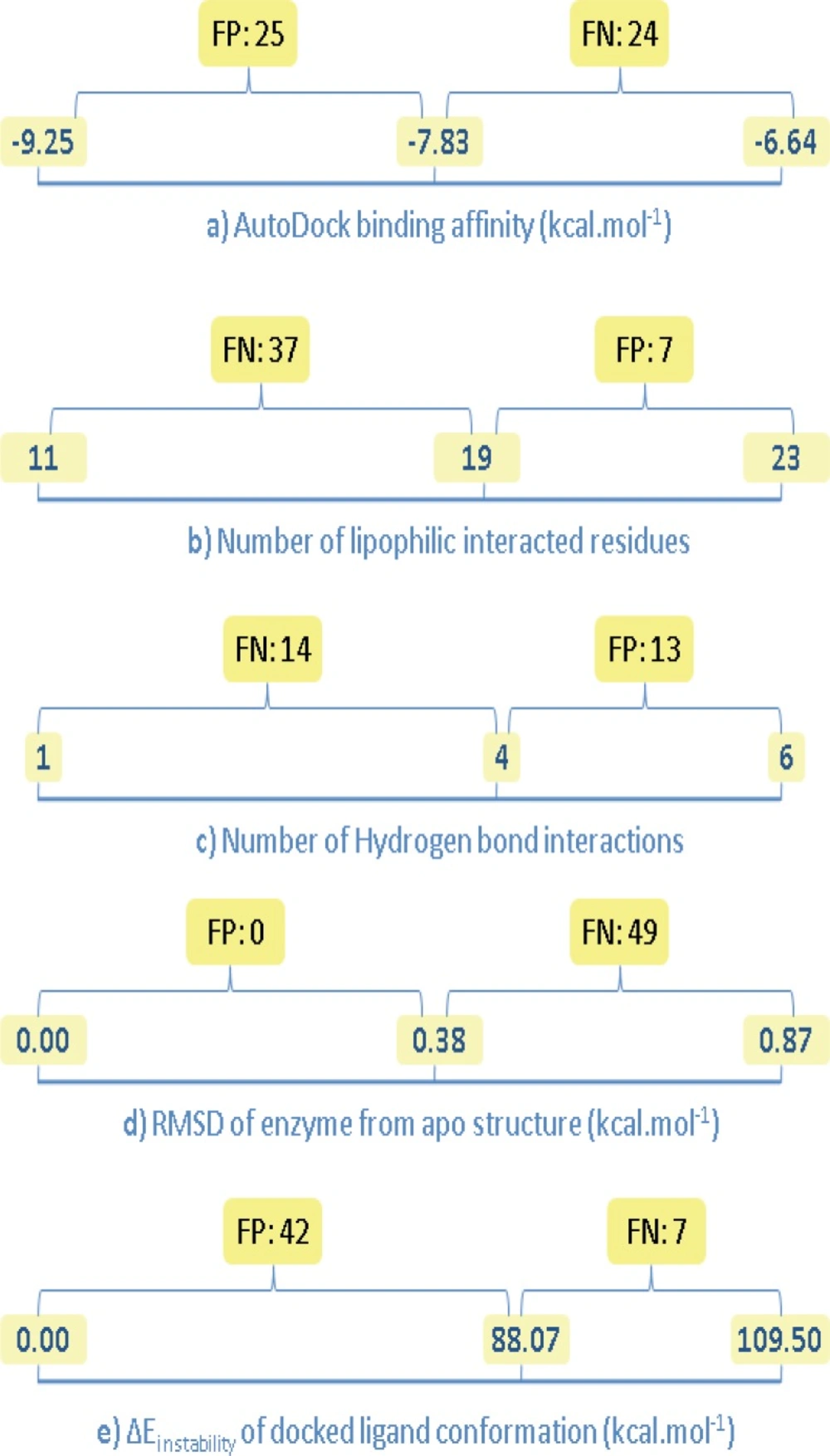
Number of false negative (FN) and false positive (FP) results for the estimated binding factors of docked Amprenavir/HIV-1 PR complexes; left side digits indicate optimum levels (except for b and c), center digits show the estimated value for Amprenavir/HIV-1 PR co-crystallographic complex, right side digits indicate non-optimum levels (except for a, d and e
Discussion
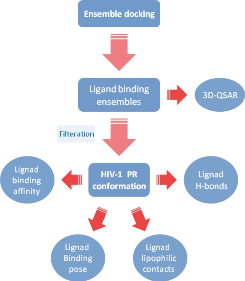
A typical flowchart representing an ensemble docking protocol for HIV-1 PR inhibitors
- Docked Amprenavir showed different hydrophobic and H-bond binding patterns in multiple conformational ensembles of HIV-1 PR active site.
- Docking results demonstrated that Asp25(A), Ile50(A), Asp25(B) and Ile50(B) were determinant residues contributing to key H-bonds (with Amprenavir) within HIV-1 PR crystallographic ensembles. For more clarification, the fluctuation of H-bond lengths between these four residues and Amprenavir in the active site of HIV-1 PR ensembles is depicted in Figure 10. H-bond distance variations ranged 1.78-2.70, 1.75-2.53, 1.69-2.81 and 1.82-2.49 Å for Asp25(A), Ile50(A), Asp25(B) and Ile50(B) residues, respectively.
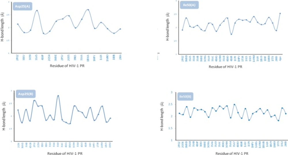
Fluctuation of H-bond lengths in binding of Amprenavir with Asp25(A), Ile50(A), Asp25(B) and Ile50(B) residues in HIV-1 PR crystallographic ensembles
- On the basis of binding results, Ala28(B) is the most important residue contributing to key electrostatic interactions in nearly all of the Amprenavir binding poses. Such priority orders emphasize the effect of target conformation on docking results.
- Comparison of different binding poses of Amprenavir revealed that the binding conformations represented by 2PSV and 2Q55 PDB codes were supported by the highest lipophilic contacts (Figure 4). For HIV-1 PR induced conformation designated by the PDB code 3SAB, minimum lipophilic contacts could be detected.
- On the basis of obtained data, the contribution of H-bond among studied HIV-1 PR conformations might be prioritized as Ile50(B) > Ile50(A) > Asp25(A) > Asp25(B). It is also notable that none of the ligand binding conformations showed all of the four key H-bonds with HIV-1 PR while sixteen binding poses (1T7J, 1MUI, 1BV9, 1AJX, 1A9M, 1HPV, 2PQZ, 3O9H, 3SAC, 3SA9, 3MXE, 3SAB, 3MXD, 4DJR and 4DJO) interacted with three out of four residues simultaneously (Table 3).
- Analysis of binding maps showed that hydroxyl group of Amprenavir contributed to the H-bond(s) with Asp25(A) and Asp25(B) while sulfonamide oxygen atoms may be involved in H-bond interactions with Ile50(A) and Ile50(B) residues of HIV-1 PR. 2D schematic representation of binding interactions between Amprenavir and HIV-1 PR structure (PDB ID: 4DJO) is depicted in Figure 11.
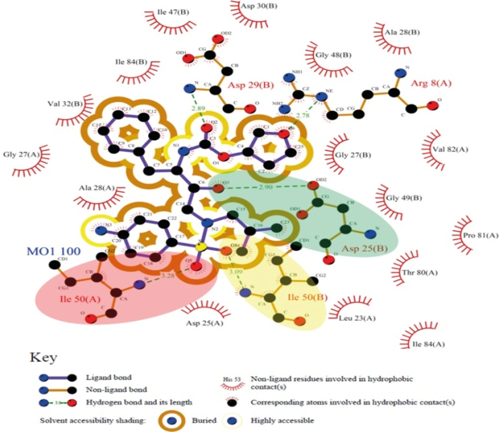
2D schematic representation of binding interactions between Amprenavir and HIV-1 PR active site (deposited PDB code: 4DJO), green, yellow and red ovals represent H-bonds between Amprenavir and Asp25(B), Ile50(B) and Ile50(A), respectively
![3D schematic representation of pair wise structure alignment between apo HIV-1 PR conformation (3IXO: orange stick) and a) 1HPV (RMSD=0.38 Ǻ) designated by blue stick, b) 1XL2 (RMSD=0.87 Ǻ) designated by blue stick containing a cognate ligand <i>i.e</i>., <i>N</i>-benzyl-2-(2,6-dimethylphenoxy)-<i>N</i>-[((3R,4S)-4 {[isobutyl(phenylsulfonyl) amino] methyl} pyrrolidin-3-yl)methyl] acetamide, the most distorted residues are highlighted by red circle in 1XL2](https://brieflands.com/journals/ijpr/articles/125306/figures/ijpr-14-785-g012-preview.webp)
3D schematic representation of pair wise structure alignment between apo HIV-1 PR conformation (3IXO: orange stick) and a) 1HPV (RMSD=0.38 Ǻ) designated by blue stick, b) 1XL2 (RMSD=0.87 Ǻ) designated by blue stick containing a cognate ligand i.e., N-benzyl-2-(2,6-dimethylphenoxy)-N-[((3R,4S)-4 {[isobutyl(phenylsulfonyl) amino] methyl} pyrrolidin-3-yl)methyl] acetamide, the most distorted residues are highlighted by red circle in 1XL2
| Descriptor | Non-optimum | Optimum level b | Estimated for IHPV system | Significance% of the estimated descriptor for IHPV system |
|---|---|---|---|---|
| AutoDock binding affinity (kcal/mol) | -6.64 | -9.25 | -7.83 | 45.6 |
| Number of lipophilic interacted residues | 11 | 23 | 19 | 66.7 |
| Number of H-bond interactions | 1 | 6 | 4 | 60 |
| RMSD of enzyme from reference structure c (kcal/mol) | 0.87 e | 0 | 0.38 | 56.3 |
| ∆Einstability d of docked ligand conformation (kcal/mol) | 109.50 f | 0 | 88.07 | 21.4 |
[object Object]
[object Object]
[object Object]
[object Object]
[object Object]
