Biological materials
C. crinita; C. sedoides and C. compressa are brown algae which were collected from the Mediterranean Tunisia coasts in various areas of the coastal region of Monastir, in June 2008, at a depth between 1 and 5 m. After collection, the seaweeds were rinsed with fresh water to remove associated debris and epiphytes. The cleaned material was then air dried to dryness in the shade at 30 °C. The dried samples were finely powdered and stored at – 20 °C until use. Identification of specimens was carried out in the National Institute of Marine Sciences and Technologies (Salamboo, Tunisia). A voucher specimen has been deposited in the Department of Pharmacology, Monastir University.
Chemicals and reagents
Carrageenan (BDH Chemicals Ltd Poole England), Acetylsalicylate of Lysine (ASL), Dimethylsulfoxide (DMSO), Dulbecco’s modified Eagle’s minimum essential medium (DMEM), Fetal Bovine Serum (FBS), 3-(4, 5-Dimethylthiazol-2-yl)-2, 5-diphenyl-tetrazolium bromide (MTT), Penicillin and Streptomycin, were purchased from Sigma Chemical (Berlin, Germany). The following chemicals, used for antioxidant activity, were purchased from Sigma-Aldrich. Chemical Co (St. Louis, MO, USA): DPPH (2, 2- diphenyl-1- picrylhydrazyl), Folin- Ciocalteu reagent, Gallic acid (GA), Trolox (6-hydroxy-2, 5, 7, 8-tetramethylchroman-2-carboxylic acid) were obtained from Sigma Chemical.
Preparation of extracts
Each Seaweed sample (400 g wet weight) was cut into small pieces mixed with a blender. Finely powdered algal material were packed in small bags (5 x 10 cm) of Whatman filter paper # 1 and all bags were sealed and macerated with water at room temperature during 24 h. The mixture was then centrifuged at 5,000 rpm for 10 min and the surnageant was filtered (Whatman No 1) to remove debris. The macerate was lyophilized during 3 days using a laboratory freeze dryer until obtaining the crude aqueous extract which was stored at 4 °C, before use for experiments.
Antioxidant activity (AOA)
Total phenolic content (TPC)
The total phenolic contents of the three aqueous extracts of the genus Cystoseira (AQ-Ccri, AQ-Csed and AQ-Ccom) were estimated by the method of Taga et al. (24). Briefly, 100 µL aliquot of sample were mixed with 2.0 mL of 2% Na2CO3 and allowed to stand for 2 min at room temperature. After incubation, 100 µL of 50% Folin- Ciocalteu’s phenol reagents were added, and the reaction mixture was mixed thoroughly and allowed to stand for 30 min at room temperature in the dark. Absorbance of all sample solutions was measured at 720 nm using spectrophotometer (Jenway 6505 UV/Vis). A calibration curve of gallic acid (ranging from 0.05 to 1 mg/ mL) was prepared, and TPC was standardised against Gallic acid and expressed as mg Gallic acid equivalent per gram of sample on a dry weight basis (DW). All determinations were performed in triplicate.
DPPH radical scavenging activity
DPPH is a chromogen-radical-containing compound that can directly react with antioxidants. DPPH has been used extensively as a free radical to evaluate reducing substances and is a useful reagent for investigating the free radical scavenging.
When the DPPH radical is scavenged by antioxidants through the donation of hydrogen to form a stable DPPH-H molecule, the colour is changed from purple to yellow. DPPH radical scavenging activity of the three aqueous extracts of the genus
Cystoseira (AQ-
Ccri, AQ-
Csed and AQ-
Ccom) was determined according to the method of Kim (
25). Each sample stock solution (1 mg/ mL) was diluted to final concentrations of 500, 250, 100, 50 and 10 (µg/ mL) in ethanol.
A total of 0.5 mL of 30 mM DPPH ethanol solution was added to 0.5 mL of sample solution at different concentrations and allowed to react at room temperature. After 30 min, the absorbance (A) was measured at 520 nm. The ability to scavenge the DPPH radical was calculated using the following equation:
Radical Scavenging capacity (RSC, %) = 1- [(A sample – A sample blank) / A control] x 100.
Where the A control is the absorbance of the control (DPPH solution without sample), the A sample is the absorbance of the test sample (DPPH solution plus test sample), and the A sample blank is the absorbance of the sample only (sample without DPPH solution). Synthetic antioxidant, Trolox was used as positive control. Concentration of extract which required reducing DPPH radicals by 50% (IC50) was calculated by linear regression of plots, where the abscissa represented the concentration of tested marine algae extracts and the ordinate the average percent of scavenging capacity from three replicates. DPPH was expressed in terms of Trolox Equivalent Antioxidant Capacity (TEAC) which was calculated based on its concentration of extract required to reduce DPPH radicals by 50% (IC50), as follows:
TEAC (mg Trolox/ 100 g) = IC50 (Trolox) / IC50 sample x 100.
Ferric Reducing Antioxidant Power (FRAP)
Ferric reducing power of the three aqueous extracts (AQ-
Ccri, AQ-
Csed and AQ-
Ccom) was determined by the method of Oyaizu (
26) (Oyaizu, 1986). Briefly, 1.0 mL of each sample dissolved in distilled water was mixed with 2.5 mL of phosphate buffer (0.2 M, pH 6.6) and 2.5 mL potassium Ferricyanide (1.0%). Reaction mixture was incubated for 20 min at 50 °C. After incubation, 2.5 mL of trichloacetic acid (10%) was added, and the mixture was centrifuged for 10 min. Finally, 2.5 mL of the upper layer were mixed with 2.5 mL of distilled water and 0.5 mL of FeCl
3 (0.1%). The solution was incubated at ambient temperature for 30 min for colour development. Absorbance of all the sample solutions was measured at 700 nm, and compared to a Gallic acid calibration curve. The data were presented as Gallic acid equivalent per gram of seaweed material (GAE/ g). A greater value of GAE related to greater reducing power of the sample.
Anti-inflammatory activity
Animals
Male adult Wistar rats weighing 150 - 170 g were obtained from Pasteur Institute (Tunis, Tunisia). They were housed in polypropylene cages and were left for 2 weeks for acclimatization to animal room maintained under controlled conditions: (a 12 h light–dark cycle at 22 ± 2 ◦C), on standard pellet diet and water ad libitum. Before the day of assay, wistar rats were fasted overnight with the free access to water.
Animal experiments are conducted in full compliance with local, national, ethical, and regulatory principles and local licensing regulations. Housing conditions and in-vivo experiments were approved according to the guidelines established by the European Union on Animal Care (CFE Council (86/ 609)). The rats were used only for the anti-inflammatory evaluation of the extracts testing.
Carrageenan Induced Rat Paw Oedema
Wistar rats were divided into groups of six animals. Oedema was induced by injecting 0.05 mL of 1% carrageenan subcutaneously into the sub-plantar region of the left hind paw (
27).
AQ-Ccri, AQ-Csed and AQ-Ccom were administered intraperitoneally (i.p.) (Doses 25 or 50 mg/Kg) and were dissolved in Saline water.
The control group received the vehicle (Saline water without the extract) (2.5 mL/ Kg, i.p.). The reference group received acetylsalicylic of lysine (ASL, 300 mg/Kg, i.p.) or Dexamethasone (1 mg/Kg, i.p.).
All drugs were administrated 30 min before the injection of carrageenan. Measurement of paw size was done by means of volume displacement technique using plethysmometer (Ugo Basile no.7140) immediately before carrageenan injection and 1, 2, 3, 4 and 5 h after carrageenan injection. Percentages of inhibition in our anti-inflammatory tests were obtained for each group using the following ratio: [(Vt −Vo) control− (Vt −Vo) treated] × 100/ (Vt−Vo) control
Where Vt is the average volume for each group at different hours after treatment and Vo is the average volume obtained for each group before any treatment. Lower and or higher doses were administered, in order to study doses dependent of the anti-inflammatory activity.
Antiproliferative activity
Cell line and culture conditions
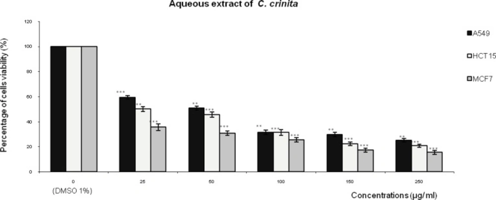
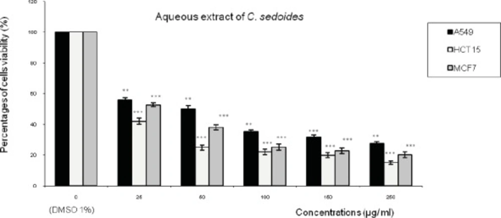
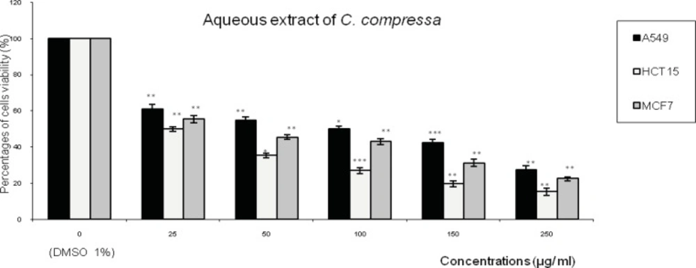
The human tumor cell lines A549 (lung cell carcinoma), HCT15 (colon cell carcinoma) and MCF7 (breast adenocarcinoma) and normal cell lines (Mardin–Darby canine kidney (MDCK) and rat fibroblast) were purchased from the American Type Culture Collection (ATCC; Manassas, VA, USA). Freshly trypsinized cells were seeded and grown in Dulbecco’s modified Eagle’s minimum essential medium (DMEM) supplemented with 10% (v/ v) fetal bovine serum (FBS), and 1% penicillin/ streptomycin, all obtained from Biochrom AG (Berlin, Germany). They were grown on Flasks (Nunc, Denmark) at 37 ˚C in a humidified atmosphere containing 5% CO2.
Cells were replicated every 2- 4 days and the medium changed once in-between.
An aliquot of each fraction was dissolved and sterilized by 0.22µm microbiological filters (Whatman, UK) and kept at 4.0 °C before analysis.
Viability assay
The potential effects on cell viability were investigated according to previously reported conditions (
28,
29) using the MTT assay [3-(4, 5-dimethylthiazol-2-yl) - 2, 5-diphenyl tetrazolium bromide, Sigma-Aldrich Chimie, Saint- Quentin-Fallavier, France] as an indicator of metabolically active cells (
30).
However, the development of this rapid colorimetric assay, which relies on the ability of mitochondrial dehydrogenase enzymes to convert 3, -4, 5 dimethyithiazol- 2, 5 diphenyl tetrazolium bromide (MTT) to a purple formazan precipitate, has simplified large scale screening of cells and drugs.
The formazan crystals are dissolved and the optical density measured using a microplate reader. The use of MTT has thus become the method of choice because of its simplicity and adaptability to automation.
Concentrations ranging from 25– 250 (µg/mL) of the AQ-Ccri, AQ-Csed and AQ-Ccom were prepared from the stock solutions by serial dilution in DMEM to give a volume of 200 µL in each well of a microplate reader (96- well).
The final concentration of DMSO in the culture medium was maintained at 1% (v/v) to avoid toxicity of the solvent. A known number of A549, HCT15 or MCF7 cells (103) were transferred into 96- well plates (Nunc, Denmark) in a volume of 200 µL of culture medium and incubated for 24 h before addition of test compounds.
After 24 h, Cells were exposed at 37 ˚C to known concentrations of the different aqueous extracts to be tested. After drug exposure, cells were washed with phosphate-buffered saline (PBS) and then reincubated in fresh culture medium for a further 48 h, then the culture medium was removed and 200 µL of MTT reagent (diluted in culture medium, 0.5 mg/ mL) was added.
Following incubation for 4 h, the MTT medium was removed and DMSO (200 µL) was added to dissolve the formazan crystals. Absorbance values were measured with a microplate reader (Bio Tek EL 340, USA) using a test wavelength of 570 nm and a reference wavelength of 630 nm.
Results were evaluated by comparing the absorbance of the treated cells with the absorbance of wells containing cell treated by the solvent control. Conventionally, cell viability was estimated to be 100% in the solvent control.
All experiments were performed at least twice in triplicate. The concentration of substance required for 50% growth inhibition (IC50 value) was estimated as that resulting in 50% decrease in absorbance as compared to control incubated simultaneously without test substances.
Data and Statistical analysis