
Introduction
Experimental
Results
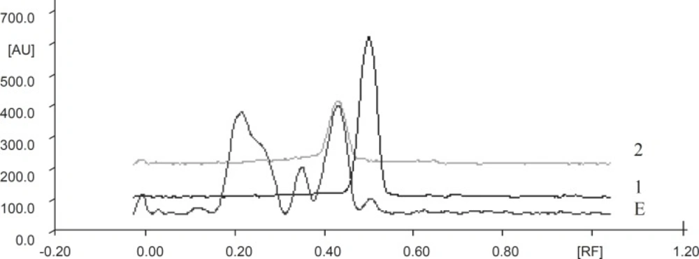
TLC densitometric chromatogram of methanolic extract of green tea with gallic acid standard and catechin standard solution. E: Extract, 1: Gallic acid, 2 : Catechin standard solution
| Extract | Solvent system | No. of spots | ||||||
|---|---|---|---|---|---|---|---|---|
| Methanolic extract | Toluene: | 8 | ||||||
| rf -values | 0.03 | 0.12 | 0.22 | 0.35 | 0.43 | 0.50 | 0.63 | 0.68 |
| Relative % | 3.30 | 1.84 | 33.03 | 15.11 | 35.09 | 4.99 | 1.27 | 1.05 |
| Groups | ST interval (msec) | QT interval (msec) | Heart rate (bpm) |
|---|---|---|---|
| Control (Group I) | 29.16 ± 1.53 | 62.5 ± 1.11 | 403.66 ± 9.51 |
| DOX (Group II) | 62.5 ± 2.14*** | 96.66 ± 3.8*** | 280.83 ± 23.28*** |
| DOX+GTE (Group III) | 33.33 ± 2.78*** | 67.5 ± 2.81*** | 395.83 ± 4.72*** |
| GTE (Group IV) | 30.0 ± 1.29 | 66.07 ± 2.06 | 389 ± 10.21 |
| f-value | 62.27 | 35.81 | 17.71 |
| p-value | p < 0.0001 | p < 0.0001 | p < 0.0001 |
| Groups | Lactate Dehydrogenage (U/L) | Creatine Kinase (U/L) | SGOT (U/mL) |
|---|---|---|---|
| Control (Group I) | 169.83 ± 4.62 | 231.16 ± 12.68 | 32.33 ± 2.0 |
| DOX (Group II) | 610.33 ± 77.66 *** | 511.5 ± 17.69 *** | 102.05 ± 5.86*** |
| DOX+GTE (Group III) | 307.16 ± 18.04 *** | 261.66 ± 17.77*** | 41.94 ± 2.35*** |
| GTE (Group IV) | 177.5 ± 6.02 | 240 ± 16.32 | 31.33 ± 2.4 |
| f-value | 26.45 | 68.19 | 91.68 |
| p-value | < 0.0001 | < 0.0001 | < 0.0001 |
| Groups | Lipid peroxidation | Reduced glutathione | Superoxide dismutase (Units/mg protein) | Catalase |
|---|---|---|---|---|
| Control (Group I) | 3.06 ± 0.16 | 9.45 ± 1.21 | 2.33 ± 0.36 | 4.02 ± 0.32 |
| DOX (Group II) | 4.75 ± 0.28*** | 5.14 ± 0.15*** | 0.6 ± 0.18** | 1.85 ± 0.18*** |
| DOX+GTE (Group III) | 2.98 ± 0.06*** | 8.40 ± 0.23** | 2.15 ± 0.27** | 4.61 ± 0.29*** |
| G TE (Group IV) | 2.75 ± 0.20 | 8.87 ± 0.59 | 2.16 ± 0.33 | 4.41 ± 0.47 |
| f-value | 21.89 | 7.77 | 7.33 | 14.24 |
| p-value | p < 0.0001 | p = 0.0012 | p = 0.0016 | p < 0.0001 |
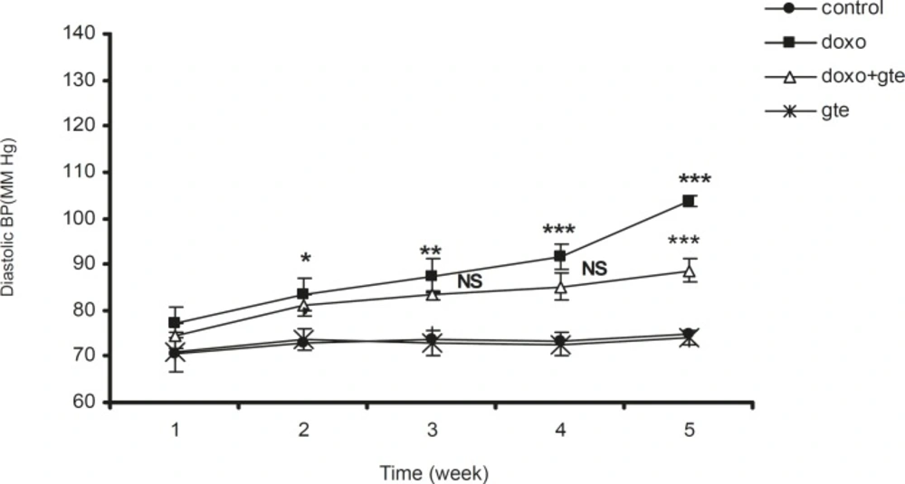
Effect of administration of DOX alone and along with GTE on diastolic blood pressure. Values are expressed as mean ± SEM (n = 6). Group II was compared with group I. Group III was compared with group II. *p < 0.05, **p < 0.01, ***p < 0.001, NS = Non significant
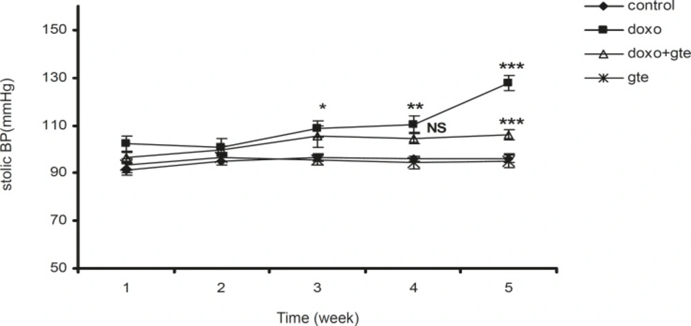
Effect of administration of DOX alone and along with GTE on systolic blood pressure. Values are expressed as mean ± SEM (n = 6). Group II was compared with group I. Group III was compared with group II. *p < 0.05, **p < 0.01, ***p < 0.001, NS = Non significant
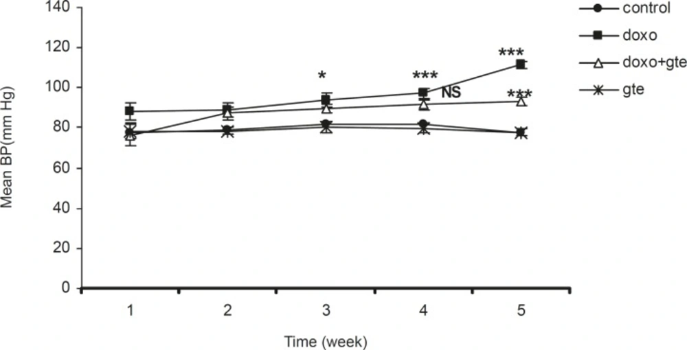
Effect of administration of DOX alone and along with GTE on mean blood pressure. Values are expressed as mean ± SEM (n = 6). Group II was compared with group I. Group III was compared with group II. *p < 0.05, **p < 0.01, ***p < 0.001, NS = Non significant
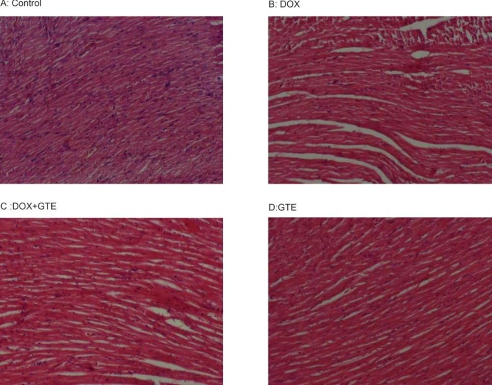
Cross sections of hearts in rats treated with DOX and along with GTE. hearts from control (A) and GTE treated rats (D) Shows normal feature of myocardium. However, hearts from a doxorubicin treated rats (B) Shows a massive necrosis of heart muscle fibres along with focal loss, marked fragmentation and disorganization of myocardial fibres. Administration of GTE along with DOX (C) Restored these changes towards normalcy
