Chemicals
RPMI-1640 medium, Dulbecco′s modified Eagle′s medium (DMEM) (Gibco, USA), Penicillin/streptomycin solutions, Fetal bovine serum, Trypsin-EDTA (Invitrogen, USA), MTT 3-(4,5-dimethyl-thiazol-2-yl)-2,5-diphenyltetrazolium bromide, Neutral red dye (NR) Sigma (St. Louis, MO, USA), Dimethyl sulfoxide (DMSO) (Sigma-Aldrich, USA), Cytotoxicity Detection Kit of lactate dehydrogenase enzyme (LDH) Pars Azmoon (Tehran, Iran), Caspase-9 colorimetric assay Kit BioVision (USA) were of analytical grade and purchased locally.
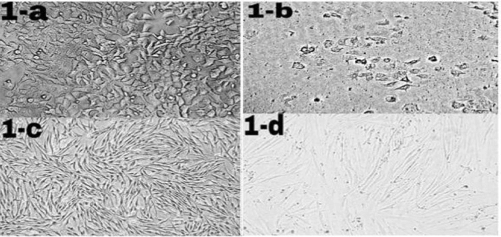
Morphological changes of ICD-85 on MCF-7 and HDF cells. MCF-7 and HDF cells (1 × 10⁴ cells/well) were cultured in RPMI-1640 and DMEM medium respectively supplemented with 10% heat inactivated fetal bovine serum and treated in the absence (control cells) or presence of ICD-85 at 80 μg/mL for 24 h at 37 °C. Morphological changes of treated cells were observed with an invert microscope and compared with control cells. Figure 1b (20X) shows granulation and cell rounding in MCF-7 cells treated with ICD-85 as compared to untreated MCF-7 cells (1-a, 20X). There are no significant morphological changes in HDF normal cells treated with ICD- 85 (1-d, 20X) as compared to untreated HDF cells (1-c, 20X)
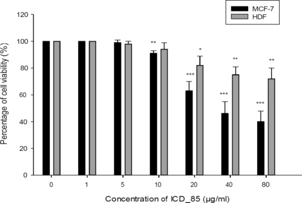
Cell viability of the MCF-7 and HDF cells after treatment with ICD-85. The cytotoxicity was evaluated by the MTT method 24 h after treatment of the cell lines with ICD-85 (1–80 μg/mL). The results were shown as mean ± SD of three independent experiments (n = 3). Doses of 10 µg/mL or greater, significantly killed MCF-7 cell compared to the control **P < 0.01
The neutral red uptake assay to assess the cell viability of MCF7 and HDF cells incubated with various concentrations of ICD-85 (1–80 μg/mL) for 24 h. The data were presented as mean ± SD (n = 3). Doses of 5 µg/mL or greater, significantly killed MCF-7 cell compared to the control *P < 0.05
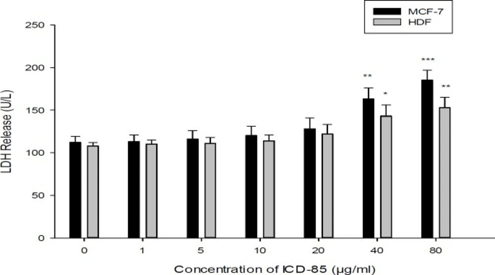
Effect of ICD-85 on LDH leakage in MCF-7 and HDF cells. The cells were treated with different concentrations of ICD-85 for 24 h. At the end of the incubation period, the LDH assay was performed to assess the LDH leakage as described in methods section. The data were expressed as the mean ± SD of three independent experiments carried out in triplicate. Significances were indicated in comparison to control.*P < 0.05, **P < 0.01, ***P < 0.001
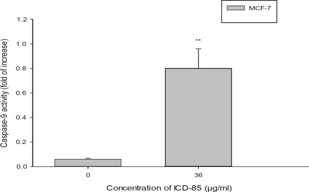
Determination of caspase-9 activity in MCF-7cell treated with ICD-85. For evaluation of caspase-9 activity, MCF-7 cell were treated in the absence and in the presence of IC50 concentration (36.45 ± 0.38 μg/mL) of ICD-85 for 24 h. The datashown are the means ± SD of three independent experiments. Significances were indicated in comparison to control. **P < 0.01
ICD-85 (venom-derived peptides)
The active fraction of ICD-85 were combinated of three peptides, ranging in size from 10000 to 30000 Da and being derived of the venoms from Iranian brown snake (Gloydius halys) and yellow scorpion (Hemiscorpius lepturus). The peptides were selected based on a study of crude venom cytotoxicity and isolation of cytotoxic peptides by two-step purification that included gel filtration and HPLC. The lyophilized peptides was stored at −20 °C and solubilized in phosphate buffered saline (PBS) immediately before its use in the tests.
Cell culture
Human breast adenocarcinoma (MCF-7) and normal Human Dermal Fibroblasts (HDF) cell lines were purchased from Pasteur Institute of Iran and cultured according to the manufacturer’s instruction. The cells were propagated in 25 mL plastic flasks in RPMI-1640 and DMEM medium respectively. The Media were supplemented with 10% heat inactivated fetal bovine serum (FBS) along with 100 U/mL penicillin and 100 µg/mL streptomycin. The culture was incubated at 37 °C with 95% humidity air that contained 5% CO2.
Mitochondrial activity
Mitochondrial function, as an indicator of cytotoxicity, was assessed by measuring the capacity of MCF-7 and HDF cells to reduce MTT to formazan (
21). Briefly, MCF-7 and HDF cells were seeded in 96-well plates at a density of 1 × 10⁴ cells per well and grew to 75% confluence. The cells were treated with different concentrations (1, 5, 10, 20, 40 and 80 µg/mL) of ICD-85 and incubated for 24 h. After treatment, 20 μL of MTT stock solution (5 mg/mL) was added to each well and the cells were incubated for 3 h at 37 °C. The solution was then discarded and 100 μL of Dimethyl sulfoxide was added into each well and incubated for 15 min in the dark to dissolve insoluble formazan crystals. Immediately, the absorbance of each well was read at 570 nm using an ELISA reader.
The percentage of the viable cells was calculated according to the following formula: Percentage of viability of each concentration = (corrected mean OD of test/corrected mean OD of control) × 100.
Neutral red uptake assay (NRU assay)
Neutral red uptake assay was done to determine the accumulation of neutral red dye in the lysosomes of viable, uninjured cells (
22). MCF-7 and HDF cells were seeded in 96-well plates at a density of 1 × 10⁴ cells/well. The cells were treated with various concentrations (1, 5, 10, 20, 40 and 80 µg/mL) of ICD-85 and incubated for 24 h. After overnight, the wells medium was replaced with a new one containing NR (40 μg/mL). After 3 h of incubation, neutral red medium was removed and the cells were washed with PBS for the remaining dye. Finally, neutral red destain solution (50% from ethanol 96%, deionized water 49% and glacial acetic acid 1%) was added to each well and the plate was gently shaken for 20 min. Optical density (OD) of neutral red extract was read with Synergy HT Microplate Reader (Bio-Tek Instruments, Winooki, VT) at 540 nm.
Morphologic Analysis Using an Inverted Microscope
Morphological studies using a normal inverted microscope were carried out to observe the morphological changes of cell death in cancer and normal cells treated with ICD-85. The untreated cells were served as the negative control.
Measurement of cell necrosis (Lactate dehydrogenase assay)
LDH is a cytoplasmic enzyme retained by viable cells with intact plasma membranes, but released from necrotic cells with damaged membranes. HDF, MCF7 cells were plated at a density of 1 × 10⁴ cells per well in 96-well cell culture plates, and incubated for 24 h. Post media changes, and treatments of the cells at ICD-85 concentrations (1, 5, 10, 20, 40, and 80 µg/mL) that the cells were incubated at 37 °C for 24 h. After incubation, the released LDH in the media was measured with the Cytotoxicity Detection Kit (Pars Azmoon, Tehran,
Iran).
Caspas-9 Assay
The Caspase-9 activity was determined by Caspase Colorimetric Assay Kit (according to the manufacturer’s instructions, BioVision, USA). Therefore, MCF-7 cells (3 × 10⁶) were harvested and lysed in 5 mL of chilled lysis buffer and then incubated on ice for 10 min. The suspension was centrifuged for 1 min at (10,000 × g) and the supernatant (cytosolic extract) was transferred to a fresh tube. The protein concentration assay was then performed by Lowry assay. 200 μg cytosolic protein was diluted to 50 μL cell lysis buffer for each assay in a 96- well plate. Each reaction buffer which containing 10mM dithiothreitol (DTT) and 5 μL of the 4 mM LEHD-pNA substrate was added into each well of the designated enzyme assay and the plate was then incubated at 37 °C for 2 h. At the end of incubation, the plate was read in a microtiter plate reader at 405 nm. The caspase activity increase was determined by comparing the results of treated samples with the level of the uninduced control.
Statistical Analysis
The values are expressed as means ± SD of three repeats in each group. The data were analyzed using student′s t-test with statistical significance for P < 0.05.