Reagents and media: Curcumin, Silver nitrate (AgNO3), MTT [3-(4,5-dimethylthiazol-2-yl)-2,5-diphenyltetrazolium bromide], Acridine orange, propidium iodide, and DAPI (4›, 6-diamidino-2-phenylindole) were obtained from Sigma-Aldrich (Poole, United Kingdom). Fetal bovine serum (FBS) and RPMI-1640 medium were purchased from Invitrogen. The High Pure RNA Isolation Kit and cDNA Synthesis Kit were also purchased from Roche (Mannheim, Germany) and Fermentas Inc. (Vilnius, Lithuania), respectively. In addition, the primers were obtained from Bioneer (Daejeon, Korea), and the commercial cisplatin was purchased from a pharmacy. Annexin V/PI and Caspase Activity Assay Kit were purchased from Abcam Company (Germany). Moreover, A2780 was obtained from Pastor Institute (Iran, Tehran). All the solutions were prepared with double distilled water and other reagents were of analytical grade.
Synthesis and Characterization of cAgNPs
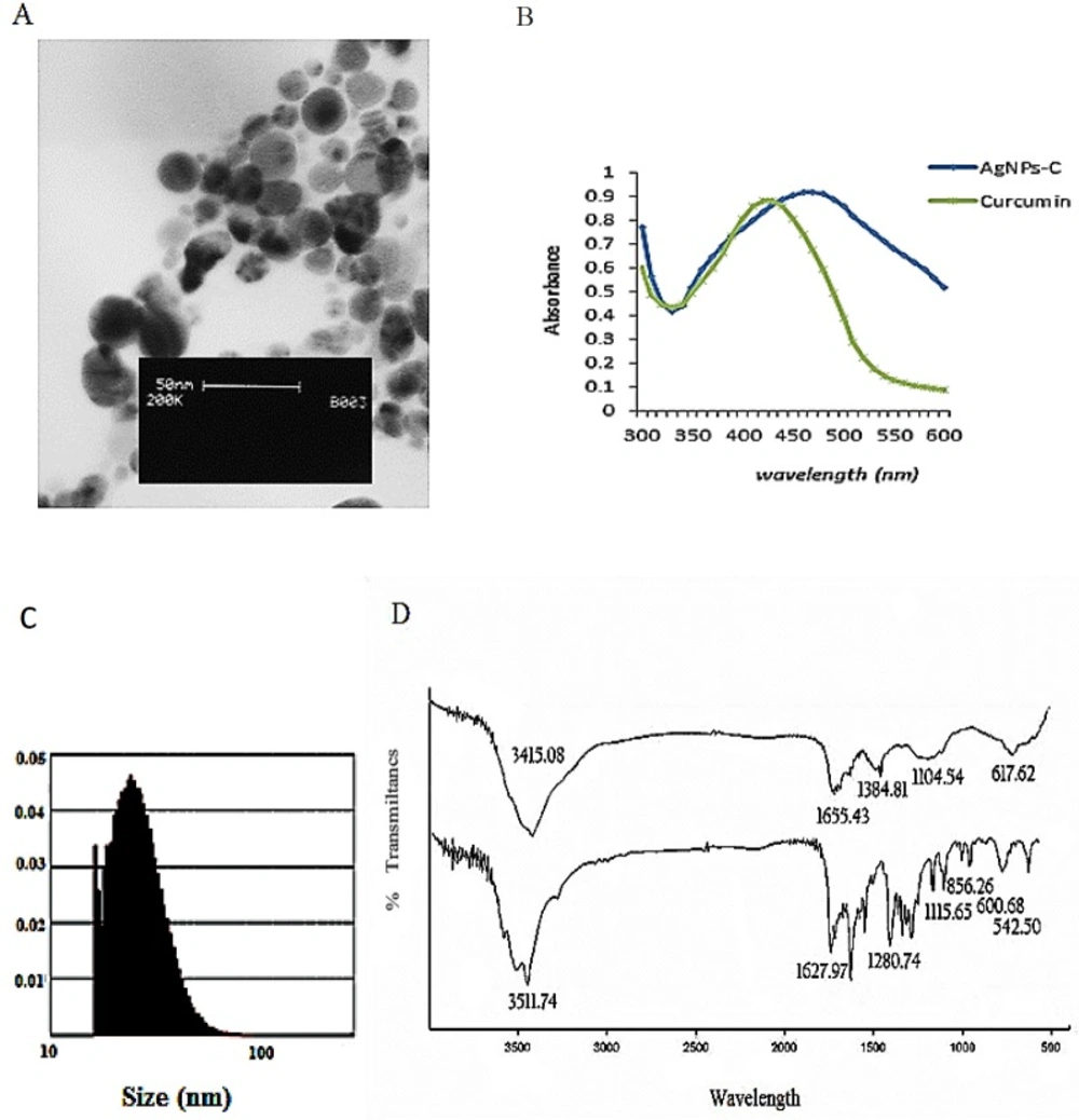
CAgNPs was synthesized using curcumin as a reducer agent, and characterized using UV-visible spectrum, Fourier transform infrared (FTIR), and transmission electron microscopy (TEM). This biosynthesized cAgNPs was used for other analyses.
Cell culture and treatment: A2780 cells were cultured in Roswell Park Memorial Institute medium (RPMI) with 10% FBS and 1% penicillin-streptomycin, followed by their incubation at 37 °C in 5% humidified CO2. When reached 90% confluence, the cells were used for other analyses. A2780 was treated with cAgNPs (1, 2, 4, 8 and 16 µg/mL), and cisplatin (5, 10, 15, 30, 60 and 120 µg/mL) for 48 h. After the calculation of IC50, the concentration below IC50 (2.5 µg/mL of cisplatin and 2 µg/mL CAgNPs) was used for the combined groups, followed by other analyses for these groups.
Evaluation of cytotoxicity
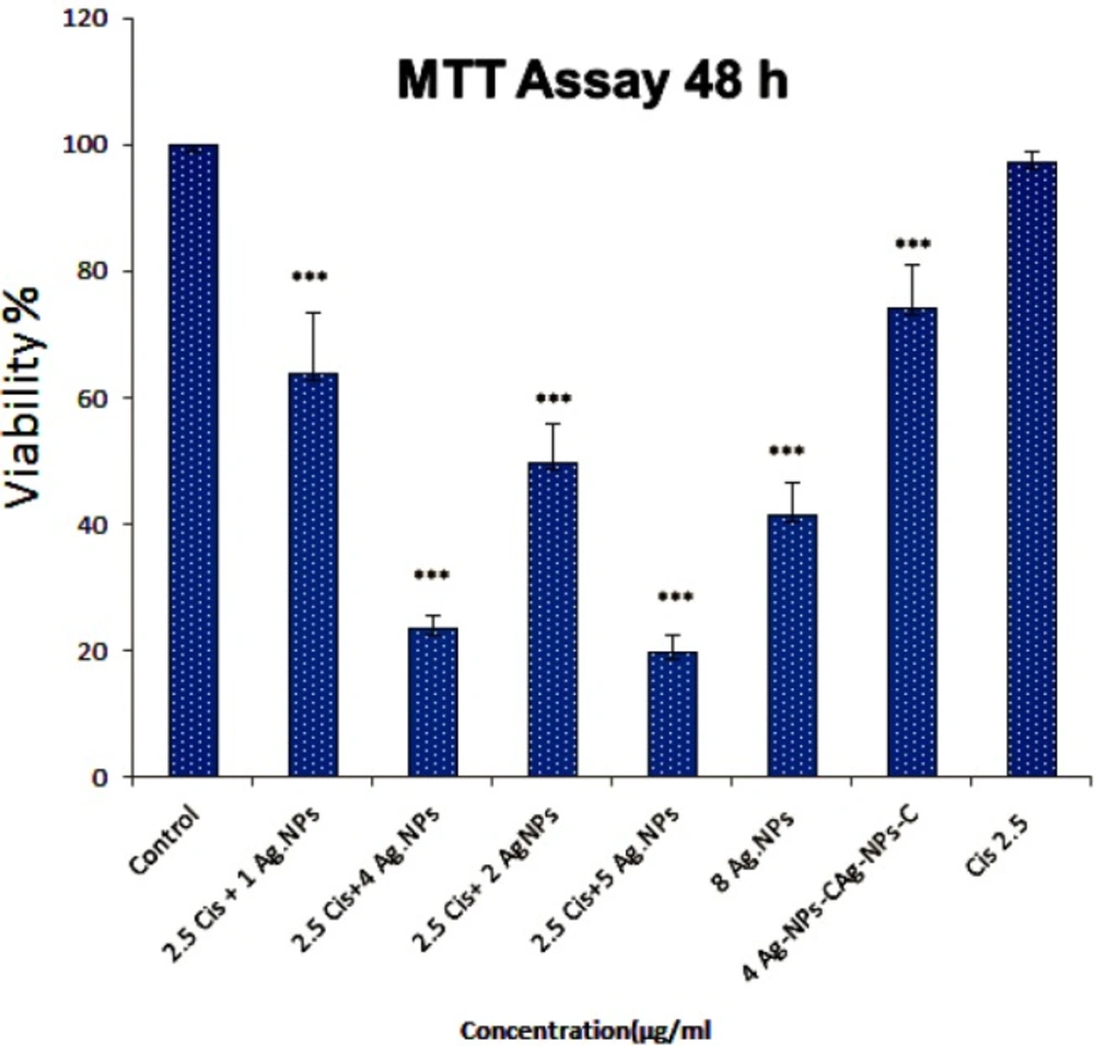
The cells were seeded and treated as describe after 48 h. Afterwards, 50 µL MTT solution (5 mg/mL) was added to each well and incubated for two hours in 37 °C. Following that, 100 µL dimethyl sulfoxide (DMSO) was added to solve formazan crystals. The absorbance was read at 570 nm using plate reader and spectrophotometer. The cell viability was calculated using the following equation:
cell viability (%) = (Atreated/Acontrol) 100, where A treated and A control are the absorbance of the treated and untreated cells, respectively (
9).
Apoptosis induction assay
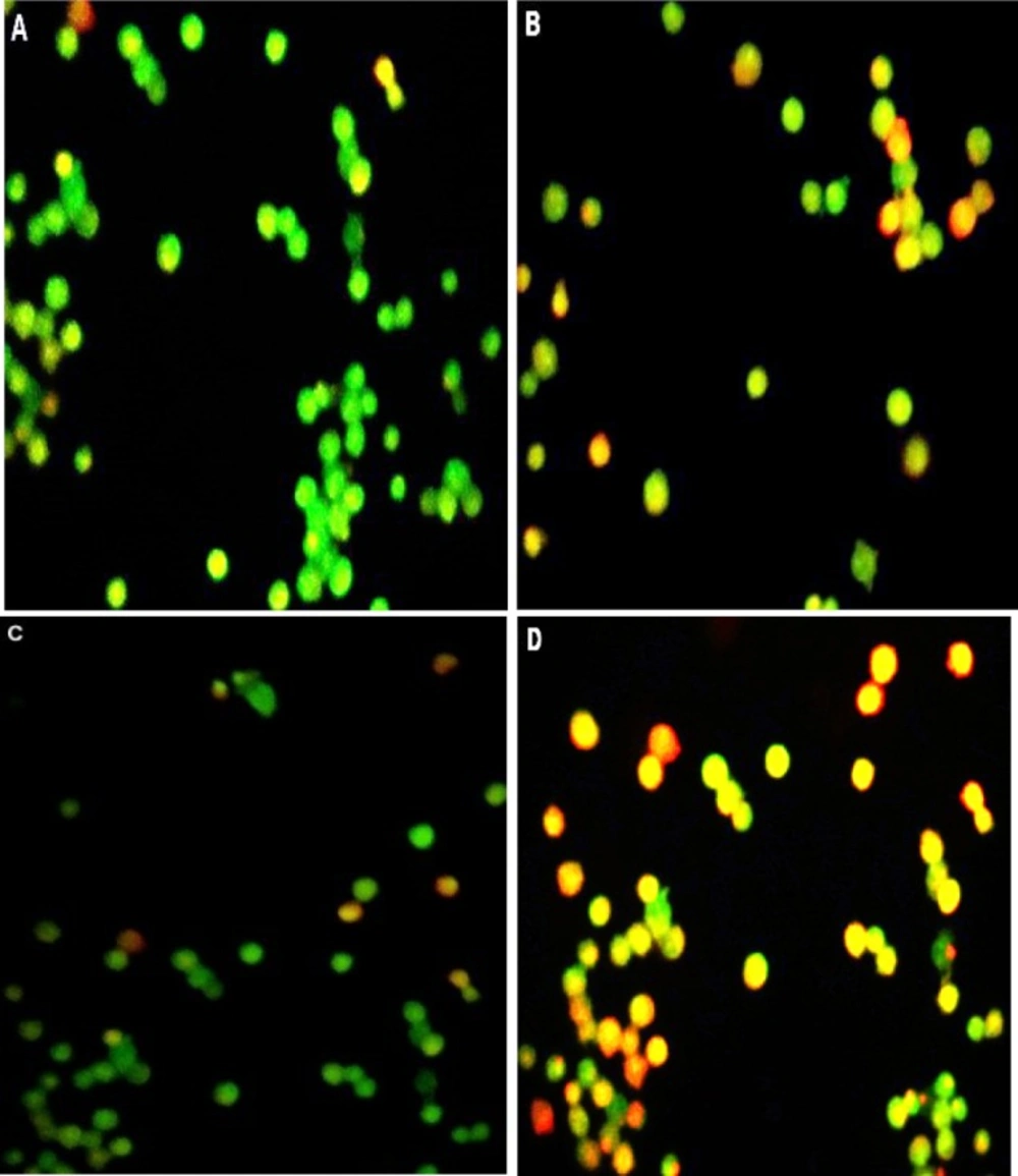
AO/PI staining: the cells were seeded and treated. Forty-eight hours after the seeding, AO/PI staining 10 µL of the live cell sample and 10 µL of AO/PI staining solution were combined and analyzed under the fluorescent microscopy.
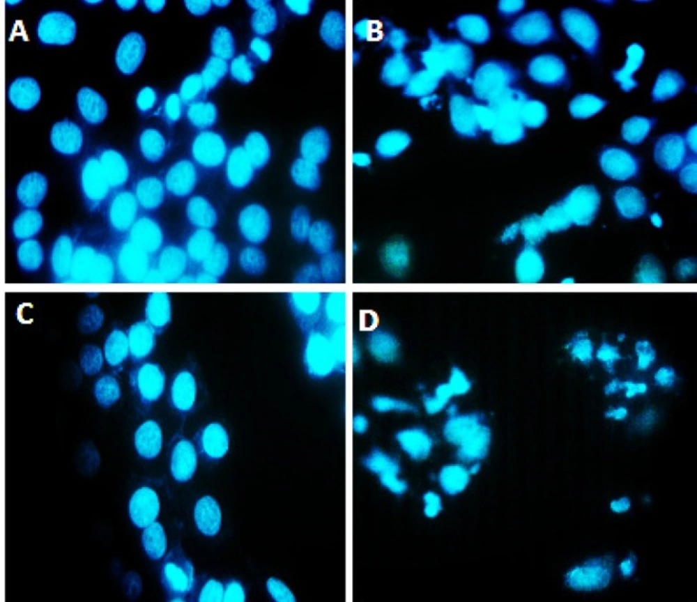
DAPI staining: About 5000 cells were cultured in plate containing gelatin-coated coverslips. After 24 h, the culture medium was removed from each well, and the cells were washed with PBS twice. In the next stage, the cells were treated with cAgNPs (2 µg/mL), cisplatin (2.5 µg/mL), or a combination of cAgNPs and cisplatin for 48 h. Afterwards, the cells were fixed with methanol and were stained with DAPI solution (1 mg/mL) for 10–60 sec. Following that, the wells were washed with cold PBS twice. Finally, the comounds were observed under fluorescent microscopy.
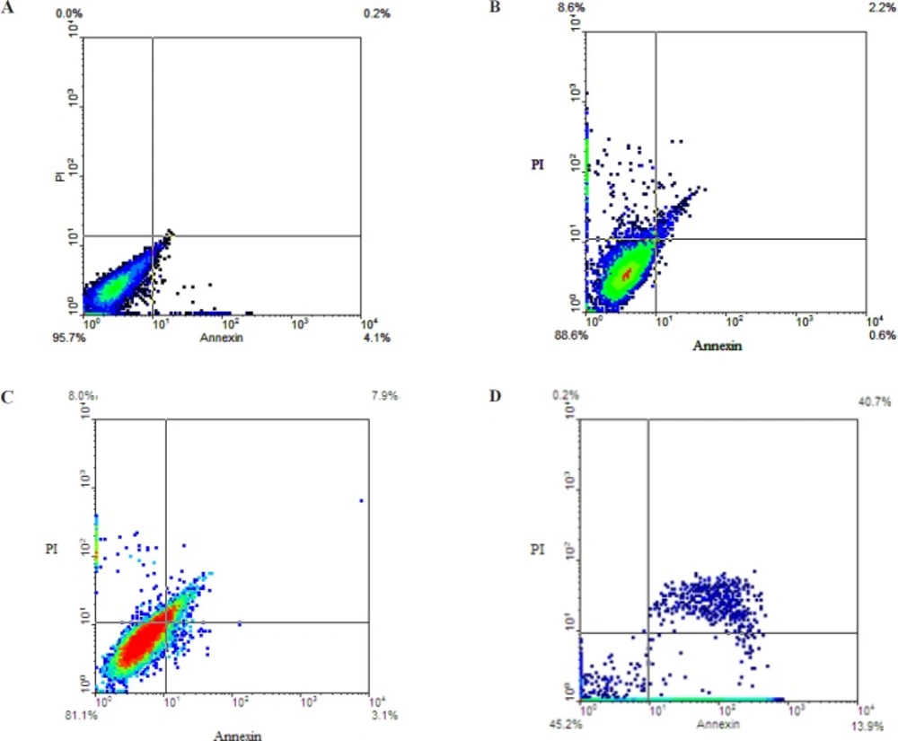
Annexin V/PI staining for apoptosis detection
Percentage of early and late apoptotic cells induced with cAgNPs, cisplatin, and a combination of both on A2780 cell were determined by Annexin-V-FITC/PI staining. According to the manufacturer’s instructions, the cells were treated with cAgNPs, cisplatin, or both for 48 h. Afterwards, the cells were harvested and centrifuged at 200×g and suspended in appropriate buffer. Following that, 5 µL Annexin-V-FITC labeling and 5 µL PI solutions were added to the mixture, which were then incubated for five minutes at 25 °C and analyzed with flow cytometry (Bd, UK).
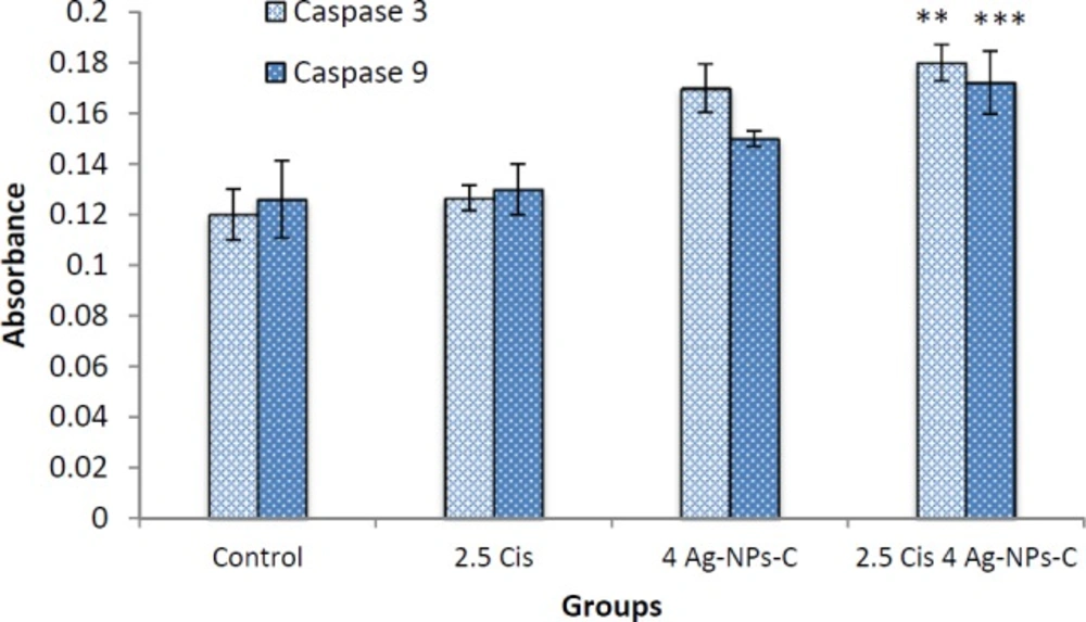
Caspase 3/9 activation assay
Caspase 3/9 activities were assessed using the Colorimetric Protease Assay Kit according to the protocol of manufacturer. Briefly, the cells were treated with AgNPs. Apoptosis was induced in the cells treated with cAgNPs and cisplatin for 24 h. After that, the 1-5 × 10⁶ cells were pelleted and re-suspended in 50 μL of chilled cell lysis buffer before centrifuging for one minute. The protein concentration was assayed using the Biuret method. For each assay, 100 μg proteins were diluted with 50 μL cell lysis buffer. Finally, the DEVD-p-NA substrate was added and the samples were read at 400 or 405 nm using a microtiter plate reader (Epoch, US). The fold-increase in caspase-3 activity was determined through comparison with the control groups.
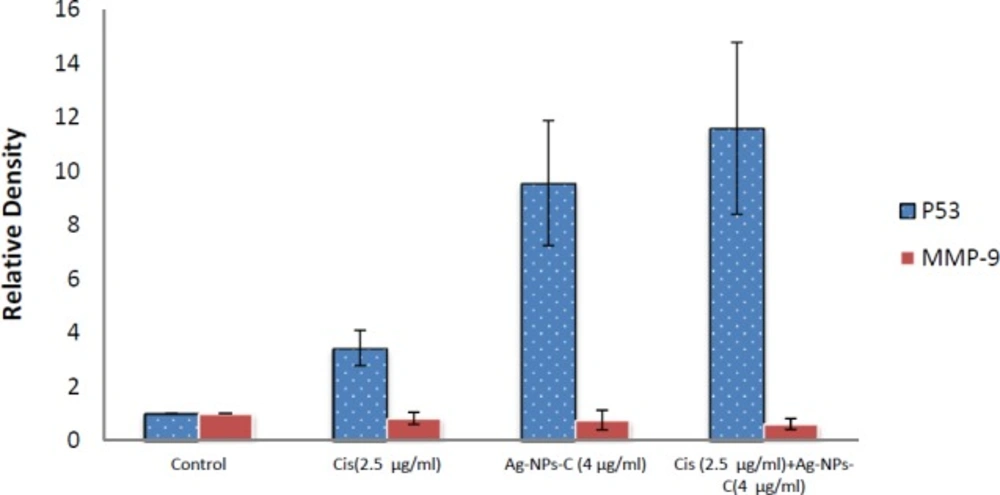
Semi-quantitative analysis of p53 AND MMP-9 genes expression
Changes in the expression of MMP genes were assessed using semi-quantitative reverse transcription polymerase chain reaction (RT-PCR). A2780 cells were treated (according to previous descriptions) for 48 h, and total RNA was extracted (Rouch, Germany). cDNAs were synthesized by reverse transcriptase and amplified by polymerase chain reaction (PCR) with specific primer pairs, as observed in
Table 1, using Fermentas PCR Kit (Fermentas, US). The PCR products were analyzed by 1.5% agarose gel electrophoresis, and the gels were observed using gel documentation (UV TEC Cambrege, UK). The tests were repeated in triplicate, and the relative band densities of cDNA.
Statistical analysis
Statistical evaluation of the data was performed using one-way analysis of ANOVA, Tukey test. The results were shown as mean ± SD and p < 0.05 was calculated as the minimum level of significance.