General Experimental Procedures
Agilent 1290 Infinity LC-6520 Accurate-Mass QTOF-MS (Agilent Technologies, PaloAlto, CA, USA); Bruker DRX-400 (400MHz for 1H and 100MHz for 13C) spectrometer (Bruker, Rheinstetten, Germany); α-Glucosidase (E.C. 3.2.1.20) from Saccharomyces cerevisiae, p-nitrophenyl a-D-glucopyranoside (PNPG) and glutathione (GSH) were purchased from Sigma-Aldrich (St Louis, MO, USA). CNBr-activated SepharoseTM 4B was purchased from Phamacia Biotech AB (Uppsala, Sweden). Distilled water was purified by a Milli-Q water purification apparatus (Millipore, Bedford, MA). All other reagents were of analytical grade.
Plant material
20 kinds of traditional Chinese herbs (including C.rotundus) were collected from Yangguang drugstore, Dalian, China. The specimens were identified by Prof. Zhai Yanjun, Liaoning University of Traditional Chinese Medicine, China. Voucher specimens were deposited at College of Pharmacy, Liaoning University of Traditional Chinese Medicine (Voucher No. from LNU-DSR-2015-1 to LNU-DSR-2015-26).
Immobilization of a-glucosidase
a-Glucosidase (0.20 mg) was dissolved in coupling buffer (0.1 M NaHCO3 and 0.5 M NaCl at pH 8.3) at a concentration of 2.0 mg protein/mL. The protein solution was mixed with CNBr-activated SepharoseTM 4B gel at room temperature in a ratio of 100 L protein solution to 5 mg SepharoseTM powder, which would swell to about 20 mL of wet gel. The gel slurry was rotated at room temperature for 1 h to ensure the enzyme bound to the beads. After protein coupling, the resident reactive sites on the gel were blocked by reacting with 0.01 M Tris-HCl buffer at pH 8.0 for 1 h at room temperature. After alternatively washed with 0.1 M CH3COONa buffer (pH 4.0 containing 0.5 M NaCl) and 0.1 M Tris-HCl buffer (pH 8.0 containing 0.5 M NaCl), the gel was kept in affinity buffer for further use. Control supports were prepared in the same manner but with no AGH being added during the immobilization step.
Enzyme immobilization yields were determined by comparison of the amount of free enzyme in the solutions before and after coupling to the gel according to the method described by Bradford using BSA as standard protein, giving 9.5 mg AGH/mL gel.
The specific activity of immobilized AGH was determined as 18.4 U/mg protein (175 U/mL gel).
Preparation of affinity solution of C. rotundus
200 mg ground C. rotundus powder was extracted in 20 mL of 80% aqueous methanol by ultrasonic extraction for 30 min. The extract was filtered and freeze-dried, then redissolved in 10 mL affinity buffer (67 mM KH2PO4 (containing 5% methanol) at pH 6.8) and filtered through a 0.45 μm membrane as the tested extract sample.
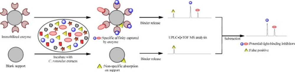
AGH inhibitors screening by immobilized AGH affinity fishing
C. rotundus extracts (400 mL) was mixed with 20 mL immobilized AGH gel and gently stirred at 37 °C for 20 min. After incubation with the extracts, the gel were washed with 400 mL affinity buffer (67 mM KH2PO4 (containing 5% methanol) at pH 6.8) four times to remove any unbound components and then treated with 200 mL desorbed solution (50% methanol aqueous solution at pH 3.3) to release the captured potential inhibitors of AGH. The released components were collected and concentrated to 10 mL under N2 atmosphere at 40 ºC, then applied to UPLC-QTOF-MS for peak identification. Elution from the control supports was collected and analyzed in the same manner.
UHPLC-QTOF-MS analysis
UHPLC-MS was performed on an Agilent 1290 Infinity LC-6520 Accurate-Mass QTOF-MS (Agilent Corporation, CA, USA). Analytes eluted from the AGH coupling supports and control supports were separated on a poroshell 120 EC-C18 column (3.0 × 50 mm, 2.7 µm , Agilent Corporation, MA, USA). The solvent system consisted of solvent A (menthol) and solvent B (0.5% formic acid in water, v/v). The solvent A content of the mobile phase was gradient increased from 5% to 100% linearly in 20 min.
| Scientific name | Parts | Inhibitory (%)
|
|---|
| 0.1 mg/mL | 0.5 mg/mL |
|---|
| Astragalus membranaceus (Fisch.) Bunge var. mongholicus (Bunge) P. K. Hsiao | Root | 1.2 | 5.1 |
| Salvia miltiorrhiza Bunge. | Root | 4.5 | 8.0 |
| Rheum palmatum L. | Root | 3.0 | 12.6 |
| Taraxacum mongolicum Hand.-Mazz. | Herb | 5.6 | 19.2 |
| Schisandra chinensis (Turcz.) Baill. | Fruit | 5.0 | 25.5 |
| Trigonella foenum-graecum L. | Seed | 15.0 | 38.5 |
| Cinnamomum cassia Presl | Bark | 19.6 | 40.0 |
| Cuscuta chinensis Lam. | Seed | 14.5 | 40.1 |
| Atractylodes lancea (Thunb.) DC. | Root | 9.4 | 40.5 |
| Aucklandia lappa Decne | Root | 25.7 | 43.4 |
| Angelica sinensis (Oliv.) Diels | Root | 22.5 | 47.0 |
| Pueraria lobata (Willd.) Ohwi | Root | 14.0 | 48.6 |
| Anemarrhena asphodeloides Bunge | Root | 20.0 | 48.8 |
| Gardenia jasminoides Ellis | Fruit | 40.0 | 72.8 |
| Peonia lactiflora Pall. | Root | 43.8 | 81.0 |
| Canavium album Raeusch. | Fruit | 74.5 | 87.9 |
| Morus alba L. | Root-bark | 86.9 | 93.3 |
| Sophora japonica L. | Flower | 92.8 | 97.9 |
| Glycyrrhiza uralensis Fisch. Ex DC. | Root | 81.6 | 98.7 |
| Cyperus rotundus L. | Root | 95.1 | 99.5 |
| Compounds | alpha-glucosidase inhibitory
|
|---|
| IC50 (µM) | Kinetic mode (Ki, µM) |
|---|
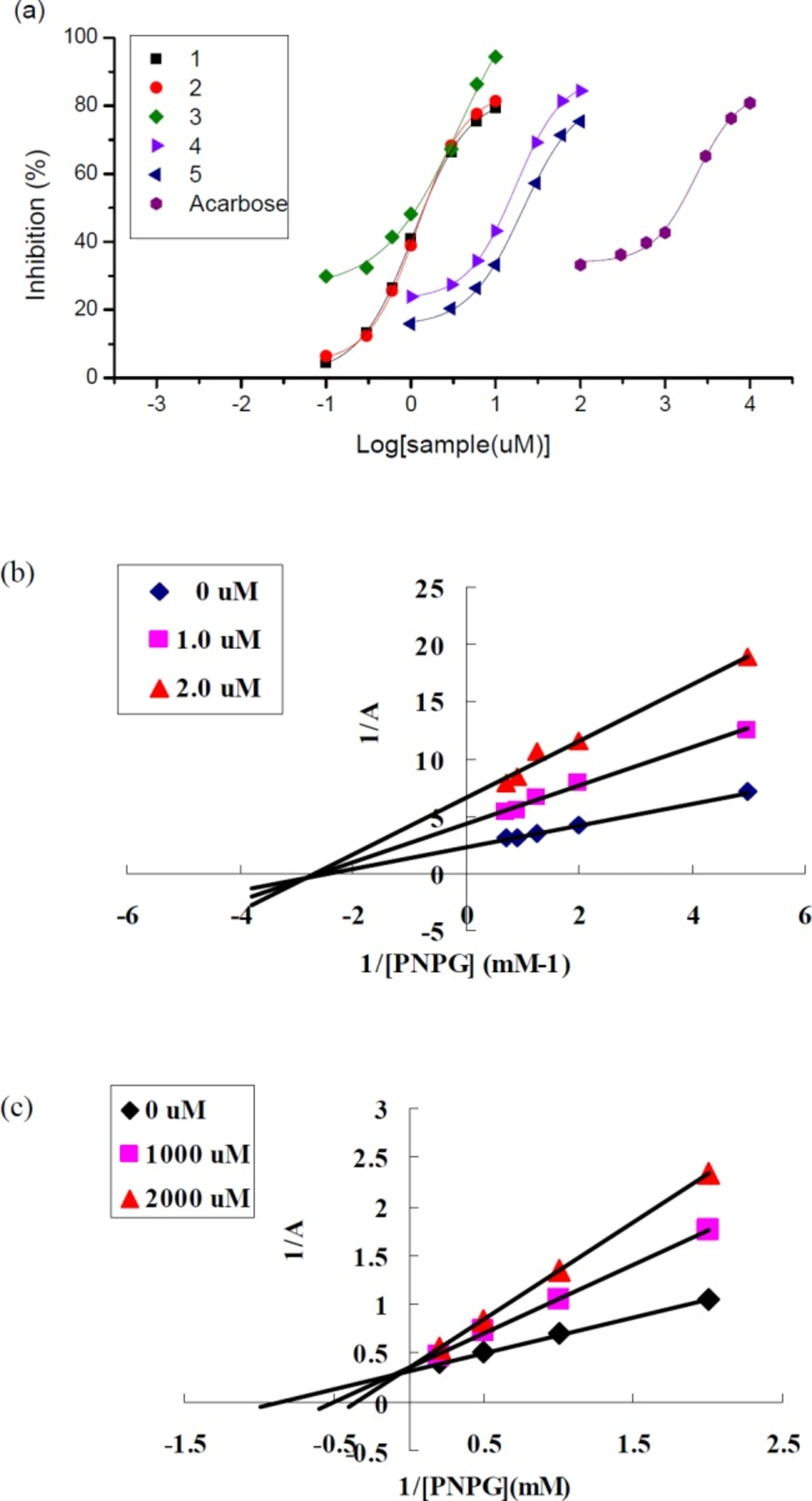
| Cyperusphenol A (1) | 1.43 ± 0.11 | Noncompetitive (11.2) |
| Mesocyperusphenol A (2) | 1.44 ± 0.17 | Noncompetitive (10.7) |
| Cyperusphenol D (3) | 1.18 ± 0.11 | Noncompetitive (8.63) |
| Scirpusins B (4) | 13.3 ± 1.66 | Noncompetitive (95.4) |
| Scirpusins A (5) | 21.4 ± 1.40 | Noncompetitive (136) |
| Acarbose (reference) | 1470 ± 117 | competitive (834) |
Rapid screening of natural AGHI from C. rotundus by affinity capture system
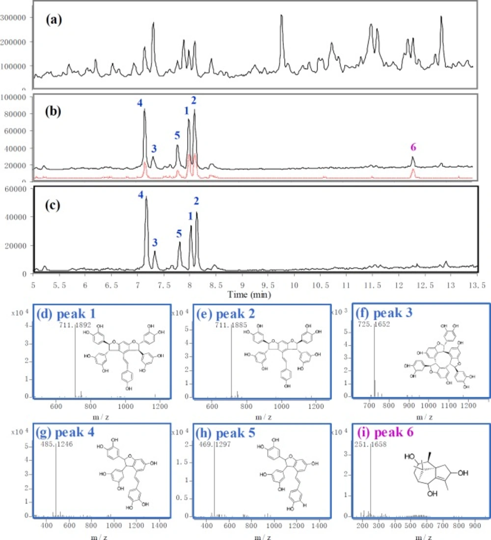
Screening results of C. rotundus by using affinity capture method. (a) TIC of C. rotundus extract. (b) TIC of the binders screened out from C. rotundus extract. The black solid line represents the binders absorbed on immobilized α-glucosidase. The red dotted line represents the binders absorbed on blank supports (false positive). (c) TIC of the highly-affinity ingredients, which was made by subtracting the signal of false positive. (d)-(i) The accurate mass spectrum and structure of peak 1-6 (cyperusphenol A, mesocyperusphenol A, cyperusphenol D, scirpusins B, scirpusins A, sugetriol)
Inhibitory effects of compounds 1-5 on -glucosidase (a) and Lineweaver-Burk plots of compound 1(b) and acarbose(c)
The flow rate was set at 0.4 mL/min. Mass analysis was performed in negative ESI ion mode. The operating parameters were as follows: drying gas (N2) flow rate, 8 L/min; drying gas temperature, 325 oC; nebulizer, 30 psig; capillary, 3500 V; skimmer, 65 V; fragmentor voltage, 100 V. The mass range was set at m/z 100–2000. The system was operated under Agilent MassHunter workstation acquisition software, version B.02.01 (Agilent Corporation, MA, USA).
Isolated procedure of 5 natrual AGHIs from C. Rotundus
The shade dried C. rotundus (1 kg) was powdered and extracted with 80% aqueous methanol (3 × 2 L) by ultrasonic extraction for 60 min at room temperature. The solution was filtered and concentrated under reduced pressure on a rotatory evaporator, yielding 102 g of crude extract. The entire methanol crude extract (102 g) was suspended in 1 L of water and then partitioned sequentially with equal volumes of n-hexane, EtOAc, and n-BuOH. The methanol extract of four fractions were detected by UHPLC-MS, respectively. Amnog them, EtOAc fraction (23 g) was found to have most of the stilbenoids oligomers and was separated over silica gel. The EtOAc fractions were eluted with n-hexane, CH2Cl2, CH2Cl2/MeOH (9:1 ® 7:3 ® 5:5 ® 3:7 ® 1:9 ® only MeOH) to give eight fractions (F1-F8). F4 (70% CH2Cl2, 2.8 g) was found to be the aboundent part by LC-MS. Then the F4 fraction was subjected on Prep-RP-HPLC with 35% aqueous MeOH as an elunt to give compound 1 ((E)-cyperusphenol A, 24 mg), 2 (mesocyperusphenol A, 11 mg), 3 (Cyperusphenol D, 6.6 mg), 4 (Scirpusins B, 19 mg) and 5 (Scirpusins A, 65 mg).
Preparation of crude extracts of 20 herbs
Each of the dried ground powder (3 g) was extracted in 30 mL of 80% aqueous methanol by ultrasonic extraction for 60 min at room temperature. After centrifugation at 5000 rpm for 5 min, the combined extract was evaporated under reduced pressure to dryness. To each herb extract, DMSO was added to get 5 mg/mL solution, respectively.
α-Glucosidase inhibitory activity assay in vitro and kinetics analysis
The α-glucosidase inhibitory activity was determined spectrophotometrically on 96-well microplate reader. In brief, to a total of 310 μL of reaction mixture containing 265 μL of 67 mM phosphate buffer (pH 6.8), 10 μL of 3 mM glutathione, 25 μL of 10 mM p-nitrophenyl-α-D-glucopyranoside and 10 μL of investigated compounds in the wells was added 10 μL of 0.3 U/mL α-glucosidase and mixed. All the reagents used in this assay were dissolved in the same phosphate buffer (67 mM, pH 6.8). After incubation for 10 min at 37 oC, the reaction was stopped by adding 800 μL of 0.1 M Na2CO3 solution. Then the absorbance of sample (AS) at 405 nm was recorded (SunriseTM microplate absorbance reader, Tecan, Austria). The control was the same mixture except for the investigated sample replaced by the phosphate buffer. The sample blank and control blank were the same mixtures as sample and control, respectively, except α-glucosidase was instead with phosphate buffer, respectively.
The α-glucosidase inhibition activity (%) of test sample could be calculated as
Inhibition activity (%) = 100% × [(AS - ASB) /(AC -ACB)]
where AS, ASB, AC and ACB are the absorbance of sample, sample blank, control and control blank, respectively. The measurement was performed in triplicates. For kinetic analysis of AGH inhibition, a certain concentration of 0.6 U/mL AGH and different contents of sample compounds were incubated with a series of concentrations of substrate. The inhibitory kinetics of the investigated compounds was analyzed using the Lineweaver-Burk plot, double-reciprocal plot of the substrate concentration and velocity.