Chemicals
The chemicals used for HAp preparation were calcium nitrate tetrahydrate (Ca (NO3)2.4H2O, with molecular weight (Mwt) 236.15 g/mole, Merk, Germany), diammonium hydrogen orthophosphate anhydrous ((NH4)2HPO4, Mwt 132.06g/mole, S.D. Fine Chem. Ltd. Mumbai, India), and ammonium hydroxide (NH4OH, Mwt. 35.5g/mole, May & Baker, England). Aluminum chloride hexahydrate (AlCl3. 6H2O) was purchased from Sigma-Aldrich.
All chemicals and reagents used were of analytical grade and used without further purification.
Preparation of nano-HAp
The nano-HAp prepared according to (
16) by preparing reacting of 0.497M/L of Ca(NO
3)
.4H2O and 0.298M/L of(NH4)2HPO4which were dissolved separately under pH control. The obtained precipitates were separated from the mother liquor by filtration and dried at 90 °C and calcined at 1000 °C for 24 h. The infrared spectra of the products were carried out with Mattson Infinity Series FTIR made in USA, in the wave number range from 400-4000 cm-1 using the KBr disc technique. The samples were characterized for qualitative and quantitative phase content by X-ray diffraction (XRD) by using Shimadzu X-ray diffractometer made in Japan. An XRD analysis was performed after calcining the synthesized powder to reveal the structural of the prepared powder after heat treatment.
Pharmacological Study of Acute Toxicity
Determination of acute toxicity for intravenous treatment with nano-HAp was carried out using the method of previously published by Lorke (
17). Fifteen rats were used to determine the toxicity of the prepared nano-HAp. The rats were divided into five groups of three rats each. The rats were injected intravenously with nano-HAp at dose levels of 100, 200, 300, 500, 1000 mg/kg b. w. Mortality was recorded for near 24 h and the final LD
50 value was determined from the minimum concentration (full death) and maximum concentration (no death) of the dose according to the coming relation:
LD50 = (M0+ M1) /2
Where M0 = Highest dose of nano-HAp that gave no mortality, M1 = Lowest dose of nano-HAp that gave mortality.
Preliminary study using nano-HAp to treat brain damage
Several experiments were carried out to evaluate the pathophysiological features of brain in rats intoxicated by AlCl3 before and after treatment with nano-HAp. The selected dose of nano-HAp was examined by the intravenous injection at different time intervals to detect the optimal therapeutic results.
It was reported that after i.v. injections of 100 mg/kg b.w. nano-HAp (
12), the particles stay in the blood until they enter the reticuloendothelial system (macrophages of the liver, spleen and bone marrow). The LD
50 of this preparation was determined as 1200 mg/kg b.w. (
15). Plasma half-life time of one hour has been reported by Xie (
18). However, structural and functional changes in brain were investigated including DNA fragmentation, neurotransmitters, and oxidation status. A divided dose of 300 mg/kg b.w. nano-HAp (a quarter dose of LD
50) was applied in the present experiment.
Animals and treatment schedule
Twenty-four male albino rats were kept under hygienic conditions with a 12/12 h light - dark cycle and were allowed free access to food and water adlibitum for at least one week prior to the experiment.
The animals were assigned into three groups (n=8). Group 1: control rats. Group 2: rats administered AlCl
3 (100 mg/kg b.w.) intraperitoneally for 90 days. Group 3: rats injected intravenously with nano-HAp (dissolved in saline, 100 mg/Kg b.w.) three times/week (
11,
12) after being induced by AlCl
3 for 90 days, for one week. The blood samples were collected from orbital venous plexus of all rats 24 h post last injection of nano-HAp for biochemical estimation. The brain tissues were obtained from the animals for DNA fragmentation and histological examination.
Tissue Preparation
At the end of treatment period, the rats were anaesthetized by diethyl ether and sacrificed by cervical decapitation. The brains were dissected out by making midline incision to view the skull. A small incision from the caudal part of parietal bone and a firm cut in the anterior part of the frontal bone were made to remove the brain more easily. The isolated brains tissues were immediately taken out and washed with ice cold saline to remove blood and they were either fixed in 10% formalin for histopathological examination or stored at -80 °C, till later analysis.
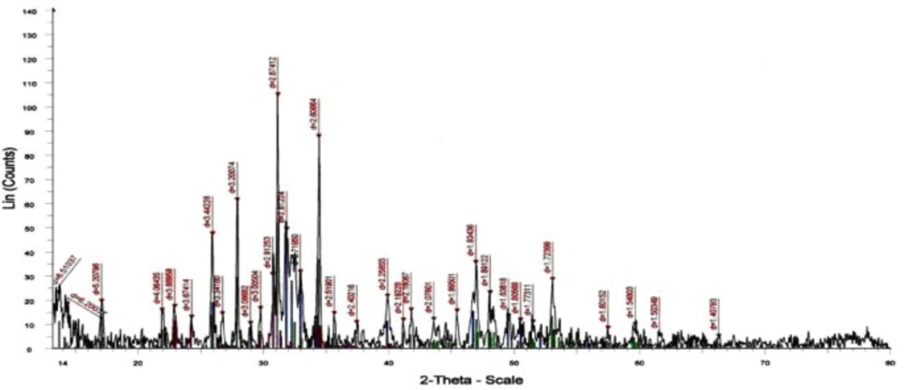
XRD patterns of the sample prepared with Ca/P molar ratio 1.67 under pH control calcined at 1000 °C
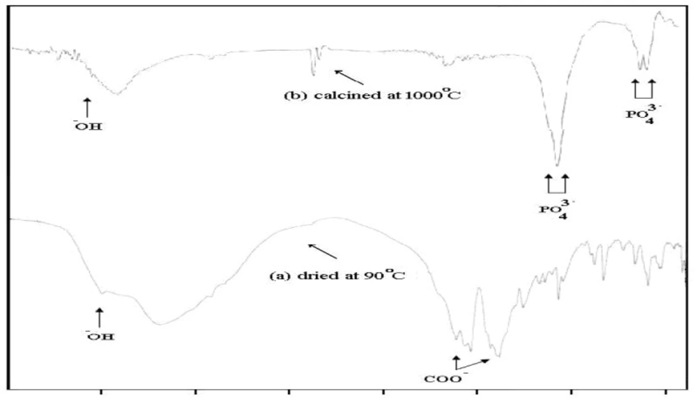
IR spectra of the sample prepared with Ca/P molar ratio 1.67 under pH control dried at 90 C and which calcined at 1000 °C
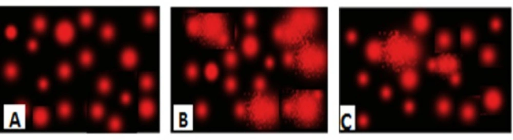
Photomicrograph represented DNA damage (Comet assay) in brain of normal rats (A), rats administered AlCl3 for 90 days(B) and rats treated with nano-HAp (C).
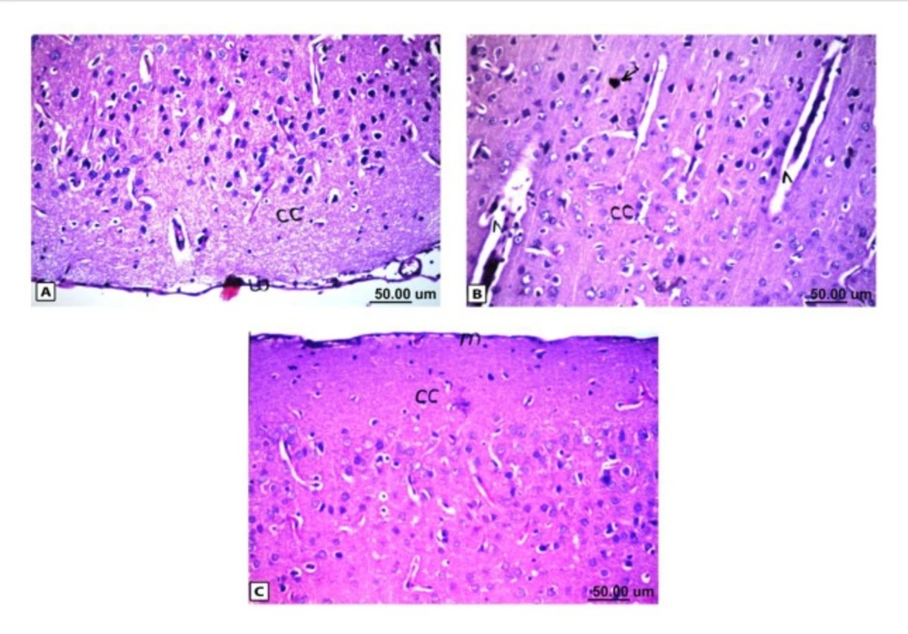
Histopathological results of rat brain area of meninges (m) and cerebral cortex (cc). (A) Normal control group: normal structure of meninges (m) and cerebral cortex (cc). (B) Rat administered AlCl3: the cerebral cortex showed congestion in the blood capillaries with degeneration in some neuronal cells. (C) Rat treated with i.v. nano-HAp after AlCl3: normal structure of meninges (m) and cerebral cortex (cc)
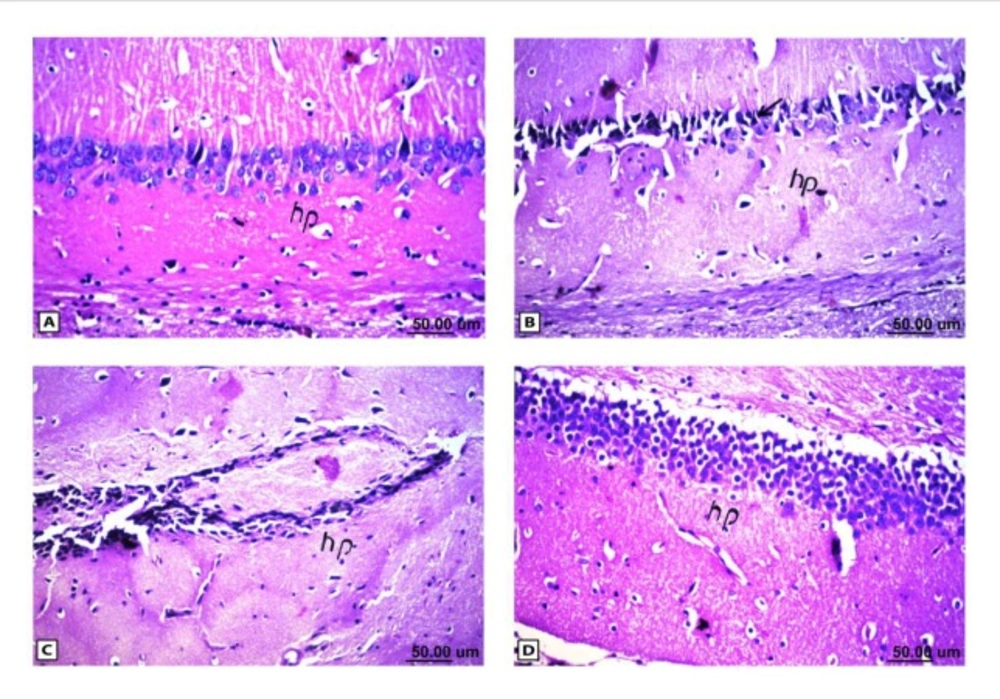
Histopathological results of rat brain area hippocampus (hp). (A) Normal control group: normal structure of the hippocampus (hp). (B & C) Rat administered AlCl3: neuronal degeneration and pyknosis (arrow) in hippocampus (hp) & atrophy hippocampus. (D) Rat treated with i.v. nano-HAp after AlCl3: normal structure of the hippocampus (hp)
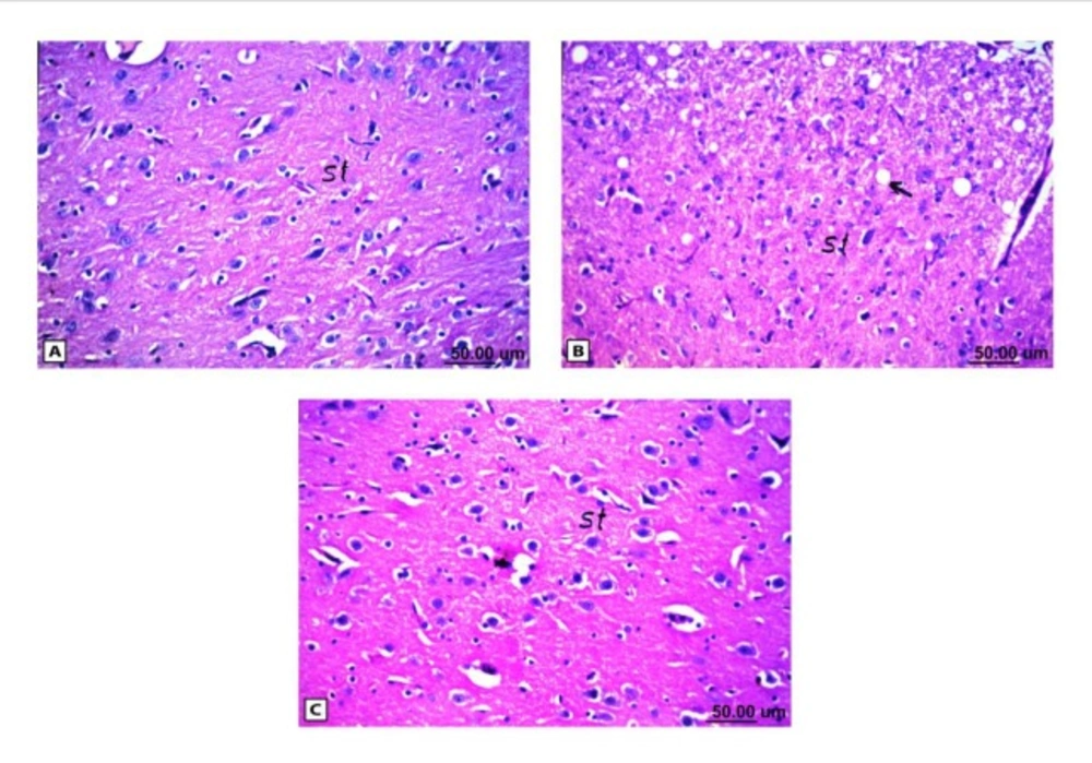
Histopathological results of rat brain area of striatum (st). (A) Normal control group: normal structure of striatum (st). (B) Rat administered AlCl3: vacuolization in the matrix of striatum (arrow). (C) Rat treated with i.v. nano-HAp after AlCl3: Normal structure of striatum (st)
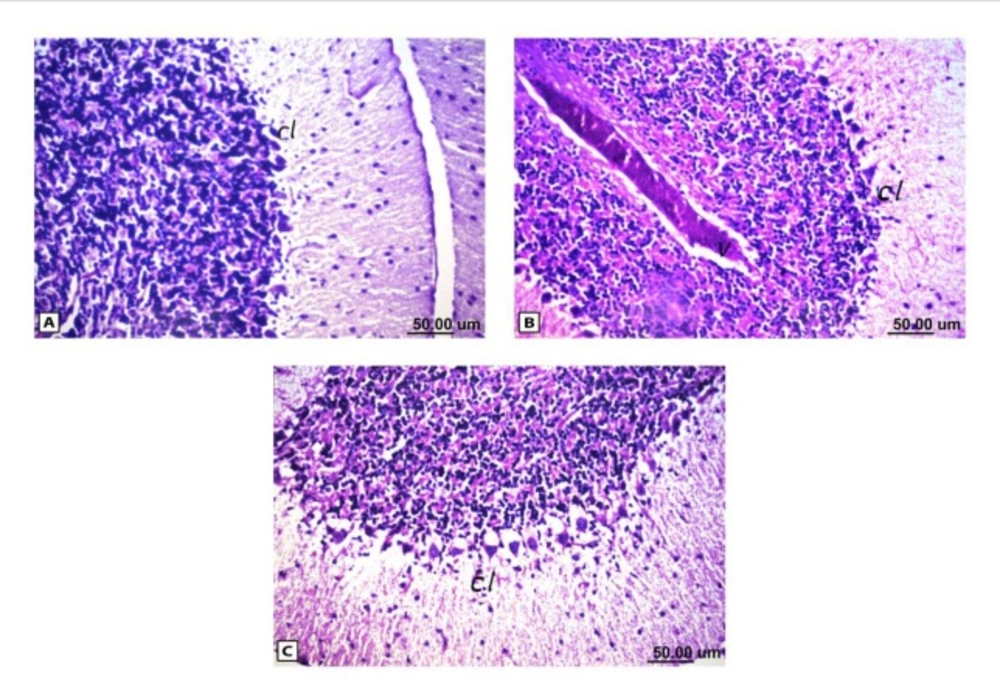
Histopathological results of rat brain area of cerebellum (cl). (A) Normal control group: normal structure of cerebellum (cl).
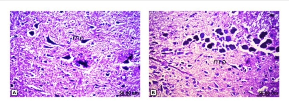
Histopathological results of rat brain area of medulla oblongata. (A) Normal control group: normal medulla oblongata structure. (B) Rat treated with i.v. nano-HAp after AlCl3: normal medulla oblongata structure
| Type of spectrum | Vibration of hydroxyl -OH υ (cm-1) | Phosphate PO 3-stretching4vibration υ (cm-1) | Phosphate PO 3- bending4vibration υ (cm-1) |
|---|
| HAp bands | 630 | 3570 | 963 | 1036 | 1091 | 565 | 603 |
| Sample dried at 100°C | - | 3570 | 980 | 1054 | 1079 | 539 | 600 |
| Sample calcined at 1000°C | 630 | 3567 | - | broad | 1180 | 569 | 603 |
| Tailed % | Untailed % | Tail length (µm) | Tail DNA % | Tail moment UNIT |
|---|
| Control | 3 | 97 | 1.22±0.04 | 1.31 | 1.59±0.05 |
| AlCl3 | 21 | 79 | 3.81±0.24 | 3.96 | 15.05±1.08 |
| AlCl3+nano-HAp | 16 | 84 | 2.89±0.11 | 2.93 | 8.47±1.01 |
| Groups | Control | AlCl3 | Nano-HAp+AlCl3 |
|---|
| Parameters |
|---|
| 5-HT(ng/100mg) | 8.56±0.17a | 6.02±0.12b | 7.90±0.22a |
| NA (ng/100mg) | 6.92±0.13a | 4.56±0.14b | 6.22±0.15c |
| Dopamine (ng/100mg) | 25.99±0.34a | 17.82±0.38b | 28.09±0.81a |
| Groups | Control | AlCl3 | Nano-HAp+AlCl3 |
|---|
| Parameters |
|---|
| Sphingomyelin(mg/gwet) | 2.83±0.07a | 1.94±0.03b | 2.82±0.10a |
| NRF1(ng/100mg) | 24.96±0.98a | 11.77±0.36b | 16.64±0.42c |
| Caspase3 (ng/100mg) | 111.95±0.72a | 168.04±1.66b | 137.23±1.69c |
| Groups | Control | AlCl3 | Nano-HAp+AlCl3 |
|---|
| Parameters |
|---|
| MDA (nmol/mg) | 0.96±0.07a | 4.27 ±0.10b | 2.04 ±0.10c |
| GPx (nmol/mg) | 37.06 ±0.73a | 19.83 ±0.54b | 30.67 ± 1.60c |
| Control | AlCl3 | AlCl3+Nano-HAp |
|---|
| - | ++++ | - |
DNA fragmentation
Brain DNA damage was determined by a single-cell gel electrophoresis (comet) assay according to the method previously published by Singh
et al., (
19). A 0.5 g of crushed brain sample was transferred to 1 mL ice-cold phosphate buffer saline (PBS). The suspension was stirred for 5 min then filtered. Cell suspension (100 µL) was mixed with 600 µL of low-melting agarose (0.8% in PBS). 100 µL of this mixture was spread on pre-coated slides, which were immersed in lyses buffer (0.045 M TBE, pH 8.4, containing 2.5% SDS) for 15 min. The slides were placed in electrophoresis chamber containing the same TBE buffer, but devoid of SDS. The electrophoresis conditions were 2 V/cm and 100 mA for 2 min. Staining was made with Ethidium bromide (EtBr) 20 µg/mL at 4 °C. The observation was reported while the samples still humid, the DNA fragment migration patterns of 100 cells for each dose level were evaluated with a fluorescence microscope (With excitation filter 420-490 nm (issue 510 nm). For visualization of DNA damage, observations were made of EtBr-stained DNA using a 400X objective on a fluorescent microscope. The comets tails lengths were measured from the middle of the nucleus to the end of the tail.
Comet capture and analysis
A total of 100 randomly captured comets from each slide were examined at 400 X magnification using a fluorescence microscope connected to a CCD camera using an image analysis system [Comet 5 image analysis software developed by Kinetic Imaging Ltd. Liverpool, UK]. A computerized image analysis system acquires images, computes the integrated intensity profiles for each cell, estimates the comet cell components, and then evaluates the range of derived parameters. To quantify the DNA damage, the tail length (TL), the percentage of migrated DNA (tail DNA %), and tail moment (TM) were evaluated. TL (length of DNA migration) is related directly to the DNA fragment size and presented in micrometers. It was calculated from the center of the cell. Finally, the program calculates TM.
The DNA damage was quantified by measuring the displacement between the genetic material of the nucleus (Comet head) and the resulting (tail).
Tail moment = Tail DNA % X Length of tail
Biochemical Estimation
The obtained tissue was analysed for the following: sphingomyelin, using a commercial quantification colorimetric assay kit purchased from BioVision (CA, USA. Catalog # K600-100), nuclear respiratory factor 1 (NRF1) and caspase-3 of brain were measured using ELISA commercial kits purchased from USCN Life Science Inc., (Wuhan, China. number SEC669 Ra and SEA626 Ra. respectively). Neurotransmitters such as catecholamine (dopamine and noradrenalin) and 5-hydroxytryptamine (5-HT) were also evaluated according to the method reported by Zagrodzka et al. (20). Lipid peroxidation products were determined by measuring the quantity of malondialdehyde (MDA) produced using colorimetric/fluorometric kit purchased from (BioVesion Research Products, USA, Catalog number K739-100). The enzyme activity of Glutathione peroxidase (GPx) was assayed using kinetic kit purchased from (BioVesion Research Products, USA, Catalog number K762-100)
Histopathological examination
For histopathological examination through the electric light microscope, the samples were taken from brain of eight rats in each group and fixed in 10% formal saline for twenty four hours then washed with tap water. Serial dilutions of alcohol (methyl, ethyl and absolute ethyl) were used for dehydration. Specimens were cleared in xylene and embedded in paraffin at 56 ⁰C in hot air oven for twenty four hours. Paraffin bees wax tissue blocks were prepared for sectioning at 4 microns by sledge microtome. The obtained tissue sections were collected on glass slides, de-paraffinzed, and stained by hematoxylin and eosin stains (
21)
Statistical analyses
All values were expressed as mean ± S.E. Statistical analysis was performed with one way analysis of variance (ANOVA) followed by Duncan’s test using SPSS program. Ρ values < 0.05 were considered to be statistically significant.