Chemicals
Dulbecco’s modified Eagle’s medium (DMEM), fetal bovine serum (FBS), trypsin-EDTA solution, penicillin/streptomycin 100 units, dimethylsulfoxide (DMSO), and all other fine chemicals were obtained from Merck, Germany. 3-(4,5 dimethylthiazole-2-yl)-2,5-dimethyl tetrazolium bromide (MTT) and trypan blue were purchased from Sigma (St Louis, MO).
Plant materials
Three endemic plants including Scutellaria nepetifolia Benth., Sc. multicaulis subsp. multicaulis Boiss., and Nepeta laxiflora Benth. were collected in mid July 2016 from Hamedan province and another plant Sc. tournefortii Benth. was collected in May 2016 from Golestan province of Iran. All plants were identified by botanists at Tehran University of Medical Sciences (TUMC) and Shahid Beheshti University of Medical Sciences, Tehran, Iran. A voucher specimen of each species is deposited at respective herbarium.
Preparation of extracts
Aerial part of each plant was shade dried, powdered, and macerated with methanol in 1 to 10 proportion, respectively. Maceration lasted for 72 h and a fresh solvent replaced the extract every 24 h. For fractionation of the selected plant, maceration was carried out with the same process except that n-hexane, dichloromethane, ethyl acetate, and methanol were used as solvents, respectively. All extracts and fractions were collected and concentrated by rotary evaporator Heidolph 4000 (Schwabach, Germany) at room temperature, to remove all solvent residuals.
Cell culture and treatment
PC12 (rat pheochromocytoma) cells obtained from Pasteur Institute (Tehran, Iran), were grown in DMEM enriched by 10% FBS, supplemented with 100 unit/mL penicillin and 100 mg/mL streptomycin and maintained at 37 °C in a humidified atmosphere (90%) containing 5% CO2.
Plant extracts and fractions were dissolved in dimethyl sulfoxide (DMSO) to make stock solutions. The stock solutions and hydrogen peroxide (H
2O
2) were diluted with DMEM to gain interested concentrations prior to use. All of the experiments involving exposure to H2O2 and measurement of NO were performed in serum-free DMEM to avoid rapid H
2O
2 degradation by antioxidants and interference with Griess assay by FBS (
12,
13).
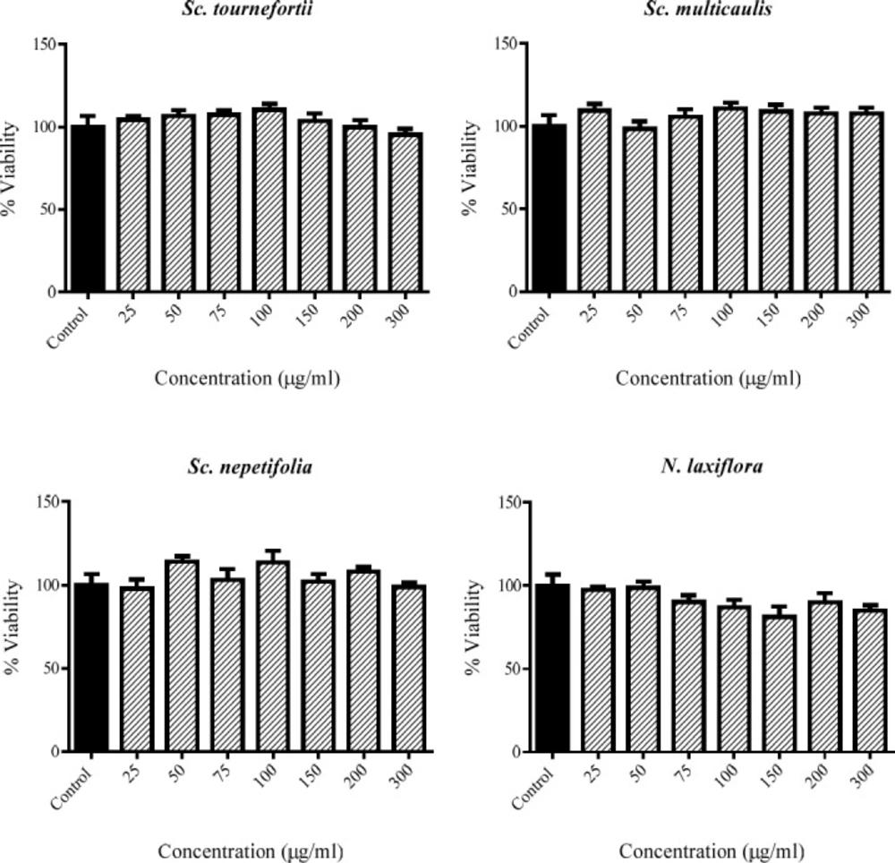
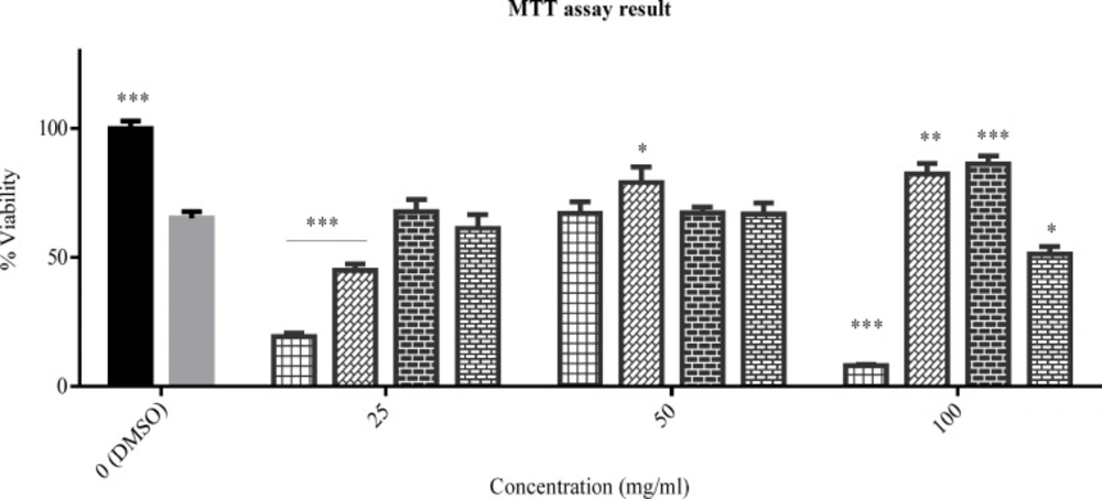
The viability percent of PC12 cells that treated with various concentration of tested plants for 24 h. Data were expressed as percentage of control group mean absorbance (Viability%) and represent as mean ± SEM (n = 6)
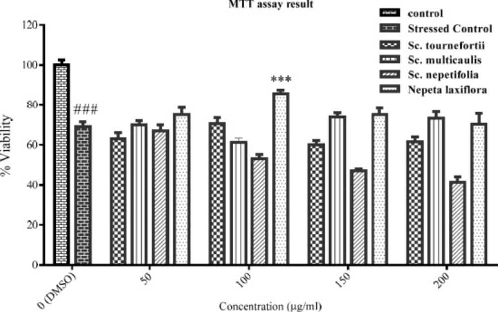
The viability percent of PC12 cells that pre-treated with various concentration of tested plants for 24 h and afterward treated with H2O2 1.5 mM for another 24 h. Data were expressed as percentage of control group mean absorbance (Viability%) and represent as mean ± SEM (n = 6). ### and ***p < 0.001 compared to control and stressed-control group, respectively
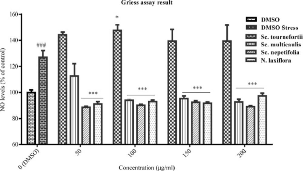
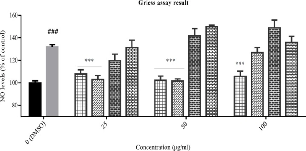
The effect of total extracts on NO production was assessed in PC12 cells that treated with H2O2 1.5 mM for 24 h. Data were expressed as percentage of control group mean absorbance (% of control) and represent as mean ± SEM (n = 6). ### and ***p < 0.001 (*p< 0.05) compared to control and stressed-control group, respectively
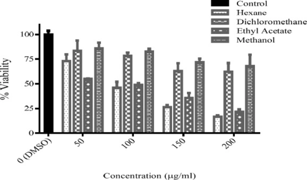
The viability percent of PC12 cells that treated with various concentration of Sc. multicaulis fractions for 24 h. Data were expressed as percentage of control group mean absorbance (Viability%) and represent as mean ± SEM (n = 6)
The viability percent of PC12 cells that pre-treated with Sc. multicaulis fractions for 24 h and afterward treated with H2O2 1.5 mM for another 24 h. Data were expressed as percentage of control group mean absorbance (Viability%) and represent as mean ± SEM (n = 6). *p < 0.05, **p < 0.01 and ***p < 0.001 compared to stressed-control group. Hex, the n-hexane fraction; DCM, the dichloromethane fraction; EtOAc, the ethyl acetate fraction; MeOH, the methanol fraction
The effect of Sc. multicaulis fractions on NO production was assessed in PC12 cells that treated with H2O2 1.5 mM for 24 h. Data were expressed as percentage of control group mean absorbance (% of control) and represent as mean ± SEM (n = 6). ### and ***p < 0.001 compared to control and stressed-control group, respectively. Hex, the n-hexane fraction; DCM, the dichloromethane fraction; EtOAc, the ethyl acetate fraction; MeOH, the methanol fraction
| Fraction | IC50 (μg/mL) | R square |
| n-Hexane | 105.5 | 0.97 |
| Dichloromethane | n/a | n/a |
| Ethyl acetate | 106.3 | 0.88 |
| Methanol | n/a | n/a |
Cell Viability Assay
The effect of extracts on cell viability was assessed by colorimetric method with MTT salt (
14). PC12 cells were seeded in 96-well plates at a density of 35000 cells/well and incubated for 24 h. Then the medium was replaced with fresh medium containing different concentrations of each extract (25-300 μg/mL) and incubated for 24 h. Negative control wells were treated with 1% (v/v) DMSO in equal volume of medium. In the next step, the supernatant was replaced with the fresh medium, containing MTT salt (0.5 mg/mL), and incubated for 24 h. In the last step, the culture supernatant was aspirated and the formazan crystals were dissolved in DMSO. ELISA plate reader from Fisher Scientific Company (Ontario, USA) measured the absorbance of wells at 570 nm. The results presented the percentage of mean absorbance of negative control wells, considered as 100% viability. The above procedure was repeated for the fractions of the selected plant in 50 to 200 μg/mL concentration range.
Inducing oxidative stress
To determine the suitable concentration of H2O2, PC12 cells were seeded in 96-well plates at a density of 35000 cells/well. After 24 h incubation, the medium was freshened and all the cells were treated by 1% (v/v) DMSO and incubated for another 24 h. Then the medium was replaced by FBS-free DMEM and the cells were treated with H2O2 (1–2.6 mM) for 24 h and the cell viability was detected by the MTT method as described previously. The concentration that produced about 50% viability (EC50) was selected for the following tests.
Cytoprotection by extracts and fractions as pretreatment
For assessing extracts and fractions effect on cell viability in stressed PC12, after 24 h incubation of cells in 96-well plate (35000 cells/well), the medium was replaced and the cells were treated by DMEM-FBS10% contains extracts (25-300 μg/mL) and fractions of selected plant (50-200 μg/mL). Two groups of negative control wells were treated with 1% (v/v) DMSO in equal volume of medium. After 24 h incubation, the medium replaced by FBS free DMEM contains H2O2 (1.5 mM), except for one group of negative control (control versus stressed-control group). Twenty-four hours later, cell viability was detected by the MTT method as described previously.
Griess assay
A standard procedure was used to determine NO production in stressed PC12 cells (
15). Briefly after seeding cells in 96-well plate and 24 h treatment with H
2O
2, as described above, equal volume (100 μL) of Griess reagent [1:1 mixture (v/v) of 1% sulfanilamide and 0.1% naphthylethylenediamine dihydrochloride in 5% H3PO4] and supernatant of each well was mixed and incubated for 15 min in dark place at room temperature. All extracts and fractions were employed in a process similar to that was described in the previous section. Subsequently, the absorbance of the mixture was determined at 540 nm with ELISA plate reader from Fisher Scientific Company (Ontario, USA).
Statistical analysis
The data were presented as the mean ± SEM. To compare the group means, one-way analysis of variance (ANOVA), followed by Dunnett’s and Sidak’s posttests were used. All Statistical analysis were carried out by GraphPad Prism software version 6.01 from GraphPad Software Inc. (San Diego, CA, USA).