


1. Background
2. Objectives
3. Patients and Methods
3.1. Study Design and Patients
| Group 1; (n = 25) | Group 2; (n = 25) | P Value | |
|---|---|---|---|
| Sex (male:female) | 14:11 | 13:12 | |
| Age at ACL-R, y | 28.8 ± 8.4 | 29.6 ± 8.2 | 0.74 |
| Age at MR examination, y | 34.8 ± 8.4 | 35.1 ± 8.2 | 0.92 |
| BMI at ACL-R, kg/m2 | 23.7 ± 2.7 | 24.3 ± 2.9 | 0.54 |
| BMI at MR examination, kg/m2 | 25.0 ± 3.2 | 25.5 ± 3.4 | 0.67 |
| Time from ACL-R to MR examination, months | 70.9 ± 9.3 | 68.2 ± 8.7 | 0.16 |
Abbreviations: ACL-R, anterior cruciate ligament reconstruction; BMI, body mass index; SD, standard deviation; y, year.
aValues are expressed as mean ± SD.
3.2. Surgical Technique
3.3. Clinical Assessment
3.4. MRI Protocol
3.5. MRI Analysis
3.6. Statistical Analysis
4. Results
4.1. Clinical Assessment
4.2. MRI Findings
| Group 1 | Group 2 | P Value | |
|---|---|---|---|
| IGSI of the IAP | 4 (16.7) | 11 (55.0) | < 0.01 |
| Proximal part | 1 (4.2) | 3 (15.0) | 0.21 |
| Distal part | 0 (0) | 2 (10.0) | 0.11 |
| Both | 3 (12.5) | 6 (30.0) | 0.15 |
| IGSI of the IOP | 17 (70.8) | 18 (90.0) | 0.11 |
| fIOP | 11 (45.8) | 18 (90.0) | < 0.01 |
| tIOP | 14 (58.3) | 15 (75.0) | 0.25 |
| Both | 8 (33.3) | 15 (75.0) | < 0.01 |
Abbreviations: fIOP, femoral intraosseous portion; IAP, intraarticular portion; IGSI, increased graft signal intensity; IOP, intraosseous portion; tIOP, tibial intraosseous portion.
aValues are expressed as No. (%).
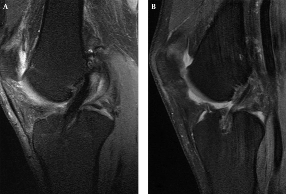
Sagittal proton density (PD) fat saturation (FS) images in (A) a 49-year-old man from group 1 and (B) a 27-year-old woman from group 2 show the difference in the signal intensity of the intraarticular and the tibial intraosseous portion of the anterior cruciate ligament (ACL) graft.
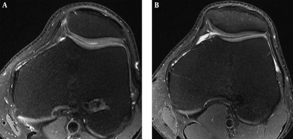
Axial proton density (PD) fat saturation (FS) images in (A) a 45-year-old man from group 2 and (B) a 24-year-old man from group 2 show the difference in the signal intensity of the femoral intraosseous portion of the anterior cruciate ligament (ACL) graft.
| Group 1 | Group 2 | |||
|---|---|---|---|---|
| IAP | ||||
| Less than 25 | 2 (50) | 2 (18.2) | ||
| 25 - 50 | 2 (50) | 5 (45.5) | ||
| More than 50 | 0 (0.0) | 4 (36.4) | ||
| IOP | ||||
| fIOP | tIOP | fIOP 18 | tIOP 15 | |
| Less than 25 | 3 (27.2) | 5 (35.7) | 7 (38.9) | 1 (6.7) |
| 25 - 50 | 4 (36.4) | 3 (21.4) | 5 (27.8) | 4 (26.7) |
| More than 50 | 4 (36.4) | 6 (42.9) | 6 (33.3) | 10 (66.6) |
Abbreviations: fIOP, femoral intraosseous portion; IAP, intraarticular portion; IOP, intraosseous portion; tIOP, tibial intraosseous portion.
aValues are expressed as No. (%).
| Group 1, ° | Group 2, ° | P Value | |
|---|---|---|---|
| CGA | 77.2 ± 8.8 | 74.6± 7.4 | 0.30 |
| SGA | 60.4 ± 5.7 | 56.5± 5.0 | 0.02 |
| Tibial tunnel, sagittal position | 62.8 ± 7.2 | 62.5 ± 8.7 | 0.93 |
| Tibial tunnel, coronal position | 58.0 ± 5.8 | 60.8 ± 6.7 | 0.70 |
| Femoral tunnel, sagittal position | 63.8 ± 8.5 | 67.4 ± 11.6 | 0.25 |
| Femoral tunnel, coronal position | 61.0 ± 7.2 | 47.7 ± 7.2 | < 0.01 |
Abbreviations: CGA, coronal graft angle, SGA, sagittal graft angle.
aValues are expressed as mean ± SD.
Abbreviations: fIOP, femoral intraosseous portion; IAP, intraarticilar portion; IOP, intraosseous portion; tIOP, tibial intraosseous portion.
aCalculated for the patients with preserved anterior cruciate ligament (ACL) graft (24 patients in group 1 and 20 patients in group 2).




