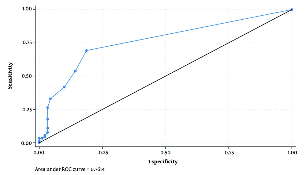
In the present study, a significant difference was found between the case group (patients with MS) and the control group (individuals without MS) in terms of the number of VRS. The number of VRS was higher in the case group compared to the control group. Our findings align with the results of studies conducted by Al-Saeed et al. in Kuwait and Etemadifar et al. in Isfahan (
17,
18).
Al-Saeed et al. (
18) reported a significantly higher number of VRS in the supraventricular region of newly diagnosed MS patients compared to controls. However, no significant differences were found in VRS frequency between the two groups in the midbrain, lenticulostriate vessels, or supraventricular WMLs. Etemadifar et al. observed a significantly higher mean number of VRS (21.4 vs. 16.8) in newly diagnosed MS patients compared to healthy controls. Our study, which included both newly diagnosed and previously diagnosed MS patients, found a lower mean number of VRS compared to Etemadifar et al.'s findings. This discrepancy may be attributed to the effects of MS treatment and warrants further investigation (
17).
A significant difference was found between the case and control groups in the number of VRS with round, oval, and curvilinear shapes. The case group exhibited a higher frequency of round, oval, and curvilinear VRS compared to the control group. Etemadifar et al. (
17) also reported a higher mean number of round and oval VRS in MS patients compared to controls. Conversely, curvilinear VRS were less frequent in the MS group. In a study by Al-Saeed et al. (
18), no significant differences were found in the prevalence of VRS in the midbrain, lenticulostriate vessels, and superior WMLs between MS patients and controls. However, dilated VRS were significantly more numerous in the supratentorial region of MS patients. These findings suggest that in patients with MS, not only is the total number of VRS increased, but also the various shapes of VRS are elevated, albeit to varying degrees.
In the present study, the size of VRS in patients with MS was observed to be larger compared to the control group. However, no statistically significant difference was found in the size of these spaces between the case and control groups. A review study by Vivash (
3) did not report an increase in the size of dilated VRS in MS patients based on the results of published studies. Conversely, Favaretto et al. (
19) found a significantly larger volume of VRS in patients compared to controls when investigating the association between eVRS and cognitive status in MS. Indeed, an increase in the volume of VRS likely corresponds to an increase in their size. The enlargement or dilation of VRS is most probably due to inflammation.
Our study builds upon previous research by Etemadifar et al. and Al-Saeed et al., confirming their findings while also providing new insights through the inclusion of a diverse population, advanced imaging techniques, comprehensive analysis, and examination of clinical subgroups. These enhancements contribute to a more nuanced understanding of VRS in MS (
17,
18).
In our study, a significant positive correlation was observed between patient age and both the total number of VRS and the number of round VRS. A notable correlation was also found between age and oval VRS. Small VRS were present in all age groups. As age increased, VRS became more frequent and appeared larger. These findings align with previous research indicating that eVRS are commonly associated with advancing age (
2,
8). Furthermore, in our analysis, after controlling for the confounding effect of age, the association between VRS and age remained statistically significant.
Our findings demonstrate that while WMLs remain a robust biomarker for MS diagnosis, VRS — particularly round-shaped VRS — also show significant diagnostic value. The moderate correlation between WMLs and VRS suggests that these markers may reflect overlapping but distinct pathological processes. Unlike WMLs, which represent areas of demyelination, VRS may indicate perivascular inflammation and glymphatic dysfunction. This distinction highlights the potential of VRS as a complementary imaging biomarker in MS.
In the present study, the mean number of VRS was found to be significantly higher in men compared to women, as revealed by logistic regression. Consistent with our findings, Etemadifar et al. (
17) reported differences in VRS characteristics between male and female MS patients, with men exhibiting a higher mean number of VRS. Similarly, Favaretto et al. (
19) demonstrated a significantly greater number and volume of VRS in male patients compared to female patients.
In the present study, no significant correlation was found between EDSS severity and the number or size of VRS in MS patients. Consistent with our findings, a systematic review by Vivash (
3) demonstrated a limited correlation between disease severity and VRS. This suggests that dilated VRS may be a non-specific marker of disease, whether neurological, cardiovascular, or a combination of both. Numerous factors influence the presence of dilated VRS, and more comprehensive studies are needed to control for these factors, not only in MS but also in other neurological and neurodegenerative diseases, to better understand the role of VRS in disease pathogenesis and presentation (
3). In a study by Ineichen et al. (
2) in the United States, at the tissue level, VRS was not strongly associated with pathological features of MS.
In conclusion, in brain MRI, the number of VRS has been shown to be a useful marker for MS, potentially holding prognostic value. Therefore, it is recommended that patients referred for brain MRI be evaluated for the number and size of VRS. However, it is important to approach these findings with caution and acknowledge the study's limitations. Our study's single-center design limits the generalizability of the findings. Additionally, the retrospective nature of data collection may introduce selection bias. The use of 1.5-T MRI, which may be less sensitive than higher-resolution imaging modalities such as 3-T MRI, could also impact the accuracy of VRS detection. Rather than overstating the implications, future research should focus on conducting prospective studies with larger, better-matched samples to validate these findings and utilizing high-resolution MRI for more accurate detection and assessment of VRS. Systematic reviews and meta-analyses are also suggested to investigate and compare the number and size of VRS in MS patients and healthy individuals.