
1. Background
Neurofibromatosis type 1 (NF1) is an autosomal dominant disorder that predisposes patients to central and peripheral nervous system tumors, including plexiform neurofibromas (PNs) (1), which can cause disfigurement, pain, motor dysfunction, and life-threatening complications (2-6). The introduction of MEK inhibitor therapy for symptomatic, inoperable NF1-associated PNs has made MRI indispensable for careful PN evaluation and longitudinal monitoring (7, 8), particularly in children because of their greater radiosensitivity and longer life expectancy (9, 10).
Current solid tumor response criteria rely on one-dimensional (1D) or two-dimensional (2D) measurements; however, PNs are typically large, irregular, and slow-growing, making three-dimensional (3D) volumetry a superior approach that is increasingly recommended in guidelines (11, 12).
Despite this theoretical advantage, critical pediatric gaps remain: 1) a lack of dedicated validation in children, despite differences in tumor behavior and growth kinetics; 2) a lack of systematic reporting of time-efficiency data in real-world pediatric settings; and 3) a lack of rigorous assessment of interchangeability between 2D and 3D measurements at the individual-patient level, which is essential for treatment monitoring.
To address these gaps, this study provides pediatric-specific validation, quantitative measurement times, and individual-level agreement analysis, with the aim of proposing a practical, stratified imaging strategy for longitudinal monitoring.
2. Objectives
This study evaluated 1D (longest diameter), 2D (product of perpendicular diameters), and semi-automated 3D volumetry for assessing plexiform neurofibromas in children with NF1 and determined whether simplified metrics can substitute for 3D volumetry in routine practice.
Specifically, this study sought to:
1) establish intra- and inter-observer reliability (ICCs) for all three methods in a pediatric cohort;
2) assess individual-level agreement between 2D estimates and 3D volumes using Bland-Altman analysis and log-log regression; and
3) quantify the time required for 3D volumetry relative to simplified measurements to inform clinical feasibility.
Based on these findings, we aimed to propose a stratified imaging strategy that balances precision with time efficiency for longitudinal surveillance and therapeutic trial endpoints, particularly in resource-limited settings where simplified measurements are commonly used.
3. Patients and Methods
3.1. Ethical Review
This retrospective study was approved by the Institutional Review Board and conducted in accordance with the 1964 Declaration of Helsinki and its later amendments (Ethical Approval Number: SCMCIRB-K2025257 - 1). Informed consent was obtained.
3.2. NF1 Diagnostic Criteria and PN Definition
All patients included in this study met the National Institutes of Health (NIH) Consensus Conference diagnostic criteria for NF1 (13, 14). A detailed description of these criteria is available in the referenced guideline.
PNs arise from nerve sheaths and exhibit diffuse, infiltrative growth along the nerve trunk, often invading adjacent structures. The initial diagnostic approach integrates clinical evaluation with imaging features, whereas histopathology and genetic testing serve ancillary roles. Physical examination may reveal a rope-like mass or a positive "buttonhole sign." However, definitive diagnosis relies on MRI, which characterizes lesions by morphology, internal structure, depth, and relationships with surrounding tissues (15).
3.3. Patient Population
A retrospective review of institutional electronic medical records from January 2020 to March 2026 identified 81 patients meeting the 2021 NIH NF1 criteria, all of whom underwent baseline whole-body MRI. Of these, 42 were excluded and categorized into mutually exclusive groups: central nervous system (CNS) lesions or isolated optic pathway gliomas without concurrent PNs (n = 21); concurrent CNS lesions and PNs < 3 cm3 (n = 10); PNs < 3 cm3 without CNS lesions (n = 7); and poor baseline image quality (n = 4). The remaining 39 patients with MRI-confirmed PNs meeting the volumetric criteria (≥ 3 cm3 and visible on three consecutive slices) were included in the final analysis (Figure 1). The diagnosis was established by surgical pathology (n = 15), genetic testing (n = 14, including 1 with additional pathology), or clinical assessment with characteristic MRI findings (n = 10). Only baseline MRI scans obtained before therapy or surgery were included.
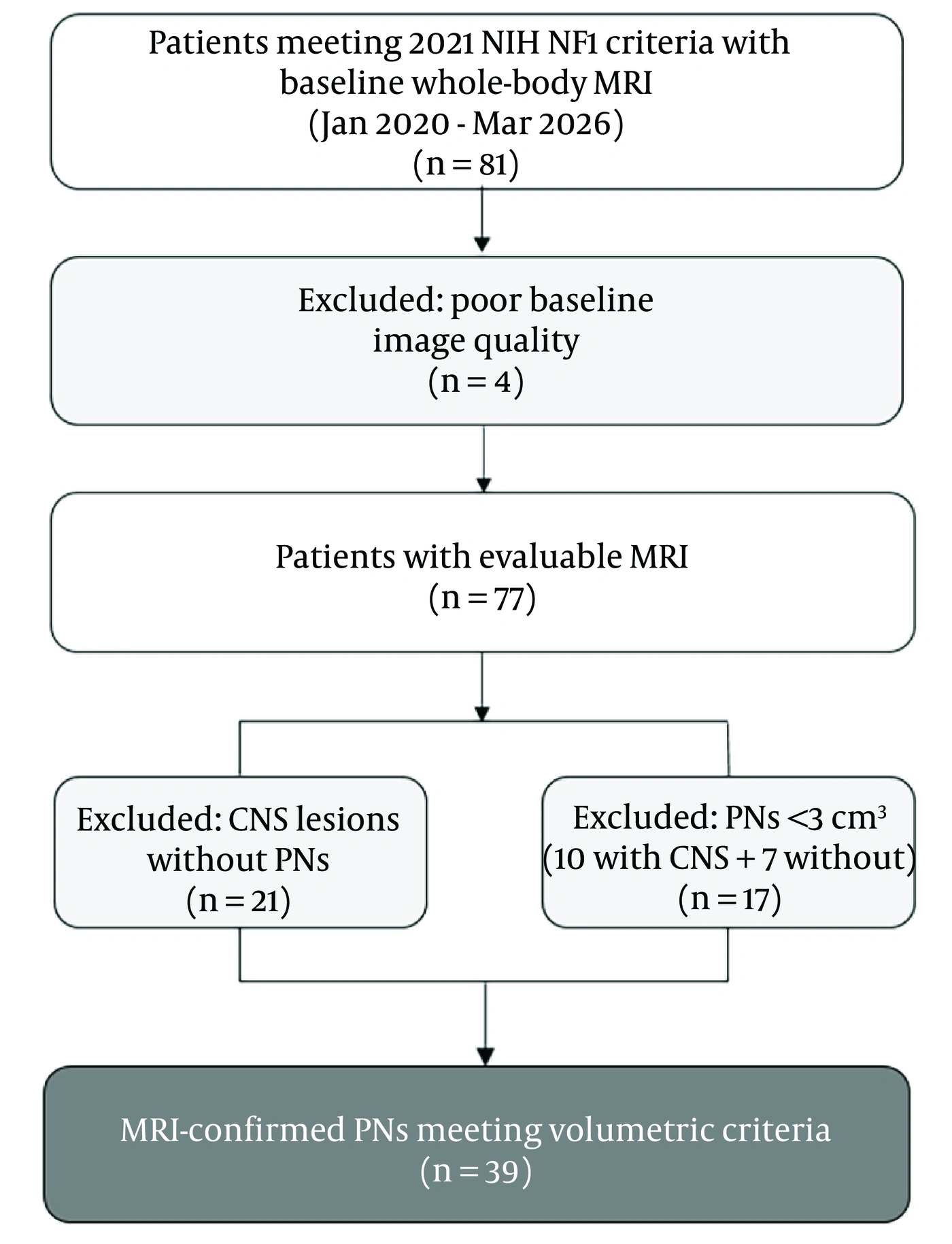
Figure 1.
Patient selection flowchart. Of 81 patients meeting the 2021 National Institutes of Health (NIH) neurofibromatosis type 1 (NF1) criteria who underwent baseline whole-body magnetic resonance imaging (MRI), 4 were excluded because of poor baseline image quality. Among the remaining 77 patients, 21 had central nervous system (CNS) lesions without plexiform neurofibromas (PNs), and 17 had PNs with a volume < 3 cm3 (including 10 with concurrent CNS lesions and 7 without). The final analysis included 39 patients with MRI-confirmed PNs meeting the volumetric inclusion criteria (≥ 3 cm3 and visible on three consecutive slices).
The most common initial clinical manifestations were palpable masses (n = 20) and café-au-lait spots with or without axillary freckling (n = 13). Other presentations, each occurring in 1 patient, included scoliosis, a heart murmur, hypertension, precocious puberty, ptosis, and limb weakness.
The cohort comprised 21 females and 18 males aged 3 to 17 years (mean ± SD, 9.30 ± 3.56 years; median, 9.00 years). According to the NF1 criteria, 4 patients met four criteria, 12 met three criteria, and 23 met two criteria. Four patients had a first-degree relative with NF1.
For each eligible patient with multiple measurable PNs meeting the aforementioned volumetric criteria, only one target lesion was selected for subsequent analysis. Selection followed predefined criteria: among all qualifying lesions in a given patient, the lesion with the largest volume or, for lesions of comparable size, the lesion with the clearest border was selected as the representative lesion. Consequently, the unit of analysis was the patient, and the final cohort of 39 lesions was derived from 39 distinct patients, ensuring statistical independence of all observations.
3.4. MRI Acquisition
MRI examinations were performed using a 3.0-T GE scanner (Discovery MR750 3.0T, GE Medical Systems, USA) with a dedicated 32-channel brain or cardiac coil. Patients were positioned supine and advanced into the magnet either head-first or feet-first, depending on lesion location. The average duration of the MRI examination was approximately 20 to 30 minutes. The imaging protocol comprised axial spin-echo T1-weighted imaging (T1WI; repetition time/echo time [TR/TE], 500 - 600 ms/10 ms), axial and coronal T2-weighted fast spin-echo imaging with and without fat suppression (T2WI and FS-T2WI; TR/TE, 3500 - 6000 ms/60 - 80 ms), and axial diffusion-weighted imaging (DWI; b value, 800 s/mm2). A slice thickness of 5 - 7 mm, adapted to lesion size, with a 1-mm interslice gap was used. All sequences were acquired without contrast administration.
3.5. Imaging Analysis
All images were retrospectively reviewed using a picture archiving and communication system (PACS) (GE Healthcare Centricity Radiology Information System [RIS] Image Workstation [IWS] 3.0). One-dimensional and 2D measurements were performed on this workstation, whereas 3D volumetric measurements were performed using ITK-SNAP version 3.7, a free, open-source software program.
All measurements were performed using FS-T2WI sequences, which provide optimal contrast between hyperintense PNs and surrounding tissues. Lesions were measured using three approaches:
1) 1D length measurement: The longest diameter (cm) was measured in the axial plane, supplemented by coronal or sagittal sequences as needed, according to RECIST 1.1.
2) 2D area measurement: According to WHO criteria, the largest cross-sectional area (cm2) was calculated by multiplying the two longest perpendicular diameters on the plane of greatest lesion extent.
3) 3D volumetric measurement: Segmentation was performed on contiguous axial slices using ITK-SNAP, with reference to coronal and sagittal sequences for accurate boundary delineation. Manual tracing on each slice enabled automatic volume computation.
For lesions abutting or encasing adjacent structures such as vessels or airways, boundary delineation for 3D segmentation was based on signal differences on FS-T2WI, concurrent reference to coronal and sagittal sequences, a conservative approach excluding indistinct boundaries, and consensus review for challenging cases.
Image analysis was performed by two board-certified pediatric radiologists (Observer AA: 29 years of experience; Observer BB: 15 years of experience). A standardized training session used 2 pilot cases that were not included in the final analysis. For each lesion and measurement method, each observer performed three repeated measurements per session, and the mean was used for analysis. Observer AA completed two sessions more than 2 weeks apart; Observer BB completed one session. Measurements were performed on anonymized, randomized scans, with the observers blinded.
3.6. Statistical Analysis
Statistical analyses were conducted sequentially. First, the reliability of all three measurement methods (3D volume, 2D area, and 1D length) was assessed. Both inter- and intra-observer reliability were evaluated using intraclass correlation coefficients (ICCs) with their 95% confidence intervals (CIs), calculated using a two-way random-effects model consistent with methodologies for tumor response evaluation (16). All ICCs were calculated using the mean of three repeated measurements per session (averaged-measurement ICCs). Intra-observer reliability was evaluated using the two measurement sessions performed by Observer AA. Inter-observer reliability was assessed by comparing the first session of Observer AA with the single session of Observer BB. ICC estimates were interpreted according to established criteria for measurement reliability: < 0.50, poor; 0.50 - 0.75, moderate; 0.75 - 0.90, good; and > 0.90, excellent (17). All 95% CIs for the ICCs were computed using the F-distribution-based method, following recent methodological recommendations (18).
Subsequently, the normality of the data distributions for each measurement type was examined using the Shapiro-Wilk test. Depending on the normality results, associations among the three measurement methods were analyzed using either Pearson correlation coefficients for normally distributed data or Spearman rank correlation coefficients for non-normally distributed data. The strength of association was reported as r or ρ, respectively. Correlation coefficients were interpreted as follows: |ρ| = 0.00 - 0.19, very weak or negligible; 0.20 - 0.39, weak; 0.40 - 0.59, moderate; 0.60 - 0.79, strong; and 0.80 - 1.00, very strong. Given the right-skewed distributions observed in all measurement indices, linear regression analyses were performed on natural log-transformed data (Ln[Volume], Ln[Area], and Ln[Length]) to satisfy model assumptions and evaluate the predictive value of 2D area and 1D length for 3D volume.
Finally, Bland-Altman analysis was performed to quantify agreement between volume estimates derived from 2D measurements and direct 3D volumetric measurements. The 2D-based estimates were calculated from the largest cross-sectional area (A) using spherical geometric modeling (19, 20). Briefly, the equivalent spherical radius was derived from the segmented area and substituted into the sphere-volume equation V = 4/3πr3. The unified conversion formula was V = (π/6) × (4A/π)3/2. Because tumor volumes spanned more than three orders of magnitude, Bland-Altman analysis was performed on natural log-transformed data; the bias and 95% limits of agreement (LoA) were then exponentiated back to the original scale to facilitate clinical interpretation. This analysis provided the mean difference (bias) and 95% LoA, thereby defining the expected range of discrepancies at the individual-measurement level. Given the wide range of tumor volumes, the robustness of these LoA estimates was further evaluated by considering the effects of sample size and data distribution (21).
All statistical analyses were performed using IBM SPSS Statistics version 26.0, and the Bland-Altman plot was generated using GraphPad Prism version 9.5.1.
A two-sided significance threshold of P < 0.001 was used for all statistical inferences, and all subsequent tests were interpreted against this criterion.
4. Results
4.1. Imaging Findings
On MRI, all 39 PNs were isointense to minimally hypointense on T1WI, hyperintense on T2WI (Figure 2A), and exhibited high signal intensity on DWI (Figure 3B). Lesion distribution was as follows: 4 maxillofacial, 6 in the neck, 11 mediastinal, 3 in the neck + mediastinum, 5 pelvic, 2 in the abdomen + pelvis, 3 in the abdominal wall, and 1 each in the lumbar region, axilla, back, hip, and thigh. Associated findings included scoliosis (n = 6, Figure 3A), intraspinal extension (n = 4), cervical spinal cord compression (n = 3), tracheal narrowing or deviation (n = 3), sphenoid wing dysplasia with exophthalmos (n = 3), and bladder or uterine invasion (n = 1, Figure 2B).
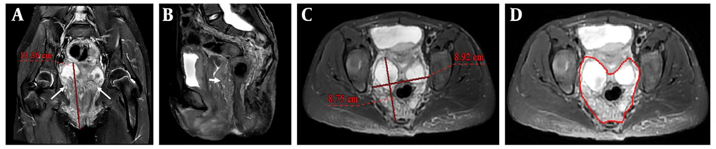
Figure 2.
A 13-year-old female with a pelvic plexiform neurofibroma (PN). A. Coronal T2-weighted fat-suppressed image demonstrates a multinodular, deep, and relatively well-defined lesion with nodular hyperintense and mildly hyperintense signal within the pelvis (long white arrows). The one-dimensional (1D) length measures 13.36 cm (red line). B. The mass is seen invading the posterior uterine wall (short white arrows). C. The maximum cross-sectional area of the mass measures 78.05 cm2 (product of the two perpendicular red lines: 8.92 cm × 8.75 cm). D. The lesion volume (546.83 cm3) was measured by manually delineating the lesion contour on each consecutive slice (red outlines) using ITK-SNAP software version 3.7. The software automatically calculated the total volume by summing the segmented areas and multiplying by the slice interval (slice thickness + gap).

Figure 3.
A 17-year-old female with a mediastinal plexiform neurofibroma (PN). A. Coronal T2-weighted image demonstrates diffuse, deep, and interdigitating lesions within the mediastinum extending into the neural foramen (long white arrow), accompanied by thoracic scoliosis (short white arrow). B. The lesion exhibits high signal intensity on the diffusion-weighted imaging sequence (DWI). C. The largest nodule was selected as the target lesion for measurement, with a maximum cross-sectional area of 46.46 cm2 (product of the two perpendicular red lines: 5.39 cm × 8.62 cm). D. The lesion volume (196.94 cm3) was derived from manual segmentation of consecutive slices (red fill) in ITK-SNAP version 3.7, which automatically calculated the total volume from the summed areas and slice interval (thickness + gap).
4.2. Imaging Classification
Based on MRI characteristics (Table 1), lesions were classified according to three criteria. Morphologically, lesions were classified as mixed type (48.7%, Figure 4A), fascicular/multinodular type (43.6%), or diffuse type. By anatomical depth, deep lesions, defined as those beneath the muscle fascia, were predominant (56.4%). Regarding relationships with adjacent tissues, lesions were classified as well-defined and circumscribed (33.3%), infiltrative and interdigitating (30.8%), or mixed (35.9%).
Table 1.
Demographic, Clinical, and MRI-Based Classification of Plexiform Neurofibromas (PNs)
| Variables | Number of Cases |
|---|---|
| Gender | |
| Male | 18 |
| Female | 21 |
| Age (y) | |
| 5 or younger | 8 |
| 5 - 10 | 16 |
| 10 - 17 | 15 |
| Locations of PNs | |
| Maxillofacial region | 4 |
| Neck | 6 |
| Mediastinum | 11 |
| Neck + mediastinum | 3 |
| Pelvic cavity | 5 |
| Abdomen + pelvis | 2 |
| Abdominal wall | 3 |
| Other sites: axilla/back/hip/thigh/lumbar region (1 case each) | 5 |
| Classification: Morphology/Internal Structure | |
| Diffuse | 3 |
| Fascicular/nodular | 17 |
| Mixed | 19 |
| Classification: Depth | |
| Deep | 22 |
| Superficial | 7 |
| Mixed | 10 |
| Classification: Adjacent Tissue Relationship | |
| Interdigitating | 12 |
| Circumscribed | 13 |
| Mixed | 14 |

Figure 4.
A 14-year-old female with a subcutaneous plexiform neurofibroma (PN) in the buttock. A. Coronal T2-weighted image reveals a diffuse, multinodular, superficial, and relatively well-defined PN within the subcutaneous tissue of the buttock, with hyperintense signal. A "buttonhole sign" is also visualized (long white arrow). B. The maximum cross-sectional area of the mass measures 520.58 cm2 (product of the two perpendicular red lines: 15.37 cm × 33.87 cm). C, D. Lesion volume (4840.42 cm3) was quantified by manual segmentation of consecutive slices (red fill), and the three-dimensional (3D) reconstruction was generated using ITK-SNAP version 3.7.
4.3. Measurement Results
Measurement results (Figure 5A) for each lesion, including the average time required per lesion, are summarized below:
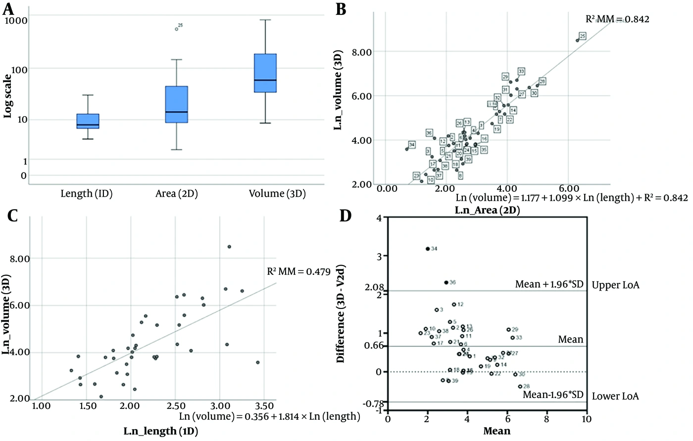
Figure 5.
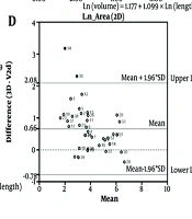
A. Comparison of the measurement data demonstrates progressively wider distributions from one-dimensional (1D) length to two-dimensional (2D) area and three-dimensional (3D) volume. B. Scatter plot showing the correlation between natural-log-transformed (Ln) 3D volume and Ln 2D area, with fitted linear regression line (R2 = 0.842; Ln[Volume] = 1.177 + 1.099 × Ln[Area]; P < 0.001). C. Scatter plot showing the correlation between Ln 3D volume and Ln 1D length, with fitted linear regression line (R2 = 0.479; Ln[Volume] = 0.356 + 1.814 × Ln[Length]; P < 0.001). D. Bland-Altman plot comparing 2D-estimated and 3D-measured volumes on the natural log-transformed scale. The bias was 0.66, with 95% limits of agreement (LoA) ranging from −0.78 to 2.09 on the log scale (n = 39).
1) 1D length: mean, 10.65 ± 6.44 cm; median, 7.83 cm (range, 3.78 - 30.85 cm). Mean acquisition time: 13.79 ± 7.70 s.
4.4. Statistical Results
Regarding reliability (Table 2), excellent intra-observer agreement was observed for all measurement methods, with ICC (3, 3) values exceeding 0.989 (95% CI range, 0.980 - 0.994). Inter-observer agreement was also excellent for both 3D volume (ICC = 0.966; 95% CI, 0.933 - 0.983) and 2D area (ICC = 0.956; 95% CI, 0.899 - 0.979). The 1D length also demonstrated excellent inter-observer reliability (ICC = 0.934; 95% CI, 0.874 - 0.965), although this value was lower than the ICCs for the other parameters.
Table 2.
Inter- and Intra-Observer Reliability of Three Measurement Methods a
| Measurement Method | Inter-observer Consistency, ICC (3, 3) (95% CI) | Intra-observer Consistency, ICC (3, 3) (95% CI) |
|---|---|---|
| 3D Volume | 0.966 (0.933 - 0.983) | 0.998 (0.997 - 0.998) |
| 2D Area | 0.956 (0.899 - 0.979) | 0.998 (0.995 - 0.999) |
| 1D Length | 0.934 (0.874 - 0.965) | 0.989 (0.980 - 0.994) |
a Note: Averaged-measurement ICCs (ICC [3,3]) were calculated using the mean of three repeated readings per session under a two-way mixed-effects model for consistency. Abbreviation: ICC, intraclass correlation coefficient; CI, confidence interval.
Because the data were non-normally distributed (Shapiro-Wilk P < 0.05 for all), Spearman rank correlation was used for all pairwise comparisons among the three measurement methods. Spearman rank-correlation analysis demonstrated a strong monotonic correlation between 3D volume and 2D area (ρ = 0.865, P < 0.001) and between 3D volume and 1D length (ρ = 0.721, P < 0.001), as well as a moderate correlation between 2D area and 1D length (ρ = 0.541, P < 0.001).
All measurement indices exhibited right-skewed distributions, characterized by means exceeding medians and wide ranges, a pattern likely attributable to the presence of several particularly large lesions within the cohort. Given this skewness, linear regression was performed on log-transformed data (Ln-transformed volume, area, and length) for statistical inference. On the log-log scale, 2D area remained a strong predictor of 3D volume, explaining 84.2% of the variance (R2 = 0.842; Ln[Volume] = 1.177 + 1.099 × Ln[Area]; P < 0.001; Figure 5B), whereas 1D length had poorer predictive value, accounting for only 47.9% of the variance (R2 = 0.479; Ln[Volume] = 0.356 + 1.814 × Ln[Length]; P < 0.001; Figure 5C).
As a necessary complement to correlation analysis, Bland-Altman analysis was performed on the natural log-transformed data to assess clinical interchangeability between 2D-derived volume estimates and the direct 3D volumetric standard (Figure 5D). The analysis revealed a mean bias of 0.66 on the log scale, with 95% LoA ranging from −0.78 to 2.09, and two data points (5.1%) falling outside the limits.
5. Discussion
NF1-associated PNs are highly variable in morphology and growth patterns, making reliable imaging measurements essential for pediatric patients receiving MEK inhibitor therapy. In recent years, selumetinib, an oral MEK1/2 inhibitor, has been approved for pediatric patients with symptomatic, inoperable NF1-associated PNs, with efficacy demonstrated in multiple trials (22, 23). In this context, MRI is essential before and during pharmacotherapy because it guides target lesion selection, response assessment, and multidisciplinary decision-making. Pediatric oncologists tailor MEK inhibitor therapy, whereas surgeons evaluate disease burden and identify signs of malignant transformation. Before treatment, whole-body MRI is recommended to assess systemic disease burden and select target lesions based on predefined criteria such as size and distinct margins (24). In addition, imaging evaluation, particularly FS-T2WI, provides excellent PN delineation without ionizing radiation, making it suitable for long-term pediatric monitoring.
The reliability and reproducibility of any measurement method are prerequisites for clinical adoption. We therefore evaluated the intra- and inter-observer reliability of 1D, 2D, and 3D measurements in pediatric NF1-associated PNs. All three methods showed excellent intra-observer agreement (ICC > 0.989). Inter-observer agreement was excellent for 3D volume and 2D area (ICC > 0.956) and also excellent for 1D length (ICC = 0.934), although the latter was lower than the ICCs for the other parameters, indicating that 1D length is somewhat more susceptible to reader-dependent variability. Overall, the 2D and 3D methods are highly reliable, whereas 1D length remains acceptable but shows slightly greater variation between observers.
Consistent with Response Evaluation in Neurofibromatosis and Schwannomatosis (REiNS) recommendations for volumetric endpoints (25), 2D area demonstrated stronger predictive value for 3D volume on the log-log scale (R2 = 0.842) than 1D length (R2 = 0.479). Despite the strong association of 2D area, Bland-Altman analysis on the log scale revealed wide 95% LoA (−0.78 to 2.09), indicating that 2D-derived estimates cannot reliably substitute for direct 3D volumetry at the individual-lesion level. This dissociation between strong predictive value and wide LoA underscores that group-level correlation does not guarantee individual-level interchangeability.
Moreover, although 1D length demonstrated excellent inter-observer reliability (ICC = 0.934), its limited concordance with 3D volumetry in regression analysis represents an important limitation. Although this metric may be sufficient for rough group-level comparisons, its weak individual-level predictive value undermines its utility for precise longitudinal monitoring in individual pediatric patients. Therefore, clinical decisions relying on 1D length alone should be made with caution.
Our pediatric-specific findings confirm this group-level correlation but suggest that the wide LoA may be even more pronounced in children than in adult populations, possibly due to greater PN morphological heterogeneity and smaller absolute volumes. Prior studies have shown that volumetric measurements provide greater sensitivity to change than linear or 2D assessments in both NF1-associated PNs and NF2-associated vestibular schwannomas (26, 27). However, most previous reports have focused on correlation or group-level sensitivity rather than individual-level interchangeability. Unlike these earlier reports, our Bland-Altman analysis directly addresses whether 2D measurements can substitute for 3D measurements at the single-patient level, a critical distinction for treatment-response assessment in children that has been less systematically examined in prior work.
Although essential for assessing irregular PNs, traditional 3D volumetric segmentation is often clinically prohibitive because it is time-consuming and relies on a manual process with limited reproducibility. To overcome this limitation, we implemented a semi-automated workflow using ITK-SNAP. Semi-automated 3D volume segmentation required 262.00 ± 162.89 seconds on average (4 min 22 s). Although this was substantially longer than 1D length measurement (13.79 ± 7.70 s) and 2D area measurement (18.36 ± 5.96 s), it remained within a practical clinical range and offered improved standardization and repeatability compared with fully manual contouring. Our experience suggests that this semi-automated approach can serve as a practical bridge toward broader clinical implementation of 3D volumetric analysis.
Nevertheless, clinical translation of this approach faces several challenges. First, the ill-defined, infiltrative borders of PNs and interobserver variability in manual initialization may affect reproducibility. Second, the large target lesion size (> 3 cm), with heterogeneous composition and multicompartment involvement, may challenge algorithm robustness. Third, segmentation accuracy depends on image quality, including motion artifacts, and therefore requires standardized protocols.
Based on these findings, we propose a stratified imaging strategy for clinical practice. Direct 3D volumetric MRI should serve as the reference standard for therapeutic monitoring and trial endpoints. Efficient 2D area measurements can support rapid surveillance and group-level trend analysis, whereas 1D length, despite good to excellent inter-observer reliability, should be used cautiously in individual patients. This framework integrates advanced MRI volumetry into routine radiologic assessment of NF1-associated plexiform neurofibromas.
Several limitations of this study should be acknowledged. First, no formal a priori sample-size calculation was performed, which represents a methodological constraint. Although our sample of 39 patients is comparable to that of similar agreement studies using Bland-Altman analysis (median sample size, 65; IQR, 35 - 124) (28), the modest sample size inevitably affects the precision of our estimates, as reflected by the 95% CIs of the ICCs. Moreover, the wide Bland-Altman LoA (−0.78 to 2.09) and skewed distribution of lesion volumes further complicate interpretation. Second, as a single-center investigation, our results reflect the specific imaging protocols and observer expertise of our institution and may not be directly transferable to other settings. Multicenter collaborations are needed to validate the robustness of our agreement metrics across diverse equipment and reading practices. Future studies with larger prospective cohorts are warranted to provide more precise and generalizable estimates.
Beyond design-related constraints, measurement-related limitations should also be noted. Manual segmentation used for 3D volumetry, although considered a reference standard, is subject to intra- and inter-observer variability, particularly at lesion margins. Similarly, 2D measurements are susceptible to plane-selection bias, which may have contributed to the wide LoA observed. To mitigate this issue, all measurements were performed by experienced radiologists, and standardized protocols were strictly followed to minimize variability. In addition, only the mean values of three repeated measurements per session were recorded; individual readings were not retained. Thus, our ICCs represent session-averaged reliability, and single-measurement ICCs were unavailable. This should be considered when comparing our reliability estimates with those from studies reporting single-measurement ICCs.
In addition to these methodological constraints, our exclusive reliance on baseline cross-sectional data precludes direct evaluation of longitudinal treatment monitoring, which inherently requires longitudinal data and is beyond the current scope. Nevertheless, this baseline-only design was a deliberate prerequisite rather than an oversight: establishing static measurement agreement is necessary before validly assessing interchangeability under dynamic conditions because any longitudinal bias would otherwise be uninterpretable. The right-skewed size distribution in our cohort further suggests that future longitudinal efficacy analyses should incorporate size-stratified baselines. Thus, our baseline-focused design provides the methodological groundwork for subsequent longitudinal monitoring studies. Given the cross-sectional nature of the study, our findings suggest, but do not directly prove, that 3D volumetry may be advantageous for monitoring treatment response. We therefore frame this recommendation as an informed hypothesis rather than a definitive conclusion. Longitudinal studies with repeated measurements are warranted to confirm whether this cross-sectional advantage translates into superior performance in tracking drug-induced changes over time.
Despite the absence of longitudinal data, this study offers three contributions extending prior adult evidence and REiNS guidelines: 1) pediatric validation showing that 2D measurements cannot substitute for 3D measurements at the individual level; 2) time-efficiency metrics of approximately 4 min/lesion supporting feasibility; and 3) Bland-Altman benchmarks for single-patient decisions. By quantifying static measurement error, our baseline findings provide a reference framework for distinguishing true volumetric response from measurement bias in 2D-3D discrepancies during treatment.
However, longitudinal multicenter cohorts are needed to track volume dynamics alongside clinical symptom improvement. Advances in AI-driven automated segmentation, once further validated in heterogeneous pediatric NF1-PN cohorts, may reduce both time costs and observer variability, facilitating integration of 3D volumetry into routine workflows (29). Ultimately, a comprehensive framework coupling quantitative imaging metrics with functional outcomes remains the definitive goal.
In conclusion, this study provides pediatric-specific validation that 3D volumetry, unlike 1D and 2D metrics, demonstrates sufficient individual-level agreement with true lesion volume to serve as the reference standard for baseline assessment of NF1-associated PNs in children. Although 2D area measurements may support rapid surveillance and group-level trend analysis, reliance on 2D-derived estimates for individual treatment-response monitoring remains unjustified without longitudinal validation. Our proposed stratified imaging strategy, using 2D measurements for screening and 3D measurements for confirmation, offers an evidence-based and time-efficient pathway for integrating advanced volumetry into routine radiology and improving individualized disease evaluation while longitudinal corroboration is awaited. Larger-scale studies are needed to further refine these agreement estimates and validate the proposed stratified strategy across diverse patient populations.