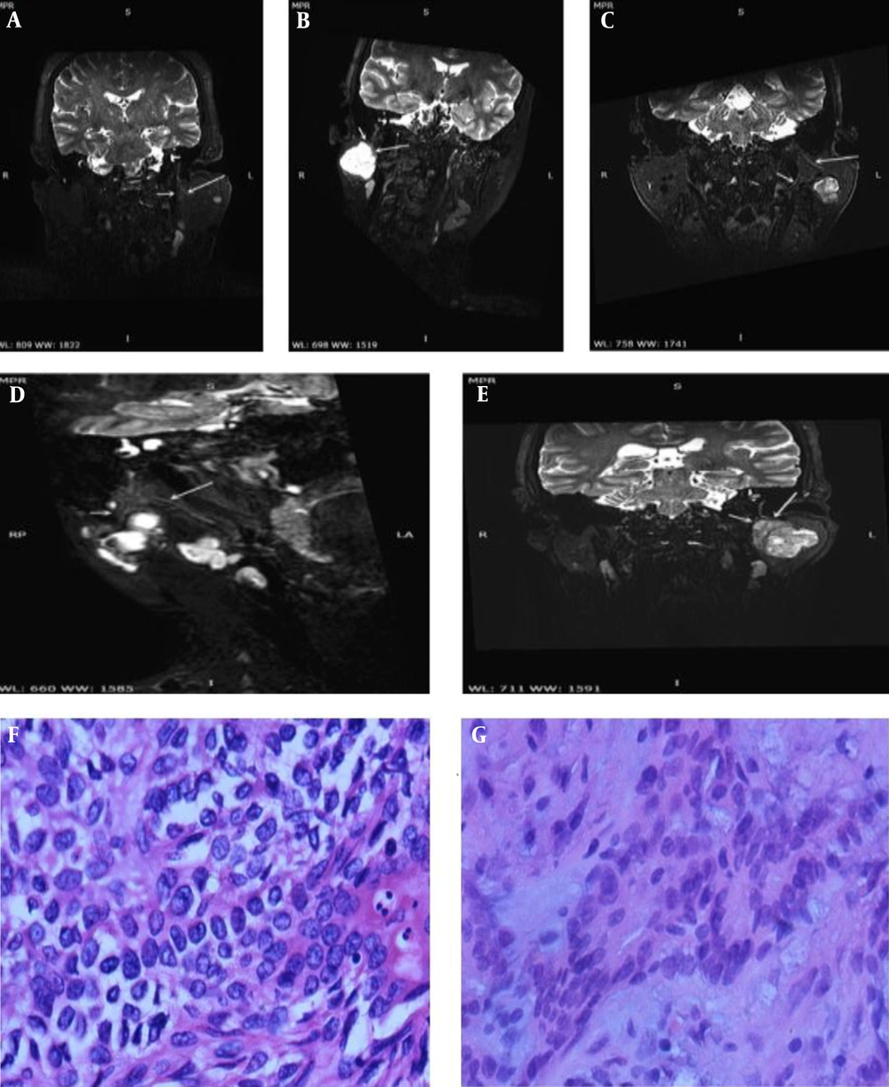
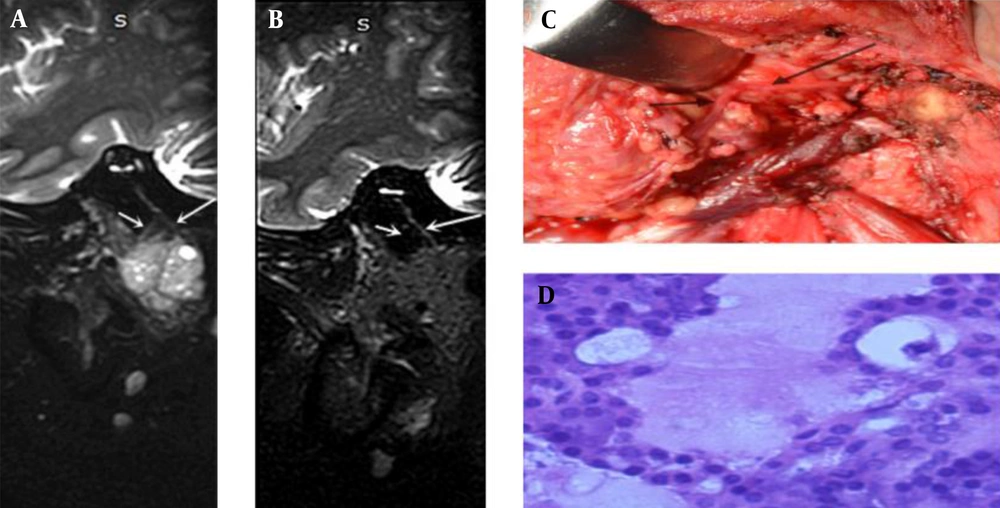
Knowledge of the anatomy of the facial nerve in the parotid gland can be helpful in surgical planning. Different strongly T2- or T2*-weighted volume 3D MRI sequences have been advocated for evaluating the peripheral nerves, including 3D gradient-recalled acquisition in steady state (3D-GRASS), 3D constructive interference in steady state (3D-CISS), and 3D fast imaging employing steady-state acquisition (3D-FIESTA) (
9-
11). The peripheral nerves showed a low signal intensity, while the cerebrospinal fluid showed a high signal intensity in these sequences. Therefore, the main trunk of the facial nerve and its main branches (temporofacial and cervicofacial trunks) were not surrounded by a fluid material, resulting in the insufficient signal contrast of the image. The facial nerve branches in the parotid gland parenchyma were particularly thin in diameter, and it was difficult to distinguish them from the peripheral vascular signals.
Because diffusion-weighted imaging (DWI) exerts an inhibitory effect on signals from the peripheral fat and blood flow, the sequences generated by DWI and T2WI may better show the anatomy and lesions of peripheral nerves. Today, the most studied sequence is 3D fast imaging with steady-state precession and diffusion-weighted imaging (3D-PSIF-DWI). Studies on healthy volunteers show that the intraparotid facial nerve is well displayed by this sequence (
5). However, the theoretical composition of the sequence was complex, and imaging was very difficult; also, this study was limited to healthy volunteers.
The 3D-SHINKEI sequence can visualize the nerve sheath due to the reduction of signals from fat, vessels, and muscles. The present findings showed that the image quality score of the intraparotid facial nerve in the 3D-SHINKEI sequence was higher than that of the 3D-T2-FFE sequence in healthy volunteers. The accuracy of representing the relationship between the main trunk of the facial nerve and the main first-level branches in the 3D-SHINKEI sequence was higher than the 3D-T2-FFE sequence. The relationship between the main trunk of the facial nerve and the main first-level branches in parotid tumors was classified into six types.
The 3D-SHINKEI sequence is a 3D-TSE sequence in combination with short tau inversion recovery (STIR) and iMSDE pulse. The main function of the iMSDE preset pulse is to inhibit the blood flow signals in vessels accompanying the nerve, as it can inhibit the blood flow signals in any direction, especially for perfusion and eddy current liquid signals (
12,
13). In this way, the fat, blood flow, muscle, and other soft tissue signals in the area of the parotid gland are suppressed to the greatest extent, and the nerve signals become more prominent.
The iMSDE has been shown to improve the visualization of nerves in different anatomical regions. In a previous study, the brachial plexus, lumbosacral plexus, and trigeminal nerve of five healthy volunteers were displayed clearly with the iMSDE sequence (
8). Researchers have also suggested this MRI sequence for the lumbosacral plexus in case of chronic neuropathy (
7,
14). Visual evaluation of the celiac plexus can be achieved using respiratory- and cardiac-triggered 3D-SHINKEI-magnetic resonance neurography (MRN) (
15).
In terms of scanning time alone, the 3D-T2-FFE sequence was significantly shorter than the 3D-SHINKEI sequence. Our results showed that both SIR and CNR were higher on the SHINKEI sequence compared to the 3D-T2-FFE sequence. The image quality score of the SHINKEI sequence was also higher than that of 3D-T2-FFE, indicating a significantly better image quality. In visualization of the main trunk of the facial nerve, there were insignificant differences between the 3D-SHINKEI and 3D-T2-FFE sequences. However, for the temporofacial and cervicofacial trunks, the 3D-SHINKEI sequence showed obvious advantages. According to the mentioned results, the main trunk of the facial nerve and its first-level branches were related to the location of benign tumors (in five types). The 3D-SHINKEI images were superior to 3D-T2-FFE images in terms of signal strength and signal uniformity. Overall, 3D-SHINKEI showed significant advantages in indicating the anatomical relationship between the intraparotid facial nerve and tumors.
In a normal parotid gland, the intraparotid facial nerve, parotid ducts, and blood vessels have relatively fixed positions. If tumors develop in the parotid gland, their position may change. Once a tumor grows in the parotid gland, its position and shape will transform due to the squeezing and invasion of the tumor. Surgery is the most important treatment for the parotid gland tumors. Therefore, accurate imaging of the intraparotid facial nerve in surgical planning is particularly important for the effective protection of the facial nerve during surgery. Overall, the 3D-SHINKEI sequence can display the relationship between the tumor and the intraparotid facial nerve position before surgery. It is also helpful in protecting the facial nerve during surgery, facilitating the functional recovery of the facial nerve after surgery, and improving the patient’s quality of life.
Among 36 patients with parotid gland tumors, seven had malignant tumors. An important feature of a parotid gland malignancy is invasion to the facial nerve (
16). In 3D-SHINKEI images of all seven patients with malignant tumors, four showed type 5 and 6 involvement; involvement of the main trunk and/or its first-level branches showed thickening, signal enhancement, and non-uniformity. Although removal of the intraparotid facial nerve is essential in malignant parotid tumors, inappropriate intraparotid facial nerve removal can be harmful, causing many problems for the patient. Suspicion of the intraparotid facial nerve invasion can be shown by 3D-SHINKEI images, and the surgeon can preoperatively inform the patient of the risk of intraoperative removal and subsequent loss of function. However, there is no literature on the signs of facial nerve changes in parotid gland malignant tumors using 3D-SHINKEI.
The present study had several limitations. First, the 3D-SHINKEI scanning time is long, and patient compliance is not simple. Second, the number of patients was small, and the pathological types were incomplete, especially in patients with malignant tumors. Third, the 3D-iMSDE sequence was not ideal for imaging the secondary branches of the intraparotid facial nerve. Finally, due to differences in the anatomical route, the tumor and the intraparotid facial nerve could not always be fully visualized together in one plane despite postprocessing; further research is needed to improve and overcome these problems.
In conclusion, the 3D-SHINKEI-MRN sequence has high spatial resolution, CNR, and SIR for the intraparotid facial nerve and the main first-level branches and can clearly show the relationship between the nerves and a parotid gland tumor. It is suggested to optimize the facial nerve protection in preoperative planning and guide the postoperative rehabilitation treatment plans. We recommend this sequence for the MRI examination of patients with parotid gland tumors.