A 19-year-old woman with a history of chest pain and exertional dyspnea from seven years ago presented with transient cyanosis in both upper and lower limbs that was improved after 15- to 20-minutes resting. She also mentioned repeated common cold with fever and hemoptysis within the recent six months. No history of epistaxis, trauma, liver diseases, transient ischemic attack, brain abscess, or brain ischemic and/or hemorrhagic events and no family history of hereditary hemorrhagic telangiectasia (HHT) was detected. We could not evaluate AVMs in other organs such as cerebral and liver properly due to patient’s dissatisfaction.
Her vital signs at the admission were stable with the systolic blood pressure of 90 mmHg, diastolic blood pressure of 60 mmHg, heart rate of 80 beats per minutes, and respiratory rate of 20 per minutes. Physical examination at rest showed a mild cyanosis, clubbing, and mild tenderness in right lower quadrant of the abdomen with no organomegaly; no mucosal or skin telangiectasis were noted. The patient gradually became bedridden during her hospitalization.
Table 1 summarizes laboratory findings and arterial blood gas parameters. A marked increased serum hemoglobin and hematocrit concentrations as well as increased red blood cell count was observed in blood analysis. Moreover, both O
2 saturation and arterial O
2 pressure were significantly decreased at the admission. Findings of two-dimensional echocardiography were normal.
| Parameter | Results |
|---|
| White blood cell count | 8 × 10⁹/L |
| Red blood cell count | 7.16 × 10¹²/L |
| Hemoglobin, g/L | 20.3 |
| Hematocrit, % | 61.5 |
| MCV, fL | 86 |
| Platelet count | 178 × 109/L |
| Urea concentration, mmol/L | 24 |
| Creatinine concentration, μmol/L | 0.8 |
| AST, IU/L | 18 |
| ALT, IU/L | 19 |
| Alkaline phosphatase, IU/L | 135 |
| Lactate dehydrogenase, IU/L | 7.85 |
| Erythrocyte sedimentation rate, mm/h | 1 |
| Arterial blood gas analysis before coiling in FIO2 ═ 21 | |
| Arterial O2 pressure, kPa | 48.2 |
| Arterial O2 saturation, % | 86.5 |
| Arterial blood gas analysis after coiling in FIO2 ═ 21 | |
| Arterial O2 pressure, kPa | 51.1 |
| Arterial O2 saturation, % | 88.5 |
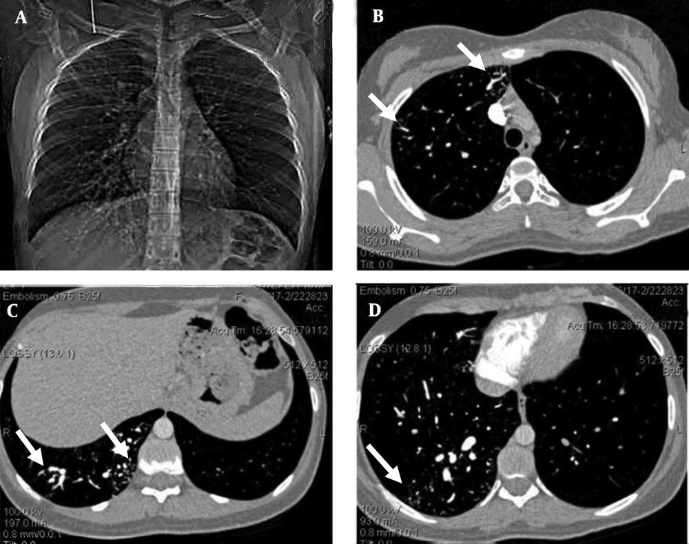
Contrast-enhanced transesophageal echocardiography confirmed extracardiac right-to-left shunt. Findings of abdominopelvic sonography were normal. The chest radiograph showed an increased vascularity of the right lung compared to the left one. The thoracic computed tomography (CT) images showed small peripherally located branching opacities and the prominent vessels in the right lung. The pulmonary CT-angiography (64-detector, Siemens, Germany) showed multiple scattered, small, dilated vessels in the right lung with a probable diagnosis of multiple AVMs or pulmonary telangiectasia (
Figure 1).
A 19-year-old woman with chest pain, exertional dyspnea and cyanosis of the limbs. The pulmonary computed tomographic angiography. A, Scout view, shows multiple dilated vessels of the right lung; especially right lower lobe. B, C, and D, Axial contrast enhanced computed tomographic angiogram source images shows scattered dilated vessels throughout right lung (white arrows); left lung is normal.
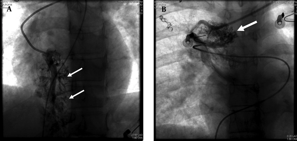
The pulmonary angiogram (GE, Innova 4200, Edison, New Jersey, USA) via right femoral vein access using Pigtail and Cobra II catheters (Torcon NB Advantage Catheter; Cook Inc., Bloomington, Indiana, USA) confirmed the former diagnosis. The morphology of the AVMs was similar to cerebral AVMs instead of usual pulmonary AVMs (
Figure 2).
A,B, Selective pulmonary angiograms. The images show multiple small abnormal vessels (white arrows) with early venous drainage (white arrow), which show a different morphology from usual pulmonary arteriovenous malformations.
We planned to coil no more than half of the lung in each session due to risk of complications such as pulmonary infarction; however, the main factor for the end point for each session was the patient tolerance; when she was restless, the embolization process was ended. The embolization was done to redistribute pulmonary flow from the AVMs to normal lung.
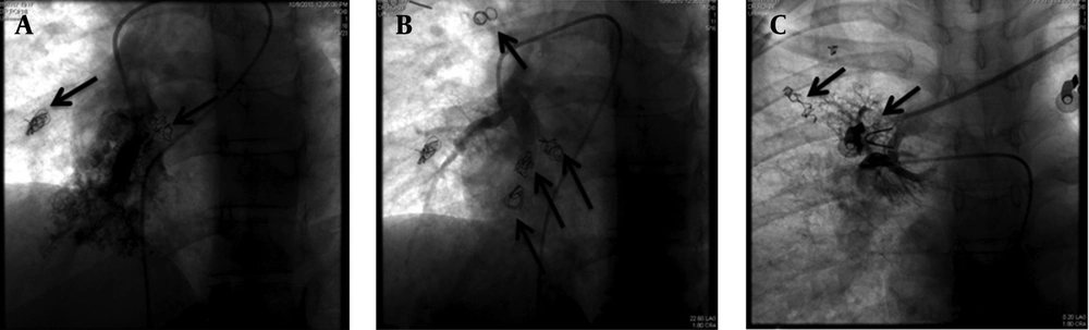
Percutaneous coil embolization of these malformations was achieved in three successive sessions with a one- to two-month interval and all detectable AVMs were occluded using 14 Pushable coils, (diameter, 3-5 mm) Tornado & Nester coils (Cook Medical Inc., Bloomington, Indiana, USA) (
Figure 3).
A-C, Multiple pushable coils are used to occlude feeders to the pulmonary arteriovenous malformations (Black Arrows).
After second embolization session, patient experienced a severe pleuritic chest pain and mild pleurisy that disappeared after two weeks. One month after the last embolization session, a noticeable improvement in the patient's respiratory symptoms was reported although PIO
2 only increased from 48.2 to 51.1 mmHg after one month of final session of coiling. One-year follow-up of patient showed that she had obtained her normal daily functions without any complain; however, because the patient was in another city and she stated that she was well and did not need any procedure, she refused to do any imaging study. Nonetheless, as seen in
Figure 3, previously occluded AVMs were still occluded in final session and no new collateral formation was noted during a three- to four-month period.