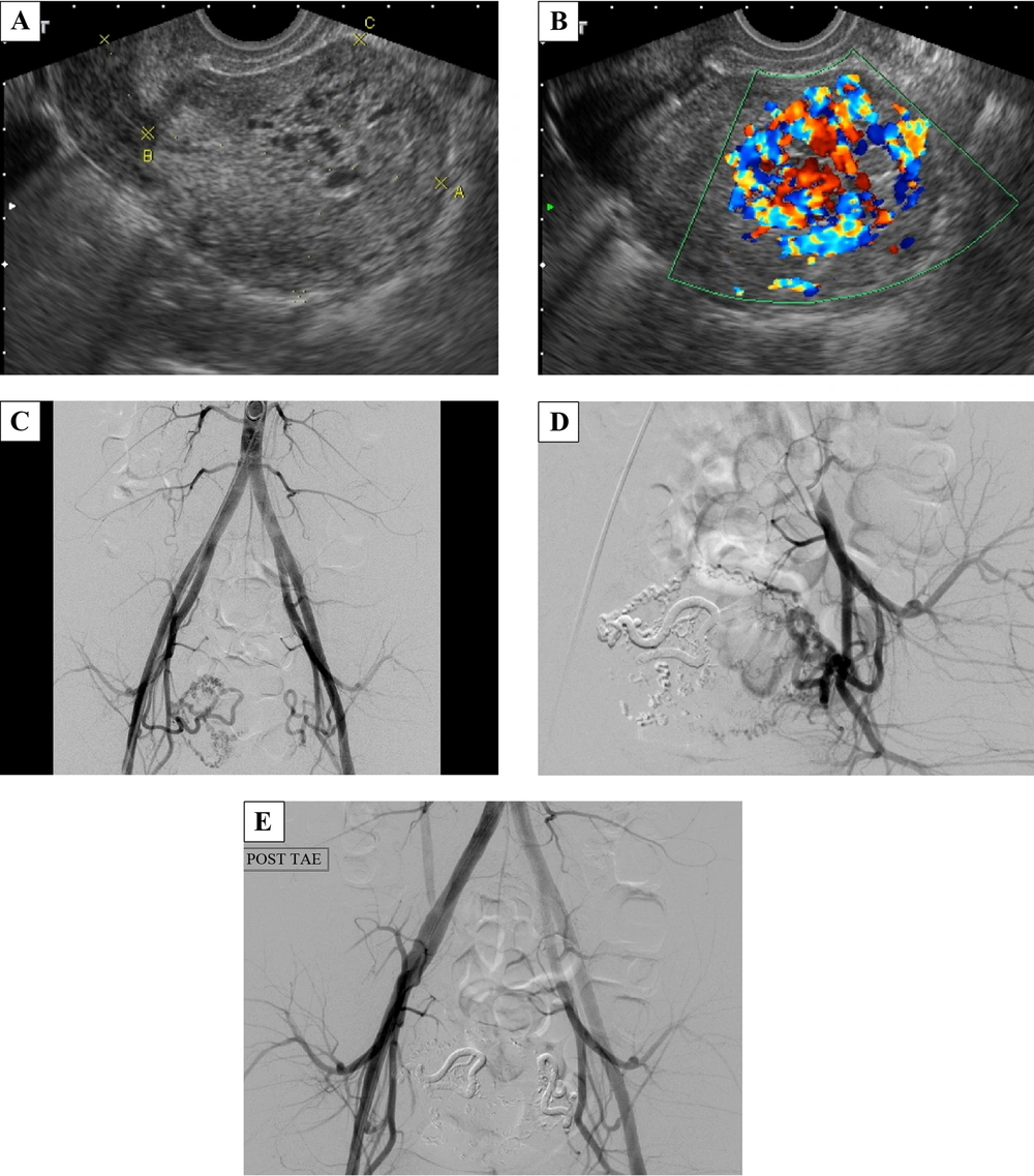
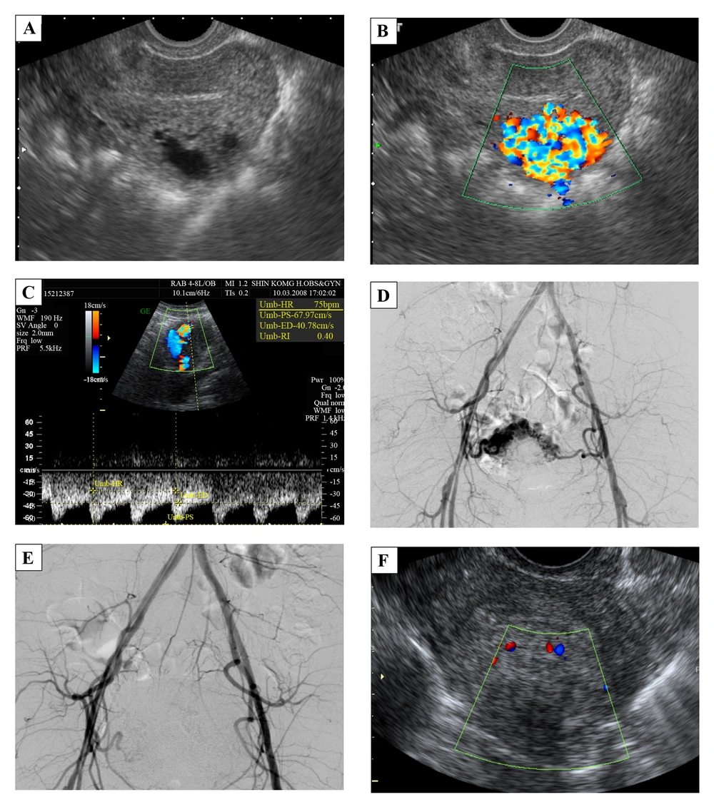
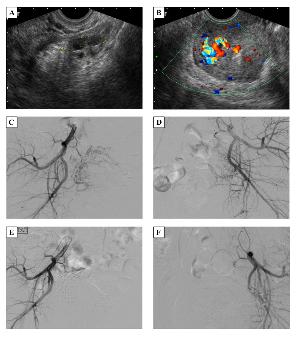
Uterine AVM is a relatively rare disorder that can cause life-threatening bleeding. They are still limited to a few case reports, a small number of series, and fewer review articles. Even though there is no large-sample analysis, O’Brien et al. report the rough incidence as 4.5% by performing ultrasonography in 464 pelvic-bleeding patients, ages 18 to 41 years (
1). Uterine AVMs may be congenital or acquired, the latter being more common. Acquired uterine AVMs may be caused by tissue damage and are followed by abnormal vascularization during the healing process. Most patients have a history of pregnancy (with delivery, spontaneous miscarriage, or surgical abortion) or dilation and curettage (D & C) (
2,
9). Each of the three cases we described had a history of multiple abortions. Uterine AVMs can also be associated with infection, trophoblastic disease, malignancy, and exposure to diethylstilbestrol (
2,
9). Hormonal change, such as pregnancy, menstruation, high-dose continuous estrogen and progestin therapy also play an important role in uterine AVMs because they may induce a bleeding episode, and most cases are in the range of 20 to 40 years of age (
6).
The common clinical manifestations include abnormal vaginal bleeding (from spotting to sudden heavy bleeding) and lower abdominal pain. Throbbing discomfort in the lower abdomen, urinary frequency or incontinence, dyspareunia, hypotension, and cardiac failure have also been described (
2). The patients in these three cases all have the common clinical presentation with a negative beta-HCG. The second patient also had a history of applying external force to her belly with a massage machine, which may have triggered her symptoms. There is no specific physical finding or sign, but pulsatile lesion, although rare, has been reported by manual pelvic examination.
Ultrasonography is a convenient imaging modality for evaluation of patients with vaginal bleeding; however, gray-scale ultrasonography does not provide powerful evidence to differentiate in this situation (
1). The color and spectral Doppler ultrasonography give us much more information to accurately approach the diagnosis of uterine AVMs. Classically, the color Doppler ultrasonography demonstrates the vessel structure with multidirectional high-velocity flow that produces a “color mosaic pattern,” and the spectral Doppler analysis typically shows high-velocity flow with a low resistance index that indicates arteriovenous shunting (
1). Nowadays, angiography is the gold standard of diagnosis. Nevertheless, it may not be used in every case unless physicians plan to do the following TAE or to use it for study purposes. Angiography of uterine AVMs shows, as is the case with AVMs at other sites, tortuous abnormal vessels between feeding arteries and early-contrast-enhancing drainage veins.
A wide spectrum of management plans have been proposed for uterine AVMs including observation, oral medications, TAE, laparoscopic bipolar coagulation, surgical ligation of uterine arteries, and hysterectomy (
7,
10,
11). The management of choice is decided according to hemodynamic stability or the need to control acute, heavy hemorrhage. Patients with asymptomatic or mild symptomatic uterine AVMs need no initial treatment but only follow-up ultrasonography, and the condition usually resolves spontaneously within months (
9). Oral contraceptive medications may also have a role in this population. On the other hand, hysterectomy is the traditional choice for stopping the troublesome bleeding of uterine AVMs. In the last decade, more and more published case reports have chosen to treat by TAE, and the clinical success rate is greater than 90% in retrospective review articles (
12,
13).
Compared with hysterectomy, TAE can preserve reproductive ability ideally, but physicians must be concerned about the embolization resulting in decreased blood supply to the uterine and whether it may lead to failed pregnancy. If the embolic materials flow into the ovarian artery, ovarian function and fertility may be disturbed. There are a lot of studies which discuss ovarian function and pregnancy following arterial embolization for postpartum hemorrhage (PPH) or uterine fibroids, but the issue is still controversial. Most articles suggest that uterine artery embolization does not cause higher risks of impairment of ovarian function (
14), fertility (
15), or major pregnancy outcome. (
14-
18). However, adverse effects, including synechia (
19), recurrent PPH (
19), miscarriage (
20), and loss of ovarian reserve (
21) are reported. The last review article suggests that uterine TAE does not have an observable effect on the ovary in most women younger than age of 45 years (
8). Berkane et al. pointed out that the smaller the embolic particles are, the higher risks of occlusion of the ovarian arteries will be, and they mention that the probable cut-off for material size is 500 μm (
19). Many case reports have transient impairment of ovarian function after embolization. They have normal menstrual cycles weeks to months later.
Currently, there is no strong evidence about TAE induced persistent ovarian failure, major complications of pregnancy, or fetal growth restriction. Case reports in this article and a handful of other documented reports describe full-term pregnancy after TAE. Searching on PUBMED, there are 21 cases that have term pregnancy after TAE for uterine AVMs (
1-
8), including one case with twin babies. The three cases in this article received TAE treatment with NBCA to control acute heavy bleeding and to protect the patients’ desire for a successful pregnancy. Varying severity of lower abdominal pain associated with post-TAE ischemia is noted in these patients and is also common in the past reports. Mild pain can be relieved by just oral NSAIDs; severe pain can be controlled well with patient-controlled analgesia use and will subside in a few days. The three cases were followed up at our gynecologic outpatient department for 2, 3, and 6 months, and there were no recurrent AVMs. Case 3 had a full-term delivery, with a gestational age of 39 weeks, three years after TAE without complications during pregnancy, delivery, or the postpartum period. The placenta had no gross abnormality.
In conclusion, TAE is a safe and effective treatment for uterine AVMs when the patient has hemodynamic instability or refractory hemorrhaging. If the patient wants to preserve her reproductive ability, TAE is the first management of choice. Our report enhances the confidence in achieving full-term pregnancy and vaginal-route delivery after superselective TAE in patients with uterine AVMs. In the future, more studies with high quality of evidence for side-effects after TAE and statistical analysis of risk factors are needed.