Multiple myeloma is uncommon in young patients. Patients under 40 years of age constitute 2.2% of all multiple myeloma cases, while those younger than 30 years constitute only 0.3% of all cases (
4).
Non-secretory multiple myeloma (NSMM) is a rare variant that accounts for 1 to 5% of all cases of multiple myeloma (
5). The disease is characterized by the absence of monoclonal gammopathy in serum and urine by immunofixation electrophoresis. NSMM is associated with longer survival, but these patients usually have more advanced disease than those with classic myeloma during initial presentation (
6,
7).
NSMM complicated with renal insufficiency is rare in view of absence of light chains in the urine (
6). In the present case, we have considered various possible causes of renal impairment. There was no ongoing renal infection, hypotensive episodes or drug related causes. Though renal biopsy was not performed, imaging studies did not show swollen kidneys, which would suggest obstructive nephropathy or plasma cell infiltration. We believe that renal impairment in this patient was caused by dehydration and hypercalcemia, as it improved with hydration and normalization of calcium level.
EM disease of MM is defined as the presence of malignant plasma cells outside the bone marrow, either as soft tissue masses spreading from bone or arising from EM organs. It is not uncommon to see intramedullary myeloma with EM extension as a result of cortical erosion and subsequent spread beyond the periosteum. However, hematogeneous spread of malignant plasma cells to the EM region is less common.This method of spread accounts for 3.4% of cases at diagnosis, 5% at relapse and during the course of disease (
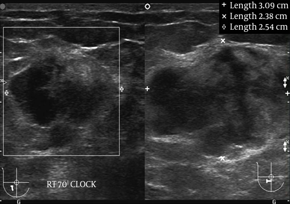
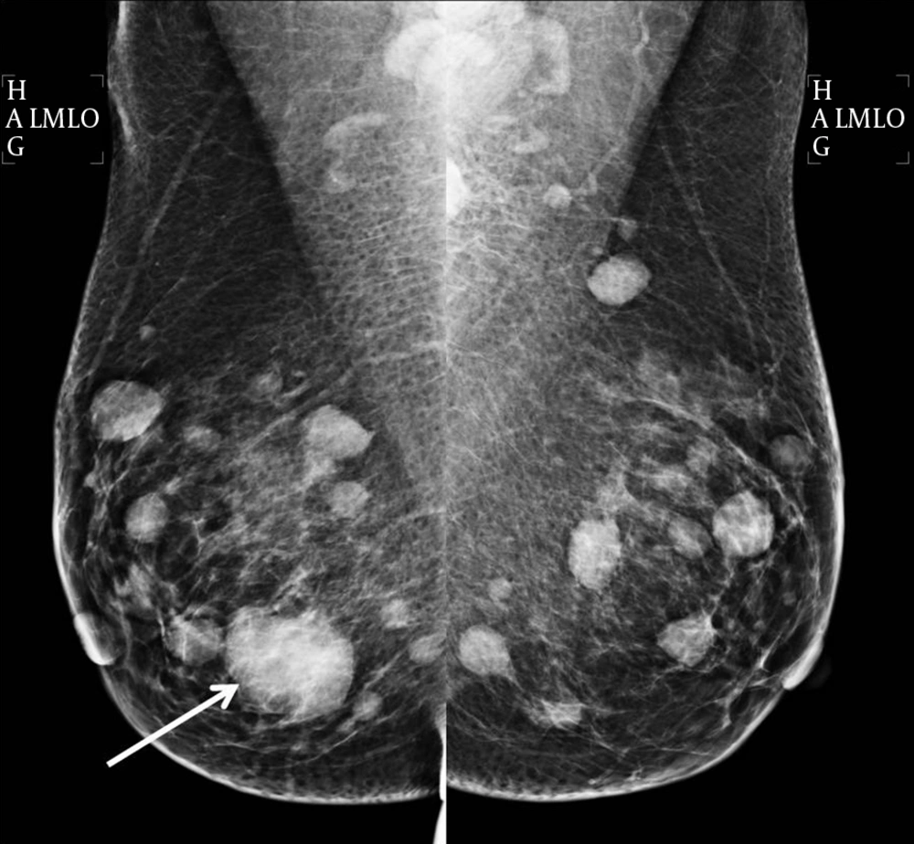
2). EM disease can affect any organ and appear as subcutaneous nodules, renal masses, breast lumps, orbital masses, lung, and pleural nodules. The imaging findings are often non-specific and often mimic other disorders.
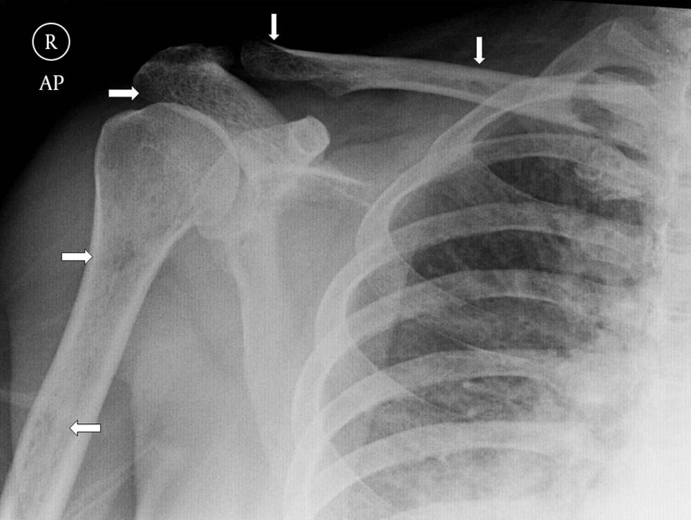
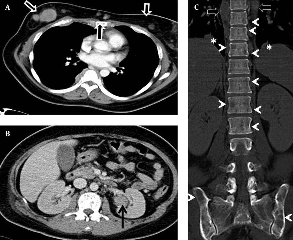
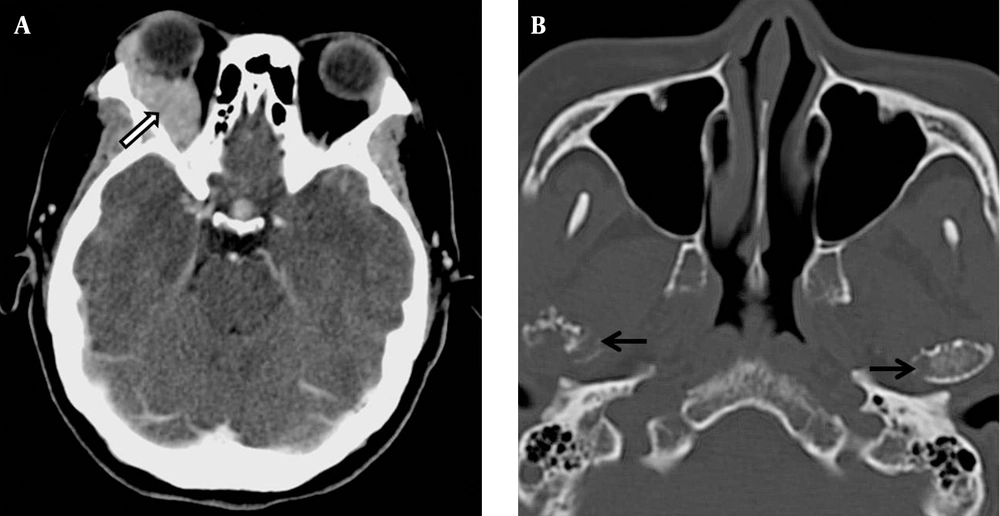
In the present case, the patient had extensive marrow and EM involvement affecting the left kidney, both breasts, subcutaneous tissue, right orbit, lung, paraspinal region, and lepto-meninges. Owing to the broad differential diagnoses, this patient’s clinical manifestations did cause a diagnostic dilemma. Our first impression was primary breast carcinoma with extensive metastases. However, breast carcinoma with advanced metastases is mostly accompanied by regional or distant lymphadenopathy. Moreover, bone scintigraphy would be expected to show extensive radio-pharmaceutical uptake or “superscan” appearance. In contrast, there was no lymphadenopathy in our patient and her bone scintigraphy was negative for osseous metastases.
Other differential diagnoses that were considered were lymphoma and soft tissue sarcoma with extensive metastases. Again, lymphoma is usually accompanied by lymphadenopathy, which was not seen in this patient. Soft tissue sarcoma should exhibit radio-pharmaceutical uptake.
Bone scintigraphy with Tc-99m is of little value in the evaluation of multiple myeloma due to excessive bone resorption and absence of characteristic osteoblastic activity (
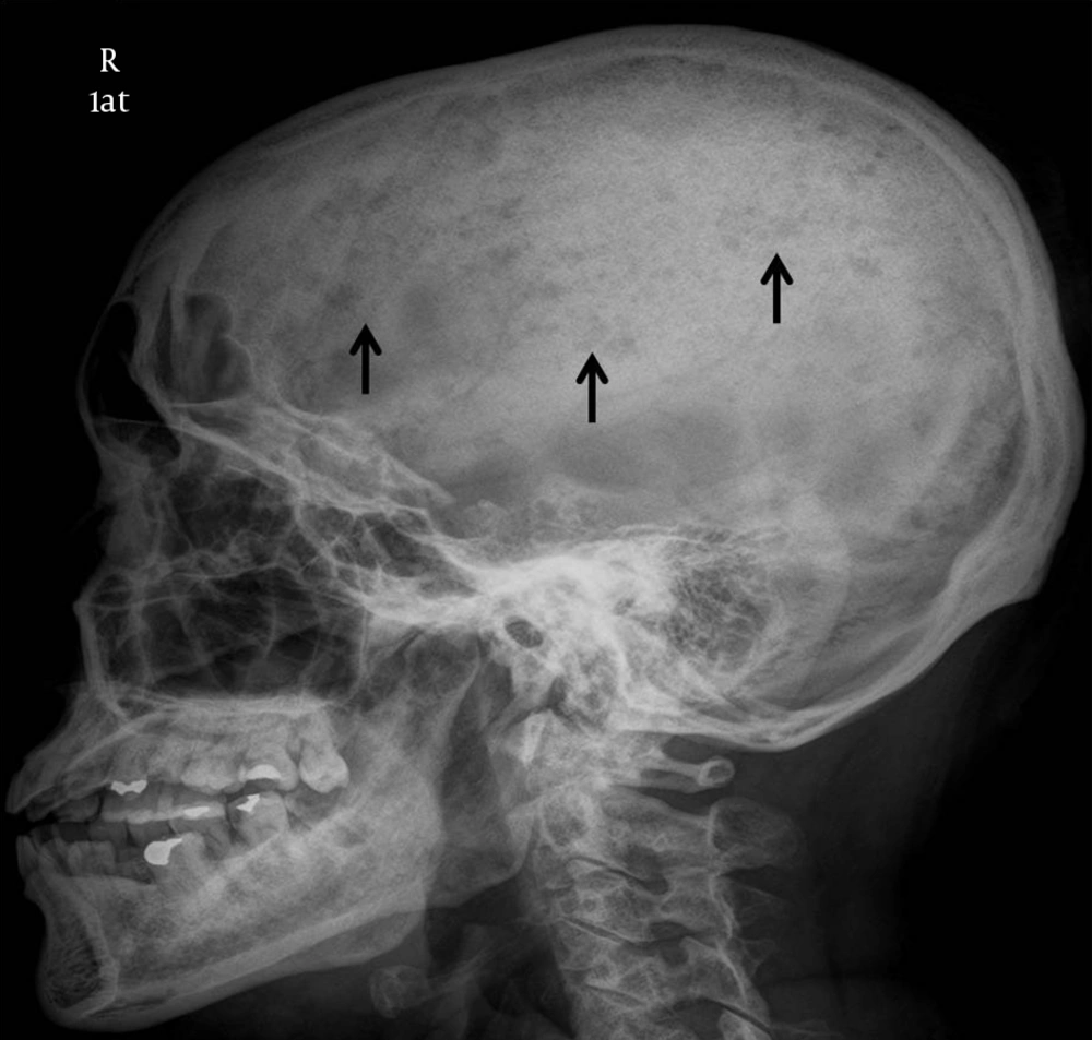
8). However, bone scintigraphy played an important role in making the radiological diagnosis in the present case. There are several tumors that are expected to show normal bone scintigraphy. These include multiple myeloma, some anaplastic tumors and pure osteolytic lesions. In the present case, although extensive lytic bone lesions were shown on CT, bone scintigraphy was negative. Mandibular involvement is common in multiple myeloma but rare in bone metastasis. Based on these two reasons, the diagnosis of MM with EM plasmacytoma should be favored. However, the young age of the patient, and the absence of monoclonal gammopathy in the urine and plasma electrophoresis prevented a straightforward initial diagnosis.
Furthermore, EM disease is associated with younger age, male gender, non-secretory subtype, advanced stage of disease (Durie-Salmon stage III), and extensive bone disease (
9). In contrast to NSMM, EM disease at the time of diagnosis shows a significantly poorer survival rate and shorter period of progression-free survival (
9).
In conclusion, NSMM with extensive EM involvement in young patients is difficult to diagnose owing to its low incidence and scant biochemical markers. The imaging features are non-specific and may mimic many conditions. However, in the case of extensive lytic bone lesions with mandibular involvement and negative bone scintigraphy, one should consider plasma cell related neoplasm as a possible diagnosis.