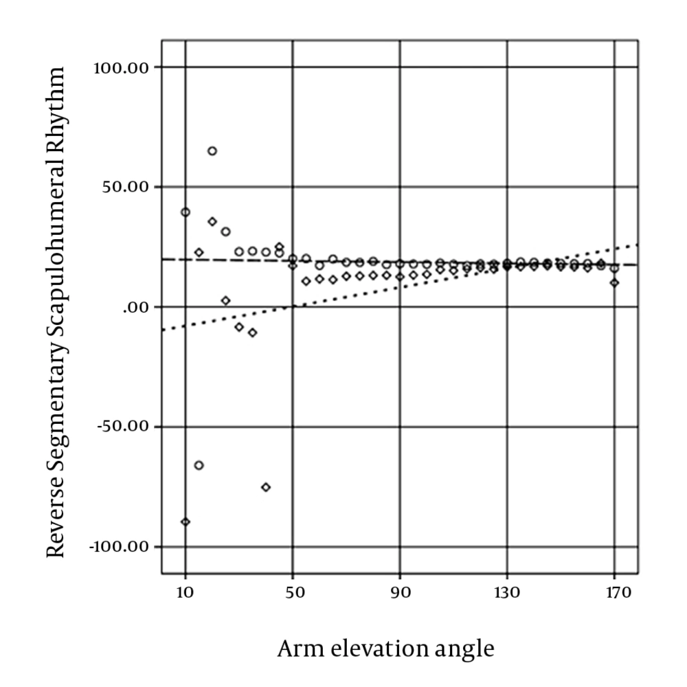
SSHR during arm elevation in the uninvolved group did not change significantly, but in the involved group, it decreased significantly. The rhythm of the involved group significantly exceeded the rhythm of the uninvolved group in the first quarter range of arm elevation (from 10o to 50o of arm elevation).
Reliability was excellent and consistent with previous studies (
22,
23). Reliability of the AC rotation variable in the middle range of arm elevation exceeded initial and final ranges of arm elevation probably due to anatomical structure overlap in the initial and final ranges of arm elevation. SEM was consistent with the finding of Massimini and colleagues (
34).
At present, the study of GH elevation on ST rotation ratio is more popular (
10-
13,
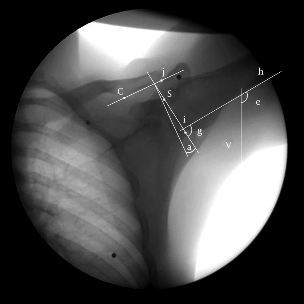
28). In the mentioned studies, the scapula and the clavicle were considered integrate in the calculation of scapulohumeral rhythm; whereas, ST upward rotation depends on the AC upward rotation, the SC elevation, and the posterior axial rotation. Reported differences between the mentioned studies could be a result of disregarding the AC internal rotation angle as a confounding variable.
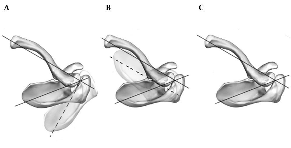
If the scapular plane was assumed perpendicular to the clavicular long axis (
Figure 4 A), elevation of the clavicle would be directly coupled with scapular anterior tilting as well as clavicular posterior axial rotation and scapular upward rotation; and if the scapular plane was assumed parallel with the clavicular long axis (
Figure 4 B), clavicular elevation and scapular upward rotation would be directly coupled as well as the clavicular posterior axial rotation and the scapular posterior tilting (
7,
28).
It was demonstrated that the AC internal rotation angle in healthy individuals was approximately equal to 75% of the assumed 90
o (
Figure 4 C); therefore, a combination of 75% of the assumed 90
o and 25% of the assumed 0
o should theoretically occur with clavicular rotation (
7). Thus, the effect of clavicular elevation and posterior axial rotation in scapular upward rotation depending on the AC internal rotation should be different in various individuals.
Teece and colleagues showed that in the AC joint, the scapula rotated upwardly 14.6º during arm elevation from 10º to 90º, and the scapula rotated upwardly 7º in the resting position (
7). Ludewig and colleagues demonstrated that the scapula in the AC joint rotated upwardly 11º during arm elevation from 20º to 120º, and the scapula rotated upwardly 5º in the resting position (
5). Based on extrapolation from their graphical results, it could be inferred that SSHR of healthy individuals increased during arm elevation.
In the present study, the scapula in the uninvolved group rotated upwardly 5.85º in the resting position, which was consistent with the two mentioned studies. The scapula in the AC joint rotated upwardly 4.86º and 8.18º during arm elevation up to 90º and 120º, respectively; which were inconsistent with the mentioned parallel studies. SSHR in 90º and 120º of arm elevation was 5.57 and 5.59, respectively, which was compatible with the last rhythm of mentioned parallel studies, respectively. However, the rhythm of the uninvolved group in the present study did not change significantly (P = 0.845); whereas, the rhythm increased in the two mentioned studies.We believe that rhythm differences between the present and the two mentioned studies probably originated from measuring methods and the differences could not be explained completely by the degree of freedom (3).
Reduction of scapular upward rotation induces the reduction of the subacromial space and it leads to the development or progression of shoulder impingement (
3,
4,
7). In the present study, in the resting position, the scapula showed an upward rotation of 5.85º in the uninvolved group and a downward rotation of 5.55º in the involved group. Less AC upward rotation angles in the first quarter range of arm elevation due to downward rotation of the scapula in the resting position in the involved group was probably an induced reduction of the subacromial space in the first quarter range of the arm elevation.
Despite what is common; prediction of the subacromial space based on the upward rotation range of the scapula is misleading. The upward rotation angle of the scapuladepends on the resting position of the scapula and the upward rotation range of the scapular. In the first quarter range of the arm elevation in the present study, the scapula in the AC joint rotated upwardly 0.3º and 7.39º in the uninvolved and involved group, respectively. Although the upward rotation range of the scapula in the involved group was more than the uninvolved group, the upward rotation angle of the scapula was 1.84º in the involved group and 6.15º in the uninvolved groupdue to the resting position of the scapula.
In order to correct the subacromial space and to reduce pain (3), probably the rhythm in the involved group showed a compensatory decrease in the present study.A rhythm up to 25º of arm elevation varied in the present study, and also sample size the groups below 20 degrees of arm elevation was disproportionate due to various starting angles of arm elevation. A rhythm below 25º of arm elevation between the two groups was not significantly different probably due to the mentioned reasons.The average of the maximum speed in the involved group decreased significantly, which was probably due to the pain in some ranges of arm elevation (
3,
4). In healthy individuals, the average arm elevation speed in the parallel studies was 33.33 (
5) and 26.67 (
7) degrees per second, while it was 32.33 degrees per second in the current study. Various speeds may lead to different motion patterns (
4). Therefore, the results must be compared according to the arm elevation speed. In this regard, the arm elevation speed in healthy athletes of this study was consistent with the study conducted by Ludewig and colleagues (
5).
Fayad and colleagues showed that rotational motion of the scapula in healthy individuals was not different between fast and slow arm elevation (18). The maximum speed of arm elevation differs considerably between the current (969º/s) and previous (90º/s) studies. In the previous study, the arm was probably elevated with feasible maximum and minimum speeds of a preferred movement pattern.
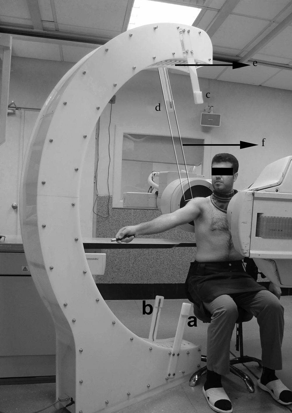
In the two dimensional study of the movement pattern, anatomical structures overlap in some ranges of the movement and little geometric transformation due to out of the plane motion are accounted for the limitations of the fluoroscopy method. On the other hand, this method has an acceptable validity (
33-
35) and reliability (
22,
33-
35). Movement pattern can be studied dynamically and functionally (
13). Fluoroscopy exposes less radiation than conventional methods without a reduction in the diagnostic accuracy (
22,
32,
36).
In conclusion, SSHR during arm elevation in the uninvolved group did not change significantly, but decreased significantly in the involved group. The rhythm of the involved group significantly exceeded the rhythm of the uninvolved group in the first quarter range of arm elevation.
A) AC internal rotation angle was assumed 90o. B) AC internal rotation angle was assumed zero. C) Normal AC internal rotation angle in the horizontal plane