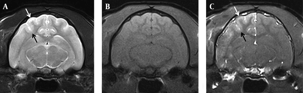
The development of a strategy for controlled, transient BBB disruption would be beneficial for basic research and clinical applications for many brain diseases, especially intractable brain tumors. Triolein emulsion has been reported to increase vascular permeability transiently when it is infused into the brain via a carotid artery. This increased vascular permeability may be observed as homogenous contrast enhancement on Gd-T1-weighted images and hyperintensity on T2-weighted images due to BBB breakdown by triolein as has been reported in the previous studies (
5,
10). However, undesirable vasogenic edema could cause unpredictable side effects and adversely affect laboratory experiments or studies on the clinical effects of treatments. Thus, an optimized triolein emulsion dosage is required that increases vascular permeability without causing undue interstitial edema.
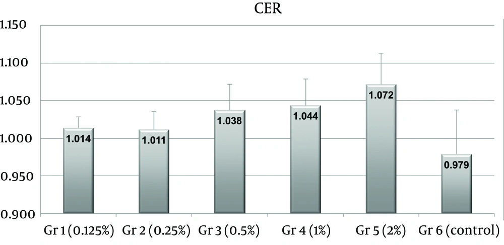
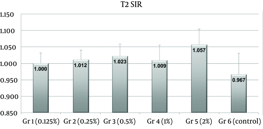
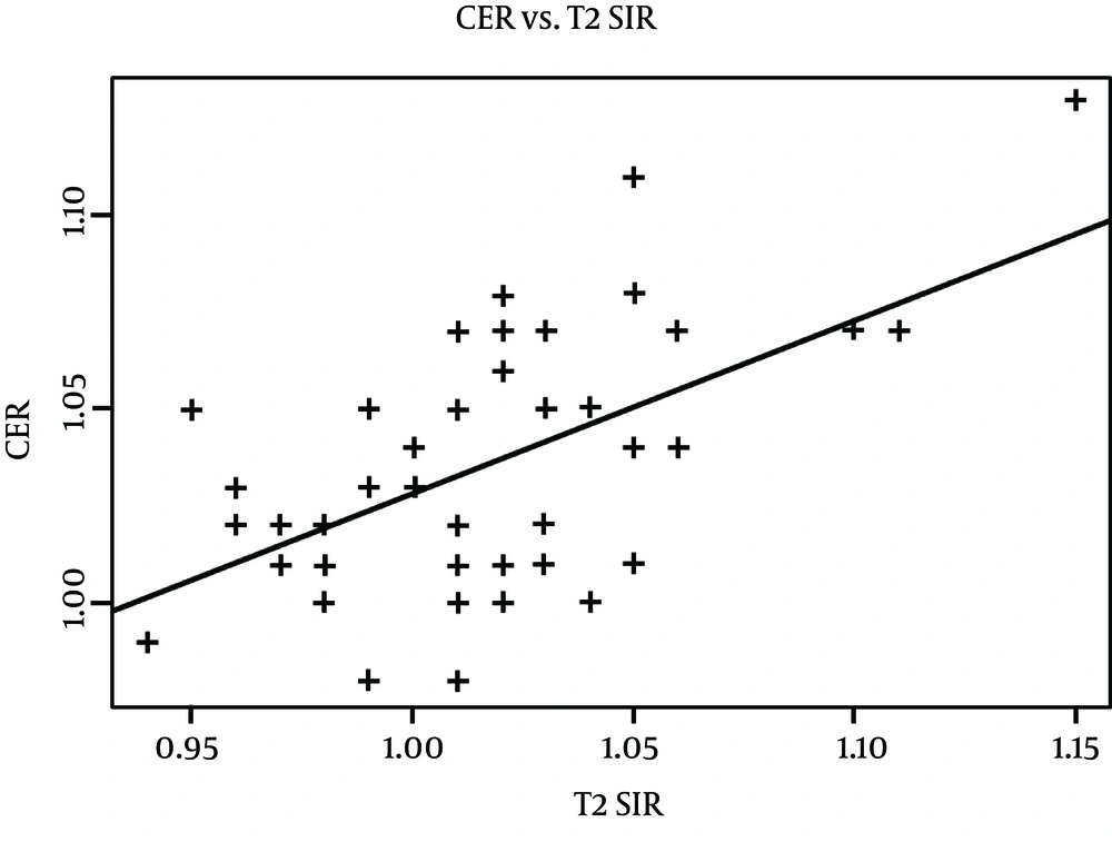
In the present study, contrast enhancement was statistically significant when triolein emulsion was infused via a carotid artery into cats at concentrations above 0.5% (0.1 mL of triolein in 20 mL of saline), and increased in a dose-dependently manner. Regarding vasogenic edema, T2-weighted images revealed no significant parenchymal edema when triolein emulsion was infused at 1%. Thus, the major finding of this study is that the minimum concentration of triolein emulsion required to increase vascular permeability adequately without brain edema is 0.5%. The lesion conspicuity was more prominent on Gd-T1-weighted image than T2-weighted image in the present study. In previous studies (
5,
7,
9), triolein emulsion has been used at 0.25-1%, but without knowledge of the optimal triolein emulsion concentration. The present study describes the relation between triolein emulsion concentration, vascular permeability, and brain edema. In fact, vascular permeability and vasogenic edema were found to be linearly correlated.
Another important consideration in studies of vascular permeability change is the total volume of triolein infused. Ryu et al. (
9) suggested that the minimum dosage of triolein emulsion (0.5%) required to achieve increased vascular permeability in the cat brain appears to be 3 mL/kg. However, the authors used only one concentration of triolein emulsion. Thus, according to the study by Ryu et al. (
9) and the present study, the optimum dosage and concentration of triolein emulsion to achieve increased vascular permeability with minimal edema in a cat model appears to be 3 mL/kg at a triolein emulsion concentration of 0.5%.
In the present study, Gd-T1-weighted images were used to detect BBB permeability changes. Contrast enhanced T1-weighted images are able to show sites of BBB breakdown (
11). In the presence of increased permeability or disruption of the BBB, gadolinium extravasates and diffuses into the brain parenchyma. In the present study, significant differences in contrast enhancement were observed between the control group and groups infused with triolein emulsion at more than 0.5%. Contrast enhancement indicating increased vascular permeability by triolein emulsion infusion into the brain is well known in experimental animal studies (
5,
7,
9). In a study of BBB disruption induced by emulsified oleic acid, nitric oxide was found to contribute to oleic acid-induced activations of matrix metalloproteinase-2 and -13 and the development of transient brain edema. After exposure to oleic acid administered intra-arterially, endothelial cells are stimulated and intercellular adhesion molecule-1 expression on the luminal surface occurs. Intercellular adhesion molecule-1 then recruits neutrophils from the blood circulation to affected brain vessels. These neutrophils stay close to vessels or infiltrate brain parenchyma and express inducible nitric oxide synthase, which produces nitric oxide and triggers the activations of matrix metalloproteinase-2 and -13 in the endothelial cells. Activated matrix metalloproteinase-2 and -13 then degrade laminin and the basal lamina structure, and ultimately, the BBB is disrupted and brain edema develops (
12). However, the mechanism of BBB disruption by triolein emulsion has not been elucidated.
Observed T2 hyperintensity has been attributed to vasogenic edema induced by BBB breakdown in the previous triolein studies (
5,
10). In the present study, a significant difference was observed between T2 hyperintensities in the control group and the 2% triolein group. In other words, no significant difference was observed in brain edema when the triolein emulsion concentration was less than 2%. A previous study of 0.5% triolein emulsion infusion into the cat brain showed that T2 hyperintensity occurred 1 hour after infusion, and that this hyperintensity diminished rapidly and approached normal 4 days after infusion (
5). In the present study, T2-weighted MR imaging showed that brain edema is not excessive when 0.5% triolein emulsion is used. However, the correlation between the amount of triolein infused and the clinical effects of increased vascular permeability or vasogenic edema have not been studied yet in an experimental animal. Furthermore, the optimal dosage may depend on the purpose of the study.
In the present study, there were contralateral paramedian lesions in two cats in group 3, three cats in group 4, and four cats in group 5. These contralateral lesions were probably due to the communication of two hemispheres similar to the Willis circle in humans. Velocity of infusion and the total amount of triolein emulsion might be related to those contralateral lesions.
Temporary BBB disruption could be useful in increasing drug delivery to tumors and adjacent brain parenchyma, and BBB disruption using hyperosmolar mannitol has been used for this purpose (
13), though its efficacy is controversial (
14). Drug delivery is most effective when a drug is administered within 5-10 min of hyperosmolar BBB disruption by mannitol (
14), but this time window appears to be inadequate for drug delivery to brain tissues. On the other hand, the time required for barrier opening is much longer when triolein is used (
5,
10). Opening of the BBB may have a substantial clinical impact on the treatment of patients with intractable brain tumors, and an adequate increase in vascular permeability by triolein emulsion infusion could make it possible to increase therapeutic response or lower the amounts of chemotherapeutic agents administered (
9).
Infarction as a side effect can also happen after triolein emulsion infusion into the brain. The size of the triolein particle in the emulsion of the present study was not uniform. This was because there had been no ideal method to make the particle uniform and to choose the particle size. Thus, if a large-sized particle of triolein enters the carotid artery, there could be occlusion of the vessel and it may result in brain infarction. Hemorrhage is another side effect after infusion of triolein (especially bolus triolein) due to mechanical disruption of the endothelial wall by triolein (
10). However, two hours after triolein emulsion infusion used in the present study is not sufficient to assess these side effects; longer follow-up time and further studies are necessary.
This study had several limitations. In the present study, the minimum concentration of triolein emulsion that significantly increased vascular permeability, but not edema was determined by MRI. Furthermore, we did not examine clinical symptoms or signs during the study, and their use could have resulted in different findings. Accordingly, we suggest that in future experiments, relations between triolein emulsion infusion concentrations and clinical outcomes should be studied.
The present study shows that the minimum concentration of triolein emulsion required to significantly increase vascular permeability with minimal edema in the cat brain at a dose of 3 mL/Kg appears to be 0.5%. T2-weighted and contrast-enhanced MR images revealed that vascular permeability and brain edema were increased by infusing triolein emulsion into a carotid artery. We believe the minimum concentration of triolein emulsion required to produce vascular permeability and brain edema when infused into the cat carotid artery as determined by the present study provides useful basic data for future studies on CNS drug delivery models.