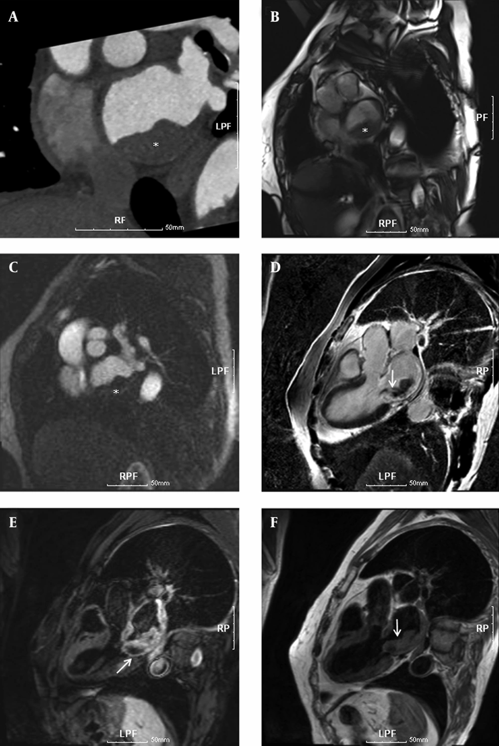
Cardiac 1H-MRS with ECG gating and respiratory motion at 3T MR system was allowed to achieve quantitative information of myocardial metabolites from small voxel volumes (< 5 mL) in the interventricular septum.
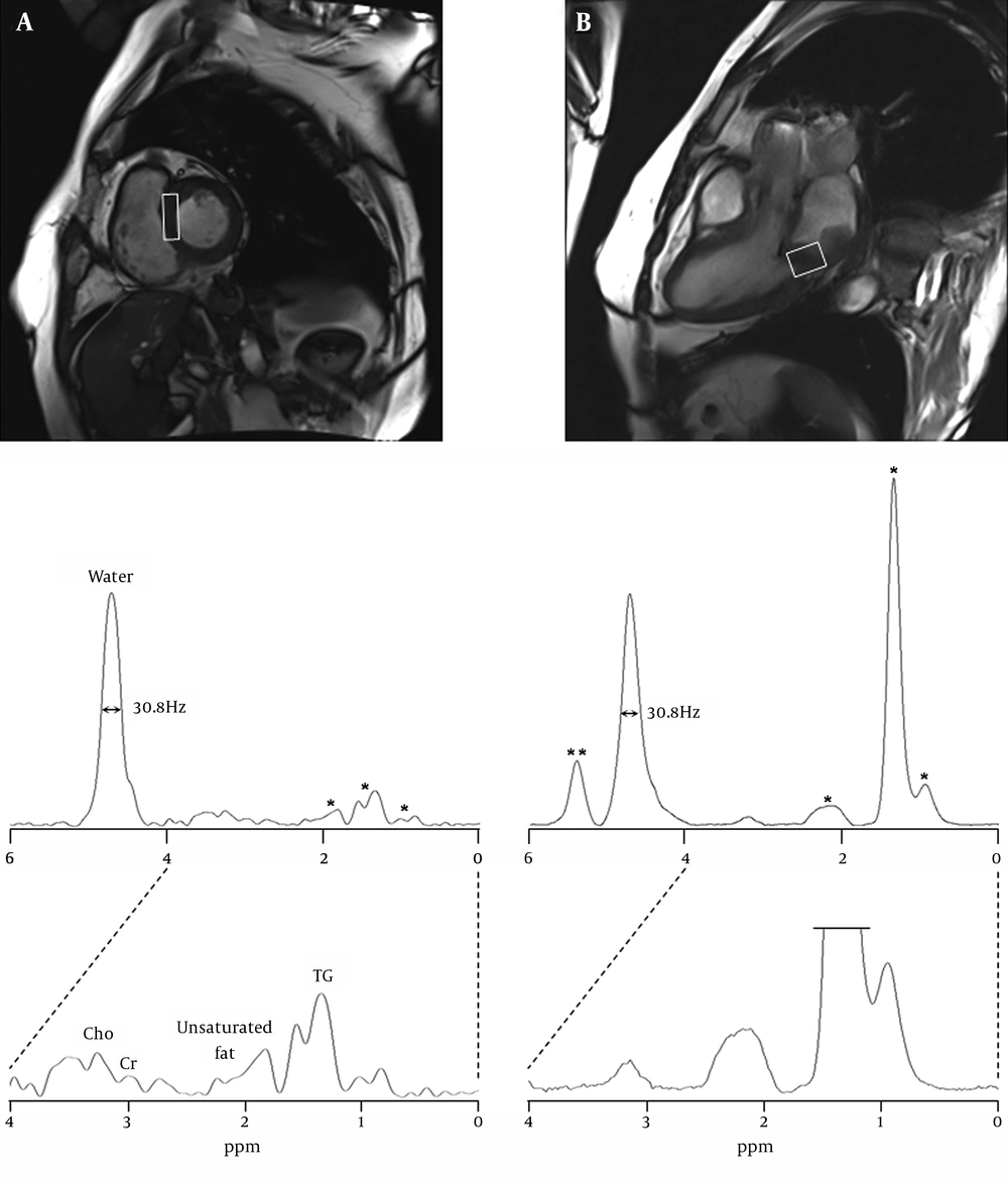
The acquisition time of a single spectrum was about 3 minutes, and the total examination time including MR spectroscopy with cardiac anatomic imaging was less than 50 minutes. With regard to spectral quality in the human myocardium, FWHM of the residual water peak was reproducible values, about 30 Hz. Accurate and precise positioning with adequate voxel size on the sarcoma of the posteroinferior left atrial wall is essential for accurate quantification of the metabolites. The MRS voxels were localized to avoid the contamination of unwanted tissues such as perimyocardium and chest muscle and the voxel sizes were optimized to cover the focal regions with minimal sizes, septum (4.5 cm
3) and myosarcoma (6.0 cm
3). In quantification of the resonance peaks in spectra of the septum and myosarcoma, the metabolic signal intensities were normalized to the residual water peak by weak water suppression as shown in
Figure 2 (
4,
10,
14).
This study was performed to compare quantitative metabolic information acquired from the normal septum and sarcoma region of the left atrial wall. The heart is the main myogenic muscular organ in the circulatory system and the spectral appearance of normal septal myocardium in this study was considerably similar to the spectral pattern in the skeletal muscle (
14). Rhabdomyosarcoma is a type of sarcoma that rarely occurs in the striated muscle. The voxel size of rhabdomyosarcoma was localized within the tumor part with a maximal thickness of 2.8 cm, and the resulting spectral pattern was similar to that of the calf muscle (voxel size of 180 cm
3) reported in Bongers’ study (
15).
Cho is a marker of cell membrane metabolism/turnover and is involved in acetylcholine (ACh) synthesis with acetyl CoA and cholinergic neurotransmission. It is well known that the Cho peak is a common biomarker for cancers with a variety of histological types (
16). Also, the Cho level is closely related to the degree of malignancy of the hypercellular tumors with rapid growth. In the present study, however, the Cho levels in both normal and lesional myocardium were in the normal range, and the concentration difference was not significant, suggesting that the sarcoma is in the early stage of malignancy (
Table 1). Myocardial Cr is associated with high-energy phosphate metabolism as an indicator of oxidative metabolism and the previous study showed that reduction of energy reserve underlies impaired contractile function in the failing heart (
3). Consequently, the tumor-specific abnormalities of Cho and Cr metabolites were not found in this case.
On the other hand, the peak at 5.4 ppm on the spectrum of rhabdomyosarcoma was assigned to C6 protons of cholesterol and/or the unsaturated groups of the olefinic region of lipids (
15,
17). In addition, the TG metabolite at 0.9 and 1.3 ppm showed the prominent difference, giving 18% in the septum and 119% in the sarcoma. In an animal study (
18), TG metabolite has shown a negative correlation with heart function, while treatment with insulin-sensitizing drugs reduced myocardial TG deposition and reversed contractile dysfunction in lipotoxic heart disease in rats with obesity. Also, myocardial TG may be a marker of myocardial viability after coronary occlusion due to enhanced esterification and/or reduced oxidation of fatty acids in ischemically insulted but viable myocardium (
19). These findings suggest that intramyocelluar TG accumulation is deleterious to the heart. Therefore, 1H MRS is a promising tool for assessing TG metabolites in human myocardial tissue.
This study included several technical limitations. First, the signal intensities on the focal regions may contain contaminations of unwanted signal from the outside of the voxel. Second, the differential voxel sizes (4.5 cc vs. 6.0 cc) on the normal septum and rhabdomyosarcoma may give rise to a quantitative error because of different signal-to-noise ratio. Third, this study used the residual water peak as a reference for quantification of the metabolites in the myocardium. However, the use of residual water signal can be prone to quantification error, due to cardiac motion and different water contents in different tissues. Fourth, the finding of increased TG content may not be specific to cardiac sarcoma because the predominant changes in TG content are frequently reported for various cardiac diseases and cancer. This study dealt with one patient and further studies are needed to confirm a more specific finding from a large population.
Although the present case report had several limitations, the advantages of 1H MRS over other cardiac imaging techniques, PET and contrast media-based MRI are that it does not expose patients to ionizing radiation and side effect of agents. Therefore, 1H MRS is widely accessible for assessing the normal myocardium and myocardial tumors (about 5 minutes in both tissues), and can concurrently provide the information of quantitative metabolic concentrations for effective therapeutic strategies in clinical settings.
In conclusion, this study, for the first time, demonstrates the feasibility that cardiac 1H-MRS would be potentially useful for the diagnosis of rhabdomyosarcoma by the use of TG level in the myocardium.