A 68-year-old man with a medical history of hypertension, aortobifemoral bypass, and femoro-femoral bypass grafting presented to the emergency department with severe diffuse abdominal pain. The patient was complaining of a vague abdominal pain for the last month that had intensified in the last couple of days. On initial examination, he was hemodynamically stable with a soft non-distended abdomen that was tender to palpation in the left lower quadrant. A slightly elevated white blood cell count of 17.79 × 10
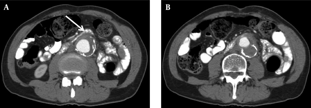
9/L and elevated C- reactive protein level of 236 mg/L (normal range, 0-5 mg/L) or 22247 nmol/L (range, 0.76-28.5 nmol/L) were measured in venous samples. Contrast-enhanced CT revealed extensive atherosclerosis of abdominal aorta and an infrarenal aneurysm 40-mm transverse diameter and 6-cm length (
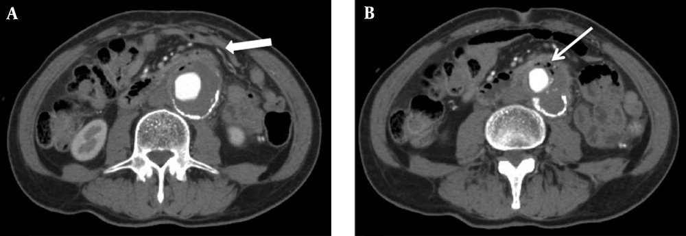
Figure 1). Aneurysm had a slightly hyperdense thrombosed crescentic portion with 11-mm diameter. In continuation with the thrombosed aneurysm, a soft tissue density was extending anteriorly. The soft tissue density was adherent to the duodenum and was slightly compressing it. With a suspected diagnosis of intramural hematoma and contained aneurysm rupture, conventional angiography of aorta was performed. Angiography confirmed abdominal aorta aneurysm and showed no contrast extravasation. The patient was prescribed pentoxifylline and coraspin and was discharged. One month later, the patient presented with mild-to-moderate hematemesis and severe abdominal pain, accompanied by tenderness to palpation with positive rebound in left lower quadrant. A nasogastric tube initially drained only gastric contents. During follow-up, the patient had an episode of melena and positive results in nasogastric lavage. A contrast enhanced CT showed an infrarenal aneurysm 50 mm in transverse diameter and 6 cm in length with the progression of mural thrombus to a diameter of 2 cm. Marked compression of small intestine, anterosuperior to aneurysm, and small air bubbles within thrombus were noted (
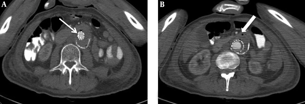
Figure 2). With a suspected diagnosis of AEF, endovascular aneurysm repair (EVAR) was planned. Two endovascular stent grafts, extending from renal arteries origins to right iliac artery, were deployed, followed by balloon dilatation of proximal graft site. Control angiography showed absence of contrast extravasation and endoleak. Prophylactic broad-spectrum antibiotic was administered. A control contrast-enhanced CT showed regression of aneurysm with a slightly decreased compression on duodenum (
Figure 3). Postoperatively, regression of mural thrombus and air bubbles within thrombus were noted. The patient was discharged on antibiotic therapy and died of myocardial infarction nine months later.