


1. Background
2. Objectives
3. Patients and Methods
3.1. Study Population
3.2. Multiplane TEE

Measurement of the myocardial grayscale intensity, velocity and displacement at the lateral side of the mitral annulus in cases without STEIRI. A. The maximal and minimal grayscale intensity in the cardiac cycle determined by grayscale intensity curve using videodensitometric analysis; B. The peak systolic velocity, the peak early diastolic velocity and the peak late diastolic velocity using tissue velocity imaging; C. The peak displacement determined by displacement curve using tissue tracking; D. Normal ECG
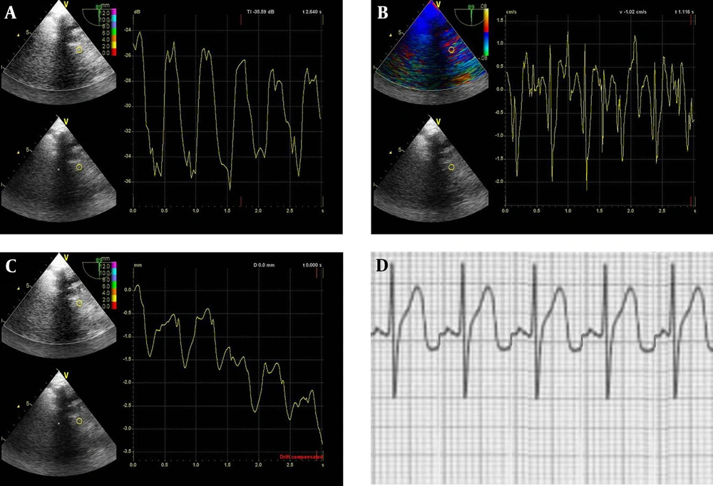
Measurement of the myocardial grayscale intensity, velocity and displacement at the lateral side of the mitral annulus in cases with STEIRI. A. The maximal and minimal grayscale intensity in the cardiac cycle determined by grayscale intensity curve using videodensitometric analysis; B. The peak systolic velocity, the peak early diastolic velocity and the peak late diastolic velocity using tissue velocity imaging; C. The peak displacement determined by displacement curve using tissue tracking; D. Representative ECG of reperfused ST-segment elevations
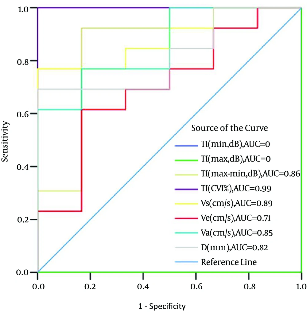
Receiver operating characteristic curve showing the performance of the myocardial grayscale intensity, velocity and displacement at the lateral side of the mitral annulus in discrimination between patients with ST-segment elevation ischemic reperfusion injury (STEIRI) or not.
3.3.Reproducibility
3.4.Statistical Analysis
4. Results
4.1. Echocardiographic Parameters
| Parameter | Normal Participants | Patients Without STEIRI | Patients With STEIRI |
|---|---|---|---|
| TI (max,dB) | -37.147±4.41 | -34.66±5.13 | -32.92±3.59 |
| TI (min,dB) | -20.49 ± 1.59 | -21.03 ± 1.34 | -23.75 ± 2.32 |
| TI (max-min,dB) | 16.66 ± 0.49 | 13.62 ± 6.11 a | 9.18 ± 2.09a,b |
| TI (CVI, %) | 44.85 ± 6.35 | 39.27 ± 5.35 a | 28.59 ± 4.52b,c |
| Vs (cm/s) | -6.16 ± 1.91 | -4.93 ± 1.38 | -2.74 ± 1.36 b,c |
| Ve (cm/s) | 6.13 ± 1.78 | 1.73 ± 0.61 c | 1.24 ± 0.59 b,c |
| Va (cm/s) | 6.30 ± 2.13 | 2.82 ± 1. 31 c | 1.34 ± 0. 73 b,d |
| D (mm) | -12.33 ± 0.51 | -4.53 ± 3.25 c | -1.81 ± 1.43b,d |
aP <0 .05
bP <0 .01, unpaired t test, compared to the values of normal participants
cP <0 .05
dP <0 .01, unpaired t test, compared to the values of patients without STEIRITI, grayscale intensity; TI (max), the maximal grayscale intensity in the cardiac cycle; TI (min), the minimal grayscale intensity in the cardiac cycle; TI (max-min), the difference between the maximal and minimal grayscale intensity in the cardiac cycle; TI (CVI, %), cyclic variation index= TI (max-min) / TI (max) × 100; Vs, the systolic velocity; Ve, the early diastolic velocity; Va, the late diastolic velocity ; D, the peak displacement in the cardiac cycle


