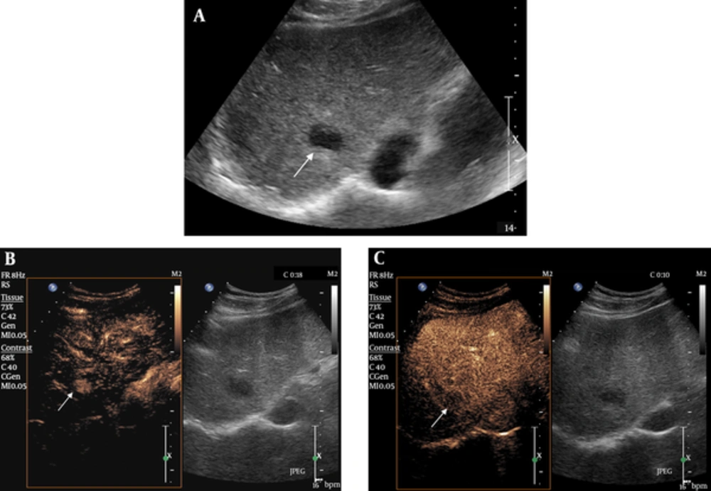
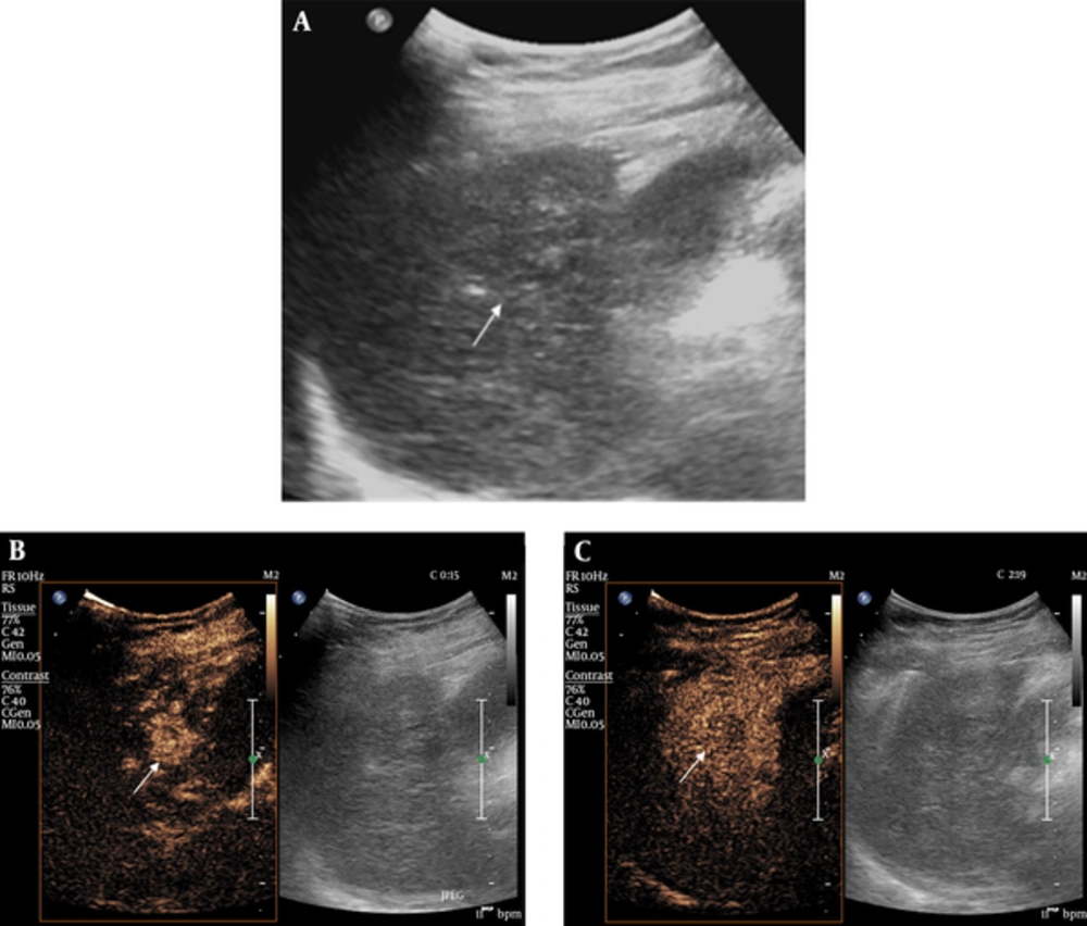
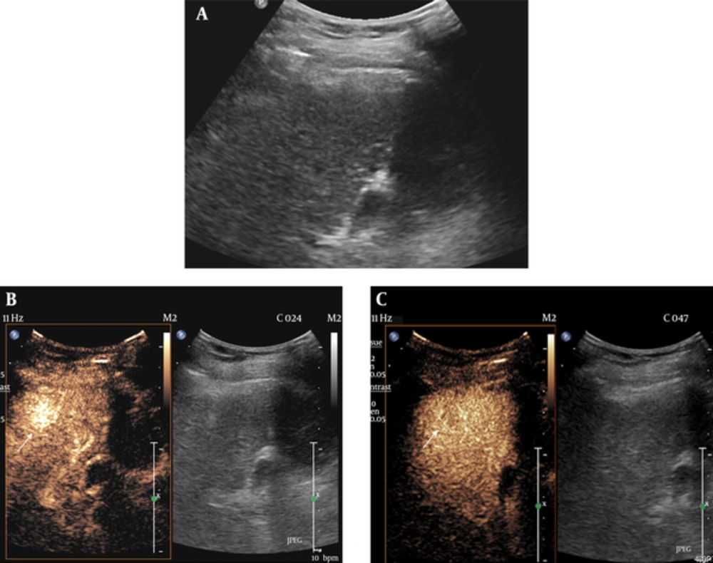
This study showed that pre-RFA evaluation with CEUS can affect the patient’s treatment plan since RFA treatment can become a treatment option for the previously undetected HCCs on GSUS. Still, surgical resection is a treatment of choice in the treatment of HCC, but the majority of HCCs are not suitable for curative resection at the time of diagnosis. Moreover, surgical resection is difficult in cases with advanced cirrhosis, tumor multicentricity, vascular, and extrahepatic involvement (
12,
13). In such cases, RFA under ultrasound-guidance is widely performed for small HCCs less than 3 cm up to three lesions or single medium-sized HCCs (
14). RFA is a widely performed treatment modality due to the ease of use, safety, reasonable cost, and applicability to minimally invasive techniques (
15). Previous studies reported that percutaneous RFA achieved the same overall and disease-free survival rates as surgical resection for patients with small solitary HCCs (
16,
17).
However, RFA requires the obvious demonstration of a focal hepatic lesion on ultrasound. For assessment of lesions that remain unclear on GSUS, CEUS is an excellent additional diagnostic tool. CEUS allows reliable and accurate tumor detection in the liver that cannot be recognized in conventional GSUS (
1-
4). The information gathered during the wash-in phase and various perfusion phases including arterial, portal venous and late phases allowed the characterization of different liver tumors.
Ultrasound contrast media is a blood pool tracer and has been used due to its ability to overcome the limitations of conventional GSUS or power Doppler US by demonstrating parenchymal microvasculature (
18). The first-generation ultrasound contrast agent (galactose 99%, palmitic acid 1%, Levovist®; Schering, Berlin, Germany) was first introduced in 1996, as air with a galactose and palmitic acid surfactant, and its main indications included heart, abdomen including vesico-ureteric reflux, and transcranial US. Sulfur hexafluoride microbubbles were introduced in 2001. It consists of sulfur hexafluoride with a phospholipid shell, and its main indications are cardiac, macrovascular, liver and breast lesions. Sulfur hexafluoride has enhanced stability and resonance behavior at low acoustic pressure compared with first generation contrast agents. Another second generation contrast agent perfluorobutane (Sonazoid®; GE Healthcare, Tokyo, Japan), is a lipid-stabilized suspension of perfluorobutane gas microbubbles that facilitates obtaining a real-time blood flow image in addition to a stable Kupffer phase, lasting up to 120 minutes after injection (
19,
20).
A number of studies evaluated the role of CEUS as a guiding tool in minimally invasive ablation therapies using ultrasound contrast agents. There were previous studies reporting the added value of CEUS in detection of focal hepatic lesions, adding strength to use of CEUS as a planning modality for RFA. Maruyama et al. reported CEUS with Levovist facilitated the application of percutaneous US-guided treatments, such as RFA and percutaneous ethanol injection (PEI), by successful localization in 75% of ultrasonically invisible hypervascular HCCs (
21). Numata et al. reported that contrast-enhanced harmonic ultrasound diagnosed 97% of viable HCCs, and 14/15 (93%) lesions not detected by conventional GSUS were successfully treated by percutaneous ablation therapy guided by CEUS (
22). A recent study by Chan et al. also showed CEUS provided fair visualization of HCCs that were either poorly visualized or not visualized on GSUS (
23). These studies strongly enhanced the need of CEUS in order to perform percutaneous US-guided treatments such as RFA or PEI.
The results of our study are in agreement with most of the previous reports on guiding effectiveness of CEUS before RFA. In our study, more than half of ultrasonically unrecognized hepatic lesions were successfully demonstrated by CEUS with sulfur hexafluoride microbubbles, therefore, added as a candidate for RFA treatment. The visualization rate of target HCC in our study is not as high as that of the previous studies, this may be due to the difference between first and second generation contrast agents, tumor size, and presence of multiple RN or DN in the cirrhotic liver background.
When the operator’s confidence about a target lesion is low on GSUS, it is practically impossible to perform US-guided RFA, especially when there are multiple similar nodules such as DN or RN adjacent to the target HCC. Therefore, when the location of the HCC is equivocal on GSUS, the operator will hesitate to perform RFA, analyzing the benefits and risks of a blind ablation. In our study, there were eight lesions with GSUS visibility score 2, which were incompatible to perform RFA after conventional GSUS. However, by performing CEUS, 7 HCCs showed marked arterial enhancement, confirming the location. Therefore, HCCs were successfully treated with RFA. CEUS cannot only depict previously invisible HCCs on GSUS but also elevates diagnostic confidence of previously equivocal hepatic nodules.
RFA can be performed under ultrasound, computed tomography, or fluoroscopy guidance. There have been studies when HCC is invisible on conventional ultrasound, CT-arteriography or iodized oil injection, or post-TACE fluoroscopy images can be helpful in the detection and guidance of minimally invasive procedures. Real-time CT fluoroscopy can be used for needle puncture guiding, and the efficacy of treatment can be evaluated directed by CT scan after the procedure is performed (
24). However, ultrasound is the most frequently used guiding modality for RFA due to its accessibility, lack of radiation, feasibility, and real-time guidance. Moreover, ultrasound offers good spatial resolution for treatment monitoring.
Recent technical expansion of real-time virtual sonography (RVS), a diagnostic imaging support system that can be synchronized with B-mode ultrasound images reconstructed from high-quality multiplanar reconstructed (MPR) CT and MR images can be used in the detection of HCC nodules that are not visualized by GSUS. Nakai et al. reported that RFA was technically feasible and local tumor control was achieved in all patients (
25). RVS displays equivalent cross-sectional MPR images as ultrasound images on the same monitor screen in real time, using volume data from CT or MRI, assisting in hepatic lesion detection. Many reports emphasized the effectiveness of RVS, RVS-guided RFA treatment and its efficacy for HCC is promising (
26,
27). However, technical difficulties remain. Real-time virtual sonography and CT may not always correlate accurately, since respiratory movements, changes in posture, and bowel peristalsis may cause distortion in images (
28). In these settings, CEUS may be helpful. Toshikuni et al. reported 55 out of 60 (90%) inconspicuous HCC nodules were identified with RVS. Moreover, by using US contrast agent, the detection rate has increased up to 96.7% (
29).
There were several limitations in our study. First, most HCCs were not pathologically confirmed. Diagnosis was made by CT or MR imaging according to non-invasive diagnostic criteria approved by EASL and AASLD of typical arterial enhancement and delay washout pattern of HCC. Furthermore, all of our cases had liver cirrhosis that supported imaging diagnosis of HCC without pathologic verification. Only one case was pathologically confirmed by percutaneous core needle biopsy due to its small size. Second, selection bias may have been present, since HCCs with a high probability of RFA treatment were requested for CEUS by a referring physician in the first place. HCC that was not suitable for RFA due to its deep, subcapsular, or perivascular location may have been excluded in the first place even without performing CEUS. Third, only a small number of cases were included in the study compared to the larger studies performed previously. Finally, eight out of 19 HCCs with GSUS visibility score 1 were not delineated on CEUS. Therefore, they could not be an RFA candidate. The possible explanation for those undelineated HCCs is due to their completely isoechoic nature, the target lesion was not precisely located on the first scan of GSUS so following CEUS could not demonstrate the lesion. In addition, short time window of arterial and delay phases of contrast agent of CEUS further limited the detection of the target tumor.
In conclusion, CEUS can demonstrate a poorly-visible or invisible HCC on GSUS by showing arterial enhancement or delay washout, thus increased number of possible candidates for RFA treatment. CEUS can be an effective planning modality for RFA especially when a target HCC is invisible or questionable on conventional GSUS.