


1. Background
Spontaneous tendon rupture is a rare injury during activity. It has been frequently associated with chronic metabolic diseases such as chronic renal failure (CRF), diabetes mellitus, systemic lupus erythematosis, rheumatoid arthritis, gout and obesity (1). Hyperparathyroidism and metabolic acidosis in CRF patients are known to be risk factors for tendon degeneration (2). In CRF patients, secondary hyperparathyroidism, osteomalacia, osteoporosis, a dynamic bone disease, soft tissue and vascular calcifications could be seen due to renal osteodystrophy. Some musculoskeletal disorders such as amyloid and crystal deposition, destructive spondyloarthropathy, avascular necrosis, and spontaneous tendon rupture could also be seen in these patients (3).
Sonoelastography (SE) is a recent ultrasound technology that provides information about elasticity and stiffness of different tissues or lesions (4). In this method, the pressure applied to the tissue provides information about the physical and mechanical characteristics of the tissue such as elasticity and hardness. These characteristics help to differentiate tissue types as well as different pathologic conditions of the same tissue (5). Recently, SE has been used to evaluate the breast, thyroid, muscle, and abdominal solid organ diseases (6-9). Previous studies have shown that SE is efficient for demonstrating alterations of Achilles tendon (10).
Shear-wave elastography (SWE) is a noninvasive elastography technique that measures soft tissue elasticity quantitatively in kilopascal units, by sending acoustic impulse to the tissue without applying pressure unlike other SE techniques (11).
2. Objectives
The purpose of the current study was to evaluate Achilles tendon thickness with B-mode ultrasonography (US) and stiffness using SWE technique in CRF patients undergoing hemodialysis.
3. Patients and Methods
3.1. Radiologic Assessments
The study was designed prospectively and approved by the research ethics committee. Signed consent forms in accordance with Helsinki declaration were obtained from patients and healthy individuals.
Thirty-four CRF patients (23 male, 11 female) undergoing hemodialysis in our hospital and 32 healthy individuals (24 male, eight female) were included in the study. The mean hemodialysis duration was 3 years (1 month - 20 years). CRF patients that had at least a one-month hemodialysis duration were included in the study. The patients with previous sports or trauma injury, lower extremity osteoarthritis, and patients who had undergone surgery or treatment, such as knee prosthesis surgery, were also excluded. The control group was formed from randomly selected healthy volunteers who were attendants of patients presented to the US unit.
3.2. Ultrasonography Technique
During the evaluation of Achilles tendon, the patients were in relaxed prone position with their feet hanging freely over the edge of the table without flexion or extension (12). As Achilles tendon is located superficially, GE Healthcare Logiq E9 device and 9 MHz frequency linear probe was used in the study. After positioning of the probe, the tendon was easily identified by following the muscle tissue to the calcaneal insertion site (13). The measurements were done by one radiologist (AH) with more than 5 years of experience in US. Each measurement was repeated three times and the mean was calculated.
Initially, both Achilles tendons were examined in transverse and longitudinal planes from calf muscles to the calcaneal insertion in terms of homogeneity, thickness, and structural abnormality. Achilles tendon thickness was measured from the distal part at the level of medial malleolus as mentioned in previous studies (14). The region of interest (ROI) was placed in the middle of the tendon and automatic stiffness was calculated in kilopascal units (Figure 1). SWE method is different from other elastographic methods for its direct measurement of stiffness of ROI without applying any pressure.
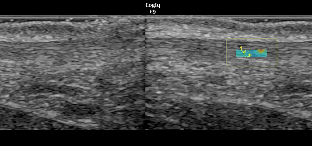
Figure 1.
A region of interest (ROI) was placed in the central area of the Achilles tendon and tissue elasticity was given automatically by machine in kiloPascal units
3.3. Statistical Analysis
Data analysis was performed using SPSS for Windows, version 11.5 (SPSS Inc., Chicago, IL, United States). Whether the distributions of continuous variables were normal or not was determined by Shapiro Wilk test. Continuous variables were shown as mean ± standard deviation (SD) or median (min - max), where applicable. Number of cases and percentages were used for nominal data. The differences in normally distributed data between control and case groups were compared by Student’s t test, otherwise, Mann Whitney U test was applied for the variables which were far from normal distribution. Nominal data were analyzed by Pearson's Chi-square test. Degrees of association between continuous variables were evaluated by Spearman’s rank correlation analyses. A P value less than 0.05 was considered statistically significant. The correlation coefficients of age and thickness and elasticity were evaluated by Pearson correlation test.
4. Results
The mean age of the patient group was 54 ± 17 (range 24 - 85) and for the control group, it was 42 ± 9 (range 20 - 56) (P < 0.05). Male/female ratio of both groups were similar (P = 0.510) (Table 1). The demographic data of the study group, the thickness and stiffness of Achilles tendon are presented in Table 1. The mean Achilles tendon thickness in the patient group was 4.14 ± 0.62 mm on the right side and 4.19 ± 0.59 mm on the left side (mean of both sides, 4.16 ± 0.59 mm). The control group had a tendon thickness of 4.79 ± 0.62 mm on the right side, 4.77 ± 0.63 mm on the left side (mean of both sides, 4.78 ± 0.61 mm). When we compared the results, the thickness of Achilles tendon was significantly lower in the patient group (P < 0.001) (Figure 2). The mean elasticity scores of Achilles tendons in the patient group was 27.10 ± 10.83 kPa on the right side, and 24.90 ± 10.39 kPa on the left side (mean of both sides, 26.00 ± 9.74 kPa). Whereas, in the control group values were 24.62 ± 7.40, 24.66 ± 7.28, and 24.64 ± 6.64 for the right, left and mean of both sides, respectively. There was no statistically significant difference in stiffness values between the two groups (P > 0.05) (Figure 3).
Table 1.Demographic and Clinical Characteristics of Control and CKD Groupsa
| Controls (N=32) | CKD (N=32) | P value | |
|---|---|---|---|
| Age, y | 42.4 ± 9.0 | 54.7 ± 17.7 | < 0.001 |
| Gender | 0.510 | ||
| Male | 24 (75.0) | 23 (67.6) | |
| Female | 8 (25.0) | 11 (32.4) | |
| Duration of hemodialysis, y | - | 3 (1 month - 20 years) | - |
| Achilles thickness, mm | |||
| Right | 4.79 ± 0.62 | 4.14 ± 0.62 | < 0.001 |
| Left | 4.77 ± 0.63 | 4.19 ± 0.59 | < 0.001 |
| Mean | 4.78 ± 0.61 | 4.16 ± 0.59 | < 0.001 |
| Elastography, kPa | |||
| Right | 24.62 ± 7.40 | 27.10 ± 10.83 | 0.362 |
| Left | 24.66 ± 7.28 | 24.90 ± 10.39 | 0.893 |
| Mean | 24.64 ± 6.64 | 26.00 ± 9.74 | 0.594 |
Abbreviations: CKD, chronic kidney disease; kPa, kiloPascal; SD, standard deviation; y, year
a Values are expressed as No. (%) or mean ±SD.
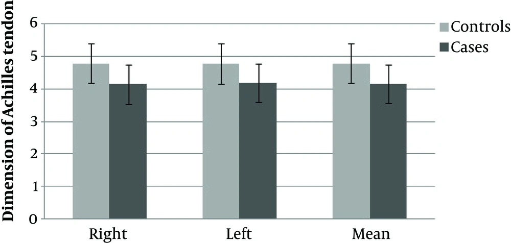
Figure 2.
Analysis of Achilles tendon thickness in patients with chronic kidney disease (CKD) and the control group
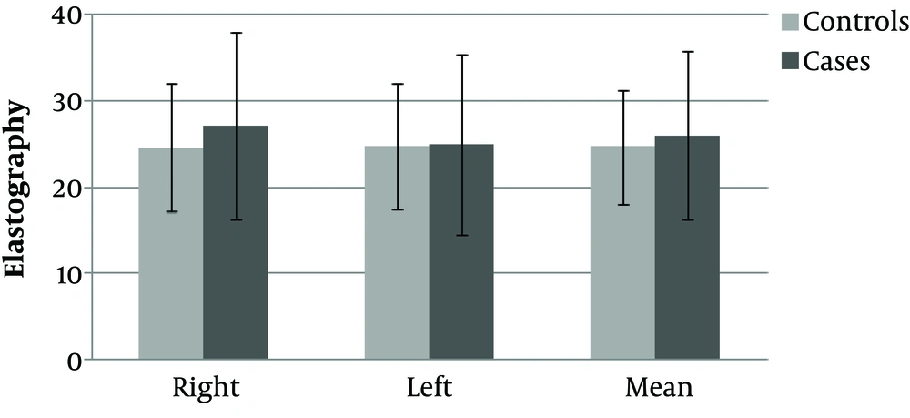
Figure 3.
Analysis of elastographic stiffness values of Achilles tendon in patients with chronic kidney disease (CKD) and the control group
There was no statistically significant difference between the duration of hemodialysis and right, left and mean stiffness values (P > 0.05) (Table 2). There were also no correlations between age and tendon thickness and elasticity both in the control and patient groups. Both tendon thickness and elasticity were mildly correlated with age (r = 0.27 and P = 0.03, r = 0.34 and P = 0.007, respectively).
Table 2.Degrees of Associations Between Duration of Hemodialysis and Elastography Measurements
| Elastography | Correlation coefficient | P value |
|---|---|---|
| Right | 0.130 | 0.464 |
| Left | 0.079 | 0.657 |
| Mean | 0.111 | 0.532 |
5. Discussion
Achilles tendon is the strongest, largest, and thickest tendon in the human body consisting of type I collagen (15). The hard structure of a healthy tendon can be affected by disease, toxin, and drugs that cause collagen defect and the tendon becomes soft and weak (16).
In CRF patients, malnutrition, accumulation of uremic toxins, secondary hyperparathyroidism, metabolic acidosis and deposition of amyloid (especially beta 2-microglobulin) was reported as a cause of weakening of the tendon and also a risk factor for tendon rupture (17, 18).
The tendon could be evaluated by B-mode sonography (19). However, B-mode sonography has limitations in differentiating early changes in the tendon as early alterations usually cannot be distinguished from the normal tendon (20). Magnetic resonance imaging (MRI) is the gold-standard imaging technique for morphological analysis of tendons however, neither US nor MRI can assess the viscoelasticity and early changes of tendons (21). SE is a more sensitive new technique for evaluation of tendon alterations (22, 23).
There are different SE methods such as compression elastography, shear-wave elastography and transient elastography. In this study, SWE technique was used. SWE was combined with conventional ultrasonographic linear transducer. In this technique, the velocity of the shear waves generated by ultrasound pulses that are sent perpendicular to the tissue, and the elasticity characteristics of the tissue are determined. Increased elasticity results in increased velocity. The quantitative measurement units are kilopascals or centimeters per second (24, 25). The shear wave velocity can be measured and used to evaluate the elasticity of the tissue. The advantages of this method are lack of manual compression requirement (22), and quantitative measurement. The limited ROI size and shape (only circle or box) are the limitations (25).
Several elastography studies confirm that a normal Achilles tendon is hard and conditions such as tendinopathy and tendon rupture cause decrease in stiffness and the tendon becomes markedly soft (8, 12, 25). Aubry et al. showed that in SWE technique, the mean velocity was decreased in the mid portion of Achilles tendon, meaning the tendon was softer in patients with tendinopathy confirmed with US (26). Their study showed that softening assessed by SWE is highly specific for tendinopathy but sensitivity was relatively low. The elasticity properties of Achilles tendon might improve the diagnosis of early tendon pathology.
There are SE studies investigating the effect of rheumatological diseases, aging and various pathologies on Achilles tendon (14, 27). In a study carried out by Teber et al. on the quadriceps tendons of 53 patients of whom the mean hemodialysis duration was 7.6 years, a decrease in the stiffness and heterogeneity in the color mapping of the tendons of patients compared to those of the healthy control group were recorded (28). In contrary with the study conducted by Teber et al. (28), we found no significant difference between the stiffness values of the patient and the control groups. The inconsistency between the results of the two studies may be caused by adoption of different techniques and/or the different mean dialysis durations of the two study populations. Teber et al. used strain elastography technique whereas we used SWE which enables quantitative measurement. It has been previously shown that tendon damage increases as duration of dialysis increases (28, 29). The mean dialysis duration of the population of the current study was shorter than that of the other study. It is within the bound of possibilities that we might have made SWE assessments early before the development of tendon alterations.
These results could also be due to the small sample size and the difference in age between the patient and control group in our study. The mean age of CRF patients was 54 ± 17; whereas, the mean age of the control group was 42 ± 9. Previously, Turan et al. (30) reported that tendon stiffness increased in elderly subjects. In this study, the higher mean age of CRF patients compared to the control group may be the reason for a relative increase in stiffness in these patients.
The results of this study showed that Achilles tendon was significantly thinner in CRF patients (P < 0.001). This result was opposed by Hussein et al., who showed that the middle and distal one third thicknesses of Achilles tendon was increased and they found positive correlation between the duration of dialysis and tendon thickness (29). Similarly, Kerimoglu et al. reported that quadriceps and Achilles tendon and plantar fascia was thicker secondary to amyloid accumulation, particularly in patients whose hemodialysis duration was over 10 years. Enthesal sites were evaluated due to significant periarticular amyloid accumulation. It was stated that tendon measurements were made at the thickest site (31). Whereas, in the current study, the measurements were taken at the middle one third of the tendon. The differences in results may be due to different sites of measurement. However, there is an agreement with Teber et al. They found that quadriceps tendon was thinner in hemodialysis patients compared to the control group (28).
We acknowledge the following limitations. First, the small sample size and the difference in age between the patient and control group. Taking into account that stiffness could also be affected by aging, the mean age of compared groups should be similar. The other limitation was the duration of hemodialysis, which was very short in some patients. Another limitation of this study was the evaluation of tendon at its middle one third portion. Evaluation of the distal part could also give useful information. Another limitation was the small-sized and limited ROI in SWE, which restricts evaluation of the tendon as a whole contrary to strain elastography. Finally, the anisotropic nature of Achilles tendon was also a limitation. In order to overcome anisotropy, the transducer should be positioned as perpendicular as possible to the tendon but it is not successfully achieved in all cases.
In conclusion, despite tendon evaluation with strain elastography in hemodialysis patients was reported before, to our knowledge no studies were performed using the shear-wave technique. The current study does not reveal any significant elastography results in the mid portion of Achilles tendons in CRF patients. However, it should be emphasized that further studies with larger patient groups is necessary.




