PPH has been classified as early hemorrhage (≤ 24 h) and late hemorrhage (> 24 h) over time by the international study group of pancreatic surgery (ISGPS) (
6). Early PPH usually occurs due to technical failure and requires immediate repeated laparotomy. Late PPH is accompanied by vessel erosion in cases involving pancreatic leak or pseudoaneurysm formation in the postoperative interval (
6-
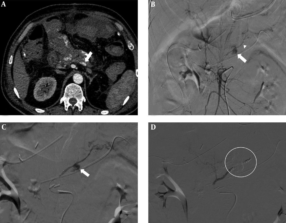
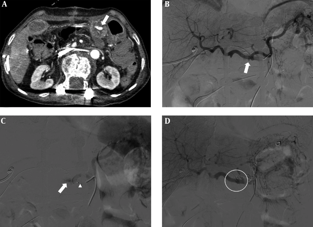
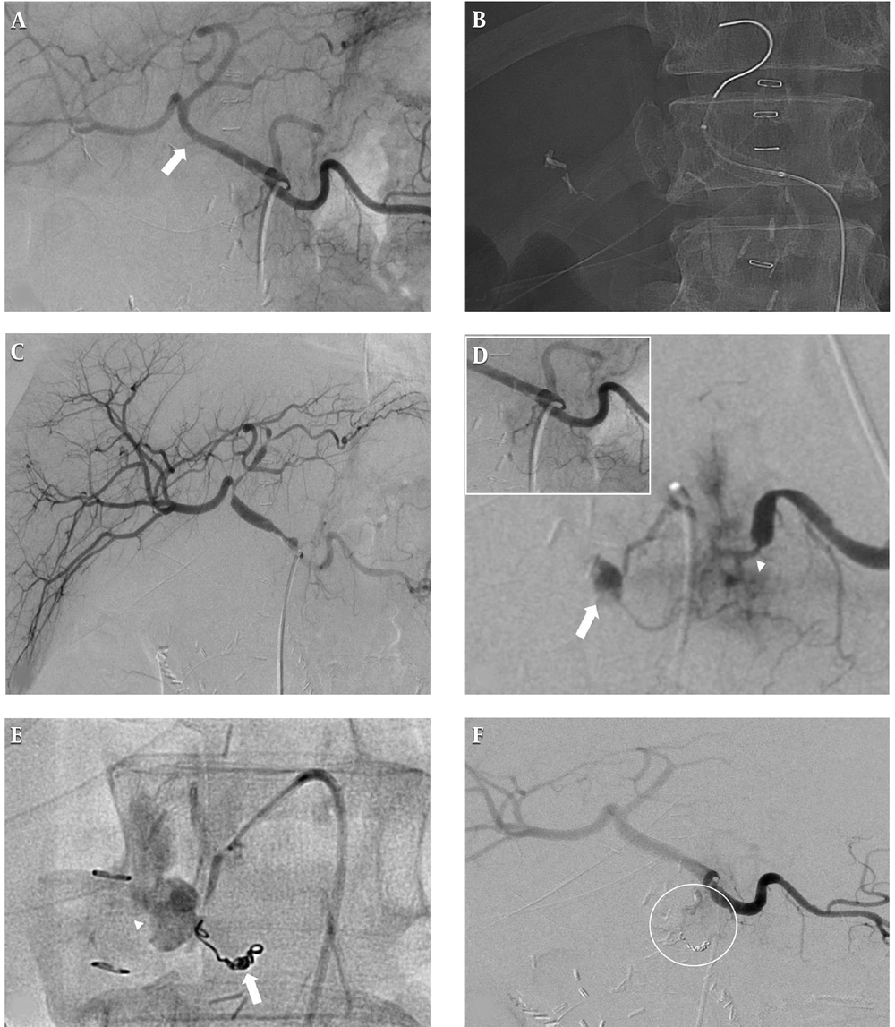
11). In our cases, late PPH with anastomotic leak from the drainage tube was identified in all patients. Local complications, such as abscess caused by anastomotic leak, potentially contributed to the development of delayed PPH and was confirmed by CT scan in two patients (cases 1 and 2), and a large hematoma in the jejunal loop was revealed on CT imaging in one patient (case 3) (
Table 1). PPHs are classified into three grades depending on the time of onset and severity of the bleeding. Grade A is defined as PPH that occurs early (≤ 24 h) and has no significant clinical impact. And grades B and C are defined as PPHs that occur late (> 24 h) and require diagnostic tests and appropriate management. A decrease in serum hemoglobin < 3 g/dL is considered a grade B while a decrease > 3 g/dL is considered a grade C. (
6). In our cases, delayed PPH occurred, on average, on postoperative day 17 (range, 15 - 21 days), and serum hemoglobin levels were reduced by more than 3 g/dL in all patients, which were classified as grade C PPHs and were treated successfully using TAE (
Table 1).
| Case | Age, y | Sex | Operation | POD | Presentation | PPH gradea | Pancreatic leak | Origin of DPA | Embolic material | Outcome | Follow-up, d |
|---|
| | | | | | | | | | Hemostasis | Death | |
|---|
| 1 | 53 | Male | Frey procedure | 15 | Melena | C | Yes | SMA | Coil | Yes | No | 1080 |
| 2 | 80 | Female | PPPD | 15 | Melena | C | Yes | Splenic artery | Coil | Yes | No | 1440 |
| 3 | 69 | Male | PPPD | 21 | Hematemesis | C | Yes | Splenic artery | Coil, NBCA | Yes | Yes | 25 |
Abbreviations: d, days; DPA, dorsal pancreatic artery; NBCA, N-butyl cyanoacrylate; POD, postoperative day; PPH, postpancreatectomy hemorrhage; PPPD, pylorus-preserving pancreaticoduodenectomy; SMA, superior mesenteric artery; y, years
aGrade A is defined as PPH that occurs early (≤ 24 h) and has no significant clinical impact. And grades B and C are defined as PPHs that occur late (> 24 h) and require diagnostic tests and appropriate management. A decrease in serum hemoglobin < 3 g/dL is considered a grade B while a decrease > 3 g/dL is considered a grade C.
The most common site for delayed PPH is known as an eroded GDA stump by pancreatic juice (
15). Less commonly, life-threatening hemorrhage may also result from injury to other arteries. Increased awareness of bleeding from unusual arteries, such as the DPA or transverse pancreatic artery from the DPA, would decrease the chances of technical failure or rebleeding rate of any radiological procedure, which necessitate re-operation. The body of the pancreas is supplied via the transverse pancreatic artery, which is the destination of the DPA. The most common origin of the DPA is the splenic artery (60%), followed by the common hepatic artery (15%), SMA (10%), celiac trunk (10%) and, rarely, others. The usual origin of the DPA is within 2 cm of the celiac terminus, courses down and right if it is from splenic, down and left if it is from the common hepatic artery, and up if from the SMA (
16). A review of the English-language literature identified only five case reports of DPA hemorrhage after pancreatic surgery (
9,
12-
14). Among them, the origins of the DPA were four splenic arteries and one common hepatic artery. Endovascular embolization was attempted as a first treatment in all cases, and technical and clinical successes were reported in four patients. Hemostasis was not achieved by embolization in one patient; therefore, a percutaneous fluoroscopy-guided direct NBCA procedure was performed, and the pseudoaneurysm was successfully treated (
13). In our cases, the origins of the DPA were one SMA and two splenic arteries, and all patients were successfully treated using TAE. To the best of our knowledge, late PPH from DPA arising from the SMA, as described in case 1, has not been reported. Our cases support the need to confirm the possibility of hemorrhage from the DPA originating from various arteries when a definite causative artery is not apparent in angiography in patients with suspected PPH.
Multi-detector CT (MDCT) is usually used as the initial diagnostic modality for detection of bleeding focus after surgery. It may help the interventional radiologist plan for the procedure and, is thus, likely to decrease the number of angiograms required to localize the bleeding site and radiation dose received by the patient. A recent study reported that MDCT found active bleeding more frequently than angiography (
9). This represents complementary roles of multiphase MDCT and angiography in postoperative management of patients undergoing pancreatectomy. The utility of MDCT in patients with unusual sites of bleeding has not been extensively studied; nevertheless, MDCT in our cases facilitated the approximate localization of the bleeding site from adjacent major vessels. In case 1, initial MDCT after surgery was negative for hemorrhage and the first angiography failed to demonstrate any area of extravasation of contrast material or pseudoaneurysm. Repeated MDCT revealed the origin of the pseudoaneurysms and the suspected branch of the hemorrhage site before angiography, which helped to find the cause of a very rare PPH, a branch of the DPA originating from the SMA. In case 2, the extravasation of contrast material in the pancreaticojejunostomy site near the splenic artery was confirmed by MDCT. Because the splenic artery itself was intact on CT imaging, it could be presumed that the branch originating from the splenic artery was damaged. Thus, MDCT would be the most useful diagnostic modality in patients with PPH and, even if there is no abnormality in the first postoperative MDCT, there should be no hesitation in performing repeat CT examination in cases of clinically suspected rebleeding.
Late PPH has been reported to occur into the gastrointestinal tract or more commonly, the abdominal cavity (
7). Endoscopy, which is the standard first-line investigative tool for gastrointestinal bleeding, may fail because of difficulties in localizing the bleeding site in the abdominal cavity and lead to delay in TAE. Therefore, endoscopy has a very limited role in the initial management of delayed PPH. In the past, the first-choice treatment for PPH has been considered surgery, and even early PPH due to the technical failure of original surgery still requires immediate repeated laparotomy (
2,
3,
7,
8,
14,
16). During re-operation, access to the bleeding vessel is limited due to postoperative inflammation, whereas endovascular treatment could be more effective in the management of PPH because rapid control of bleeding is possible. Recently, GDA stump was successfully excluded by endovascular treatments including selective embolization of the stump with coils, embolization of hepatic artery, and placement of a covered stent in the hepatic artery. Furthermore, it has been reported that endovascular treatment results in effective hemostasis with a lower morbidity and mortality than surgical care (
7,
10,
11). Okada et al. reported that selective TAE of the dorsal pancreatic artery caused only localized ischemic necrosis without clinically relevant pancreatitis on a histological change in a Swine model (
17). Therefore, percutaneous endovascular treatments are considered to be the first-line management for ISGPS grade B and C PPHs.
However, TAE also has some limitations. If the origin of bleeding is a vein, or is diffuse or intermittent, the involved vessel may not be detected on angiography, which was the scenario in the first attempt in case 1. A recent study conducted by Pottier et al. which evaluated endovascular management in patients with late PPH, reported that the rate of rebleeding after a first endovascular treatment was as high as 43% and the only predictor related to rebleeding was abstinence (no evidence of an involved artery on angiography) or technical failure in the initial session of the radiological procedure. However, almost three-quarters of the patients were successfully treated with only endovascular treatment, and 17% of them required secondary or third procedures (
7). Interestingly, cases 1 and 3 in the present study demonstrated that rebleeding occurred after the first angiography, which identified the absence of an involved artery. However, the rebleeding was successfully managed in the second endovascular procedure. In case 1, the bleeding focus was identified after increasing the volume of contrast medium on the second angiography. Although the initial angiography did not show an obvious culprit artery in case 2, the SMA, hepatic artery, and distal portion of the splenic artery could be excluded by multiple angiographies. Finally, the DPA from the proximal portion of the splenic artery was the suspected bleeding focus, which was successfully treated using TAE. In case 3, the first session and initial angiography of the second endovascular procedure revealed no evidence of active bleeding or pseudoaneurysm on celiac angiography. Fortunately, the pseudoaneurysm at the DPA arising from the proximal splenic artery was revealed by iatrogenic spasm, which was caused by catheterization of the splenic artery. Therefore, we recommend that the following points be considered when TAE is performed in patients with late PPH. First, when performing angiography, use as much contrast medium as possible, and try to perform selective angiography, such as splenic artery angiography, whenever possible. Second, clinicians should always be mindful of the possibility of an unusual bleeding focus of late PPH, such as DPA, and, even if a common bleeding site on the first attempt on angiography is not found, additional efforts must be made to find these other causes. Finally, if the initial procedure fails in patients with late PPH and the patients are hemodynamically stable, catheterization should be maintained in the femoral artery for the next 2 - 3 days. If there are signs of rebleeding, repeated endovascular procedures may be attempted before considering immediate re-operation.
In conclusion, therapeutic angiography is emerging as an effective and safe treatment strategy for PPH. The cause of bleeding after PPH is not only the well-known GDA, but also unusual origins, including the PDA, as presented in our cases, which may not be obvious in the initial angiography. Therefore, it is expected that a careful and repetitive approach, based on awareness of the various possible causes of PPH, could increase the success rate of TAE.