


1. Introduction
Henoch-Schonlein purpura (HSP) represents the most prevalent form of systemic vasculitis in children. Its global incidence ranges from 10 to 20 cases per 100,000 children annually. Notably, approximately 90% of cases occur between the ages of 2 and 10 years, with a peak incidence observed between 4 and 7 years of age (1). A higher frequency of HSP cases has been reported during the autumn and winter seasons. Additionally, the condition appears to be more common in males (2). The HSP has also been associated with a history of preceding infections, especially upper respiratory tract infections (3), and it occasionally clusters in families, suggesting a genetic component (4).
The predominant clinical manifestations of HSP include round or oval and retiform palpable purpura — primarily localized on the lower extremities — along with arthralgia or arthritis, gastrointestinal pain or bleeding, and glomerulonephritis characterized by mesangial IgA deposits (IgA vasculitis nephritis, IgAVN). Although rare, involvement of the pulmonary, cardiac, genitourinary, and central nervous systems has also been reported (5). The diagnosis of HSP is primarily clinical (4) and based on the presence of palpable purpura in conjunction with at least one of the following criteria: Diffuse abdominal pain, arthritis or arthralgia, renal involvement (manifested as hematuria and/or proteinuria), or histopathological evidence of predominant IgA deposition on tissue biopsy (6).
Treatment for mild and self-limited HSP is supportive, focusing on maintaining adequate hydration, nutrition, and analgesia. Corticosteroids are commonly used to treat significant gastrointestinal involvement or other life-threatening manifestations (4). The prognosis for childhood HSP is excellent, but some patients may develop serious gastrointestinal involvement, such as intussusception and intestinal perforation, and kidney complications (4). In addition, subcutaneous edema is a common feature of HSP, often localized to the dorsum of the hands and feet, periorbital region, lips, scrotum, or scalp (4). Lumbosacral edema, however, is a rare manifestation, with only a limited number of cases reported in the literature to date.
To learn more about the unusual manifestations of HSP, I presented a case of a 6-year-old girl with sudden non-traumatic back pain and edema, along with limited walking ability.
2. Case Presentation
A 6-year-old girl was admitted to the pediatric ward during the night shift with a chief complaint of leg pain that had begun the day prior to admission, accompanied by severe pain and swelling in the lumbar region that developed on the morning of admission. There was no history of trauma. Notably, the patient had experienced symptoms of an upper respiratory tract infection approximately one week earlier. No significant past medical history or recent medication use was reported.
On examination, the patient was afebrile, with vital signs within normal limits. Her blood pressure measured 98/65 mm Hg. During the initial examination, severe edema with a diameter of 9 × 9 cm was observed in the lumbar region (Figure 1), which was very tender and non-erythematous. Additionally, there was mild painful edema with an unclear border in the middle part of the front of the right leg.
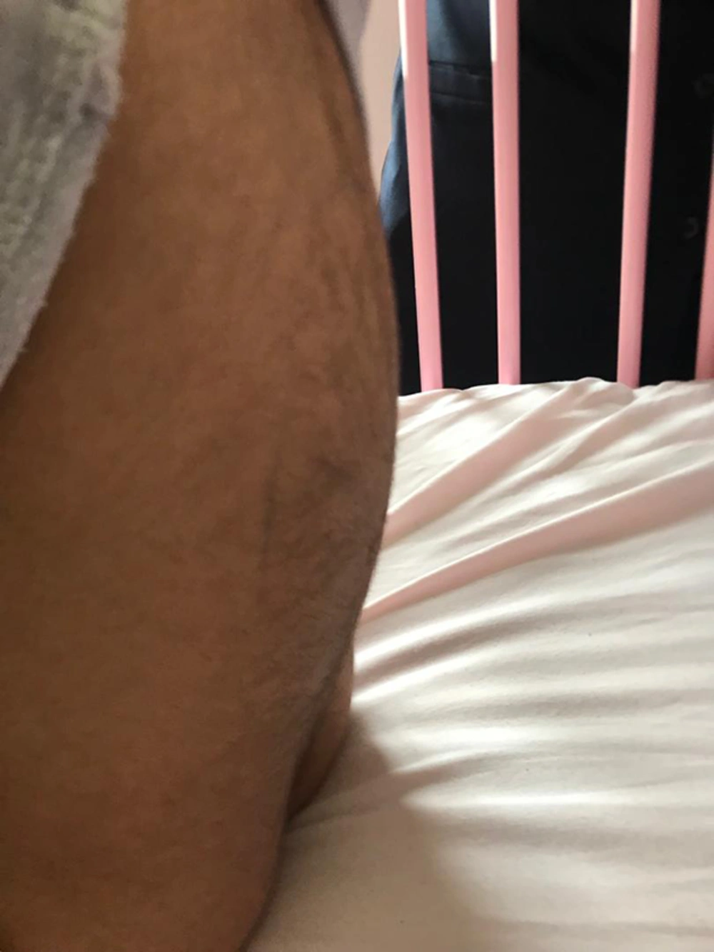
Figure 1.
Edema of the lumbar region
Furthermore, in the skin examination, a limited number of rashes in the form of petechiae and palpable purpura were seen in the lower limbs on both sides. Abdominal and neurological examinations were normal. The patient walked with hemiflexed knees to prevent an increase in back pain.
Laboratory evaluation, including CBC diff, ESR, CRP, and biochemistry, were examined and observed to be within normal limits. Platelet count and coagulation tests (PT and PTT) were within the normal range, ruling out thrombocytopenia or bleeding disorders. Urinalysis and stool tests were normal, suggesting no early renal involvement and no gastrointestinal bleeding.
A computed tomography (CT) scan of the spine was performed using a CT scanner manufactured and installed by Pars Medical Equipment Company in cooperation with Canon Company (Pars Medical Equipment Company is located at No. 23, 33rd Street, Khaled Eslamboli Street, Gandhi South Street, Tehran). The scan showed subcutaneous edema in the lumbar region without any other lesions (Figure 2).
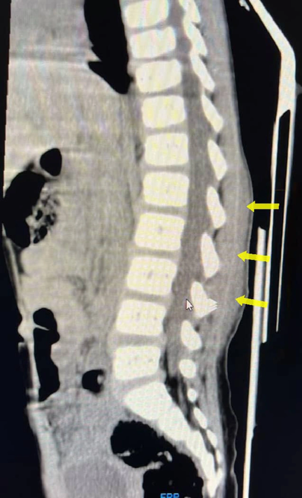
Figure 2.
Non-contrast sagittal computed tomography (CT) image of the spine shows subcutaneous tissue edema (arrows) in the lumbar region
A neurology consultation did not report any problems. Within 5 - 6 hours after hospitalization, the number of rashes increased, especially on her lower extremities bilaterally. On the second day of hospitalization, the lumbar edema decreased slightly, and the pain decreased to a great extent. On the third day of hospitalization, the rash became lighter, and the edema on the front of the right leg resolved.
Additionally, on the fourth day, the pain in the leg and lumbar region was completely resolved, allowing the patient to walk normally. The rash disappeared, and only a little edema remained in the lumbar region. In the evening of the fourth day of hospitalization, the patient developed pain and swelling in the right ankle, which resolved by the morning of the fifth day. Lumbar edema was completely resolved on the fifth day (Table 1).
Table 1.Patient's Clinical Timeline Table Since Admission
| Hospitalization Days | Symptoms | Signs | Tests/Findings |
|---|---|---|---|
| Day before admission | Onset of leg pain | - | |
| Day 1 | Severe lumbar pain and swelling | Severe edema with a diameter of 9 × 9 cm was in the lumbar region; mild painful right leg edema; few petechiae and purpura on lower limbs; walking with hemiflexed knees | CBC, ESR, CRP, biochemistry normal; normal urine and stool tests; Lumbar subcutaneous edema on CT scan |
| 5 - 6 Hours post-admission | - | Increased rash on lower limbs | - |
| Day 2 | Lumbar pain decreased to a great extent | Lumbar edema reduced a little | - |
| Day 3 | - | Fading of the rash; resolved edema on right leg front | - |
| Day 4 | Complete resolution of leg and lumbar pain | Normal walking; disappearance of the rash; persistent Mild lumbar edema | - |
| Evening day 4 | Onset of right ankle pain and swelling | Edema of the right ankle | - |
| Day 5 | Resolution of right ankle pain | Complete resolution of lumbar and right ankle edema | - |
Abbreviation: CT, computed tomography.
Due to the healing process of the pain and edema in the patient’s lumbar region and right leg from the first hours after hospitalization, drug treatment was not started, and the symptoms were resolved by resting in bed and hydration.
In this patient, several differential diagnoses were considered upon admission, including infectious causes such as lumbar abscess, traumatic injury, other vasculitides, and coagulopathies. However, due to the absence of systemic infection signs, lack of trauma history, normal laboratory findings, subcutaneous edema of the lumbar region without space-occupying lesions in this area on the CT scan, and the presence of characteristic purpura, along with arthritis and arthralgia, the diagnosis of HSP was confirmed in this case.
After the complete resolution of symptoms and signs, the patient was discharged and advised to attend follow-up appointments. During the ongoing follow-up, no recurrence of symptoms or signs has been observed.
3. Discussion
The classic tetrad of symptoms and signs in HSP includes palpable purpura, arthralgias, abdominal pain, and renal involvement (7). As there is no definitive test for the diagnosis of the disease, it is based on diagnostic criteria (8). Criteria were established by the American College of Rheumatology in 1990 and adopted by EULAR/PRINTO/PRES in 2008 (6, 9). Based on the presence of palpable purpura, along with arthritis and arthralgia in my patient, the diagnostic criteria for HSP were met. She showed no signs of renal or gastrointestinal involvement, and no skin biopsy was performed. Vasculitis affected her lumbar spine, which is an unusual situation.
The most common finding of HSP is skin involvement (10), and the predominant cutaneous finding is palpable purpura (8). Subcutaneous edema is also commonly observed and often involves the hands, ankles, and feet (11), but lumbosacral edema is very rare in HSP, and the number of previously reported cases in the literature is limited (8).
In a review of previous articles, I identified only seven previous cases of HSP with lumbar pain and edema in children. In comparison, the current patient and the previous seven patients were all in the age range of 3 to 7 years. Only one of the previous cases did not have a skin rash (7), while the other six previous cases showed a skin rash similar to my patient. Renal involvement, as in my case, was not reported in any of the documented cases. Gastrointestinal involvement was present in five of the previous cases (7, 8, 11, 12) and absent in two of them (10, 13), as in my patient. Marked arthralgia in the joints of the lower extremities was present in four of the patients (7, 10, 11), similar to my patient. Corticosteroid treatment was administered in only three of the previous seven patients (7, 8, 12); all three of them had gastrointestinal involvement, and the other four, as in my patient, did not require steroids.
The diagnosis of HSP is often straightforward when the typical rash is present. However, in at least 25% of cases, Henoch’s rash appears after other manifestations (4). Therefore, identifying atypical manifestations of the disease seems essential. Lumbosacral involvement is a rare but recognizable HSP variant. Early recognition prevents misdiagnosis in atypical presentations, particularly when purpura is delayed in appearance.



