
1. Background
Volleyball is characterized by repetitive jumping, rapid changes of direction, explosive landings, and substantial mechanical stress on the lower extremity joints (1, 2). Among sport-related injuries, anterior cruciate ligament (ACL) tears are the most prevalent ligamentous injuries in active athletes and predominantly occur through noncontact mechanisms involving rotational forces or improper landings (3-5). Anterior cruciate ligament reconstruction (ACLR) is the most common surgical intervention used to restore knee stability and improve the likelihood of returning to sport (6, 7).
Despite surgical success, evidence indicates that approximately 50% of patients fail to return to their preinjury level of sport within the first 5 years after surgery, with reinjury rates exceeding 30% in this population (4, 5). These functional limitations are attributable to persistent neuromuscular deficits, reduced lower extremity muscle strength, and altered landing mechanics, which may persist for up to 2 years after surgery (8-10). These factors highlight the complex interplay of variables influencing joint mechanics in athletic populations during this critical recovery phase (1, 11).
In recent years, modified plyometric exercises, particularly hopping protocols, have received attention as a key component of late-stage rehabilitation because they can harness the stretch-shortening cycle and induce positive neuromuscular adaptations (11, 12). Previous studies have shown that such interventions can improve dynamic balance, proprioception, and explosive power across various athletic populations (13-15). Similar impairments in kinematic parameters, dynamic postural control, and articular proprioception have also been documented after other knee arthroscopic procedures and surgical meniscal interventions, such as resection or repair, highlighting the broad impact of knee surgeries on joint function and the need for targeted neuromuscular rehabilitation (16). However, the combined effects of these exercises on clinical and functional outcomes in volleyball players undergoing ACLR remain unclear. Most previous studies have focused on isolated variables, such as balance or proprioception alone, or have examined different target populations. Given the sport-specific demands of volleyball, including repetitive jumping, single-leg landings, and sudden velocity changes that are known to induce fatigue and subsequently impair neuromuscular and biomechanical control post-ACLR (17), these athletes require a more tailored rehabilitation approach. Recent evidence also indicates that inter-limb asymmetries in dynamic balance and single-leg hop performance are strong prognostic indicators of on-court trauma among volleyball athletes, underscoring the need for targeted interventions to correct these functional deficits (18). This knowledge gap limits the ability of current rehabilitation protocols to fully address the functional needs and pain management of volleyball players post-ACLR.
Therefore, the present study was designed to evaluate the effect of an 8-week functional hopping training program on pain, muscle strength, and lower extremity function in male volleyball players during the post-ACLR recovery phase. In this randomized controlled trial, participants were allocated to intervention and control groups. All clinical and functional endpoints were assessed before and after the 8-week program using three established tools: the Visual Analog Scale (VAS) for pain intensity, a calibrated hand-held dynamometer for muscle strength, and the Triple Hop and Figure-8 Hop tests for sport-specific performance.
2. Objectives
This study primarily aimed to evaluate the effects of an 8-week functional hopping program on pain, lower-extremity muscle strength, and sport-specific functional performance in male volleyball players 6 - 12 months post-ACLR. Secondarily, it sought to determine whether integrating the functional hopping program into late-stage rehabilitation could optimize recovery protocols and enhance functional readiness. Any potential implications of these short-term functional gains for reducing reinjury risk or facilitating return to sport are beyond the scope of this trial and require further longitudinal investigation. Given the high treatment costs and prolonged absence from sport associated with ACLR, this study provides evidence-based guidance to inform late-stage clinical rehabilitation in volleyball athletes.
3. Methods
3.1. Study Design and Setting
This parallel-group randomized controlled study used baseline and follow-up assessments to examine whether an 8-week functional hopping program could improve pain, strength, and functional outcomes in male volleyball players recovering after ACLR. The study was conducted in Urmia, Iran, from April to October 2025. The local institutional review board of Shahrood University of Technology granted ethical clearance (Code: IR.SHAHROODUT.REC.1403.050), and all procedures complied with the Declaration of Helsinki. Written informed consent was obtained from all participants, who retained the right to withdraw at any time. Data were anonymized and stored securely to ensure confidentiality. Safety measures included medical supervision and immediate cessation of exercise if adverse symptoms were reported.
3.2. Participants and Eligibility Criteria
The required sample size was estimated before data collection using G*Power (v3.1), based on a medium effect size (d = 0.53) from the Triple Hop Test (15), selected for its biomechanical relevance to volleyball tasks. Although this estimate was derived from soccer players with chronic ankle instability, the source was considered pragmatically appropriate because of shared neuromuscular deficits, including proprioception, dynamic control, and unilateral power, and similar responsiveness to hopping interventions targeting the stretch-shortening cycle. Assuming alpha = 0.05, power = 80%, and 10% attrition, 30 participants (15/group) were sufficient to identify intergroup differences in the co-primary endpoint of lower-extremity isometric strength. Pain and functional tests were analyzed with equal rigor but did not inform the power calculation. Given the limited precision for detecting small effects or conducting subgroup analyses with n = 15/group, P values were interpreted alongside effect sizes (η2) and clinical relevance to support conservative inference.
The target population comprised male volleyball players aged 20 - 35 years with a history of unilateral ACLR (≤ 1 prior reconstruction) who were referred to orthopedic clinics in Urmia. Inclusion criteria were: 1) male volleyball players aged 20 - 35 years; 2) 6 - 12 months post-ACLR, consistent with standard rehabilitation protocols (19); 3) a pain score of 5 - 6 on the VAS during squatting; 4) absence of prior surgical procedures or recent trauma in the lumbar spine or lower limbs, excluding the reconstructed knee; and 5) ability to complete all functional assessments and provide written informed consent.
Participants were excluded if they missed more than three scheduled training sessions, reported exercise-induced pain during the intervention period, or voluntarily withdrew from the study at any stage.
3.3. Randomization
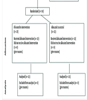
A statistician independent of the research team generated the randomization sequence using a random number table. Allocation concealment was ensured by placing group assignments in sequentially numbered, opaque, sealed envelopes that were opened only after baseline assessment and eligibility confirmation. Equal allocation (1:1) was used to assign participants to the intervention or control group. Owing to the exercise-based nature of the intervention, blinding of participants and interventionists was impractical. However, all outcome measurements and statistical analyses were performed by personnel unaware of group allocation (single-blind design). To minimize selection bias, inclusion and exclusion criteria were explicitly defined, and a purposive-random sampling approach was used.
3.4. Outcomes
The primary outcome was change in pain intensity during squatting, quantified using a 10-cm VAS. Co-primary outcomes included isometric muscle strength of the hip, knee, and ankle, assessed using calibrated hand-held dynamometry. Sport-specific functional performance, assessed using the Triple Hop and Figure-8 Hop tests, was designated as a secondary outcome. All outcomes were evaluated at two prespecified time points: before the intervention and immediately after completion at week 8, which served as the primary endpoint for analysis. No exploratory or post hoc outcomes were assessed.
Squatting pain was quantified using a 10-cm VAS (20).
Isometric muscle strength of the lower extremity was quantitatively assessed using a calibrated hand-held dynamometer (North Coast Medical, USA; model #NC15750; validity, 95 - 98%; inter-rater reliability, ICC = 0.96 - 0.99) (21), following the standardized positioning and stabilization protocol described by Kendall et al. (2005). All assessments were conducted by a single trained examiner who remained blinded to group allocation throughout data collection.
Lower-extremity isometric strength was measured using a calibrated hand-held dynamometer (North Coast Medical, USA) according to standardized protocols (22, 23). Before assessment, participants completed a standardized 5-minute warm-up that included low-intensity cycling and dynamic stretching. For each muscle group, participants were positioned according to Kendall's guidelines to isolate the target musculature and minimize compensatory movements (Figures 1-4).
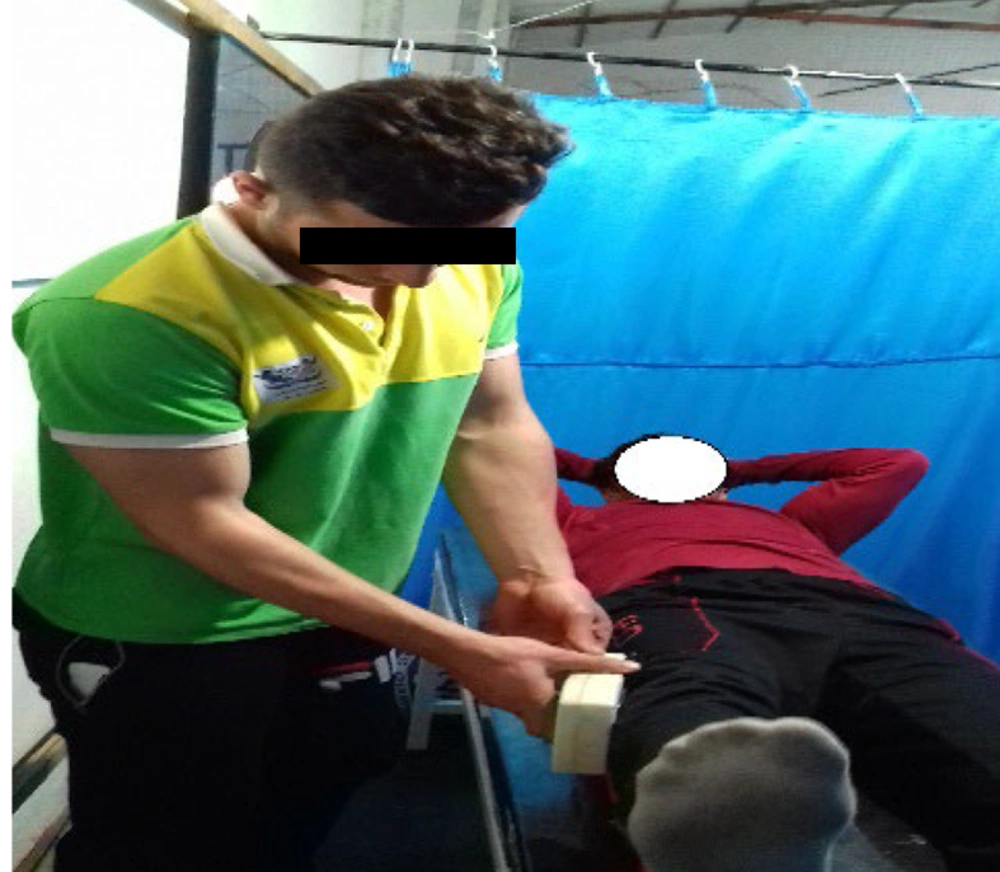
Figure 1.
Assessment of isometric hip abduction strength using a hand-held dynamometer. Participant positioned side-lying on non-test limb with pelvis stabilized; dynamometer pad placed 5 cm proximal to lateral knee joint line during maximal isometric abduction effort.
For hip abduction, participants lay in a side-lying position on the non-test limb, with the test limb in neutral rotation and 10 degrees of abduction. The pelvis and trunk were stabilized using straps. Following Kendall's stabilization protocol, the dynamometer was positioned on the distal thigh, 5 cm proximal to the lateral knee joint line, to isolate hip abductor output and minimize compensatory movements (Figure 1). Participants performed a maximal isometric abduction effort against resistance for 5 seconds (22, 23).
For hip adduction, participants lay side-lying on the evaluated leg, while the contralateral leg was bent at both the hip and knee joints and supported on a stool (Figure 2). The point of force application for hip adduction testing was standardized at 5 cm proximal to the medial femoral condyle, where the dynamometer sensor was firmly positioned. Participants performed a maximal isometric adduction effort while maintaining pelvic stability (23, 24).
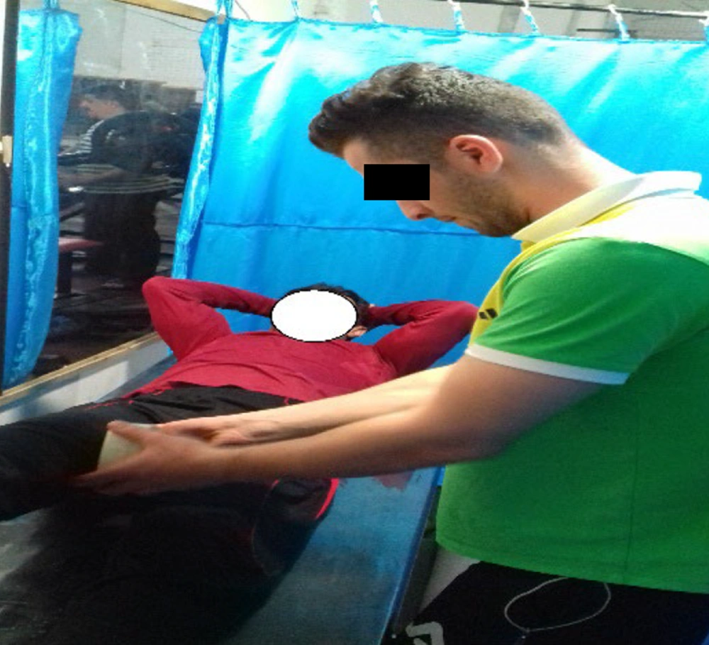
Figure 2.
Assessment of isometric hip adduction strength. Participant lies side-lying on test limb with contralateral hip and knee flexed and supported. Dynamometer positioned 5 cm proximal to medial femoral condyle during maximal isometric adduction.
For knee flexion, participants were placed in a prone position with the knee flexed to 90 degrees. The thigh was stabilized with straps, and the dynamometer pad was placed on the posterior aspect of the distal tibia, 5 cm proximal to the lateral malleolus (Figure 3). Participants performed a maximal isometric hamstring contraction (23, 24).
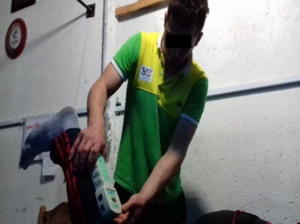
Figure 3.
Measurement of isometric knee flexion strength. Participant positioned prone with knee flexed to 90° and thigh stabilized. Dynamometer pad placed on posterior aspect of distal tibia (5 cm proximal to lateral malleolus) during maximal hamstring contraction.
For knee extension, participants were positioned supine with the knee flexed to 90 degrees and the thigh stabilized (Figure 4). The dynamometer pad was positioned on the anterior aspect of the distal tibia. Participants performed a maximal isometric quadriceps contraction (23, 25).
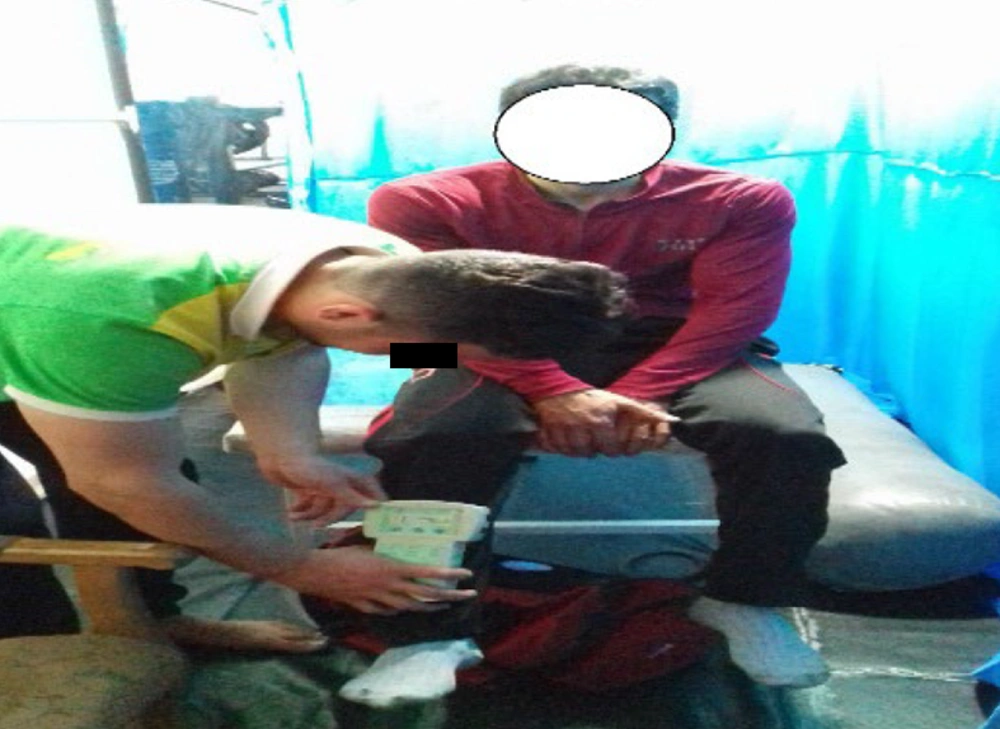
Figure 4.
Measurement of isometric knee extension strength. Participant positioned supine with knee flexed to 90° and thigh secured. Dynamometer placed on anterior aspect of distal tibia while participant performs maximal isometric quadriceps contraction.
For ankle dorsiflexion and plantarflexion, participants were seated with the knee in full extension and the ankle in neutral alignment. The point of force application for both ankle motions was standardized proximal to the metatarsophalangeal joints: plantarly for plantarflexion and dorsally for dorsiflexion assessments. Participants performed maximal isometric efforts in each direction (23, 25).
Three 5-second maximal isometric efforts were recorded for each target muscle, separated by 30 seconds of rest between repetitions and accompanied by standardized verbal encouragement to optimize performance. Peak force (kg) was recorded for each trial, and the mean of three trials for the reconstructed limb was used for analysis. Participants completed one familiarization trial per movement before testing. The device was calibrated before each testing session according to the manufacturer's guidelines, and examiner reliability was confirmed during piloting (ICC > 0.95). Participants abstained from caffeine and vigorous exercise for 24 hours before assessment to minimize physiological variability.
Lower-limb functional capability was evaluated using the Triple Hop and Figure-8 Hop tests, validated field measures of unilateral power, dynamic stability, and neuromuscular control (15, 26). The distance-based Triple Hop assessment was performed on a 6-m tape fixed to a nonslip surface. Participants hopped consecutively three times on the test limb, maintained the final landing for ≥ 3 seconds without contralateral support, and the greatest total distance (cm) from two trials was recorded (15). The Figure-8 Hop Test evaluated agility and dynamic postural control on a 5 x 1-m cone-configured course. Participants completed two circuits at maximal speed with hands on the iliac crests; the fastest time, measured with 0.01-second precision, from two timed trials was retained (15). Both tests demonstrate excellent inter-rater reliability (ICC = 0.99) in athletic populations (15), supporting their sensitivity for detecting clinically meaningful functional changes after rehabilitation. Standardized warm-up, practice trials, and verbal feedback on landing mechanics were provided to ensure consistency and safety (15).
3.5. Intervention Protocol
The intervention group completed an 8-week functional hopping program based on the protocol developed by Mohammadi Nia Samakosh et al. (15). Sessions were held three times per week and lasted approximately 40 minutes each, including a 5-minute warm-up, a 30-minute main intervention, and a 5-minute cool-down, under the supervision of a certified corrective exercise specialist and an orthopedic physician.
The program comprised six progressive hopping exercises: lateral hops, forward-backward hops, zigzag hops, square-pattern hops, figure-8 hops, and single-leg hops. Training volume and intensity were systematically increased across weeks by modifying repetition schemes; altering support conditions, progressing from bilateral to unilateral support; and adjusting arm positioning, progressing from free arm swing to hands on chest and then to hands behind head. Rest intervals were standardized at 30 seconds between sets and 1 minute between exercises. Participants received verbal feedback to maintain proper landing mechanics, avoid dynamic knee valgus, and ensure controlled foot-ground contact (Table 1).
Table 1.
Eight-Week Functional Hopping Program
| Weeks and Exercises | Sets x Reps | Support Condition | Arm Position | Rest (Between Sets/Exercises) | Progression Focus and Coaching Cues |
|---|---|---|---|---|---|
| 1 - 2 | |||||
| Lateral Hop | 2 x 10 | Bilateral | Free | 30 s / 1 min | Land softly with knees aligned over toes; maintain trunk stability |
| Forward-Backward Hop | 2 x 10 | Bilateral | Free | 30 s / 1 min | Control deceleration; avoid excessive forward trunk lean |
| Zigzag Hop | 2 x 8 | Bilateral | Free | 30 s / 1 min | Smooth direction changes; minimize ground contact time |
| 3 - 4 | |||||
| Square-Pattern Hop | 3 x 8 | Bilateral to dominant-leg support | Hands on chest | 30 s / 1 min | Maintain square geometry; emphasize quick, controlled turns |
| Figure-8 Hop | 3 x 6 | Bilateral to dominant-leg support | Hands on chest | 30 s / 1 min | Follow cone path precisely; avoid dynamic knee valgus |
| Single-Leg Hop (straight) | 2 x 8 | Unilateral (reconstructed limb) | Hands on chest | 45 s / 1.5 min | Stable single-leg landing; hold final position ≥ 3 s |
| 5 - 6 | |||||
| Lateral Hop (unilateral) | 3 x 10 | Unilateral | Hands on chest to behind head | 45 s / 1.5 min | Increase lateral displacement; maintain pelvic alignment |
| Zigzag Hop (unilateral) | 3 x 8 | Unilateral | Behind head | 45 s / 1.5 min | Faster tempo; emphasize eccentric control on direction change |
| Figure-8 Hop (timed) | 3 x 4 circuits | Unilateral | Behind head | 60 s / 2 min | Maximal speed with quality; record time for feedback |
| 7 - 8 | |||||
| Multi-Directional Hop Circuit | 4 x 6 each | Unilateral | Behind head | 30 s / 2 min | Sport-specific sequencing; simulate volleyball landing scenarios |
| Single-Leg Triple Hop | 3 x 5 | Unilateral | Behind head | 60 s / 2 min | Maximize distance while maintaining landing stability ≥ 3 s |
| Reactive Hop (coach cue) | 3 x 8 | Unilateral | Behind head | 45 s / 1.5 min | Respond to verbal/visual cues; enhance neuromuscular reactivity |
3.6. Control Group
Participants allocated to the standard-care cohort continued their prescribed clinic-based standard-of-care rehabilitation appropriate for the 6 - 12-month post-ACLR phase. This program typically included progressive resistance training (2 - 3 sessions/week, 45 - 60 minutes/session), neuromuscular control drills, balance exercises, and sport-specific return-to-sport conditioning, as directed by the treating orthopedic surgeon and physical therapist. Although total training exposure and therapist contact varied according to individual clinical prescriptions, all participants followed standard institutional protocols for late-stage ACLR recovery. Compliance was monitored using weekly self-reported training logs and biweekly telephone follow-ups to ensure adherence and prevent contamination; no structured hopping or high-impact plyometrics were permitted. All control participants maintained their routine rehabilitation schedule throughout the 8-week study period. Given the pragmatic nature of the control condition, total rehabilitation dose and supervision frequency were not strictly matched between groups. However, the design intentionally evaluated the additive clinical and functional effects of integrating a structured functional hopping program into standard late-stage ACLR rehabilitation. After post-intervention assessments, the control group was offered the functional hopping program as a crossover intervention.
3.7. Statistical Analysis
Using SPSS version 26 (IBM Corp.), data were analyzed as follows: Baseline characteristics were summarized as means ± SD or frequencies (%), and normality was assessed using the Shapiro-Wilk test. Initial between-group comparisons were conducted using independent t tests or Mann-Whitney U tests, depending on data distribution. Primary between-group comparisons for each outcome were conducted using one-way analysis of covariance (ANCOVA). Specifically, the post-intervention score was set as the dependent variable, allocation group (intervention vs. control) was the fixed predictor, and the pre-intervention value was the sole covariate; no additional covariates were included in the model. Intragroup changes were analyzed using paired t tests. Effect sizes (partial eta squared, η2) quantified intervention magnitude. The alpha level was set at 0.05 for all statistical evaluations. Given the high physiological intercorrelation among lower-extremity strength measures and the pragmatic nature of this rehabilitation trial, formal multiplicity adjustments, such as Bonferroni or Holm corrections, were not applied to avoid inflating type II error across clinically related endpoints. Instead, statistical significance (alpha = 0.05) was interpreted alongside partial eta squared (η2) effect sizes and clinical relevance. Findings across multiple strength and functional outcomes are reported with conservative, effect-size-driven interpretation to align with CONSORT recommendations for trials with correlated endpoints.
4. Results
Data were analyzed using a complete-case approach, equivalent to both intention-to-treat and per-protocol analyses, because all 30 randomized participants completed the 8-week intervention and post-assessments without exceeding three absences or withdrawing. Therefore, no missing data imputation was required. Baseline characteristics and outcome variables were analyzed as described above.
Independent-samples t tests comparing baseline characteristics between the functional training and control groups (Table 2) revealed no meaningful differences in any demographic or clinical variable examined (P > 0.05 for all comparisons). Specifically, mean age was comparable between the functional group (27.20 ± 4.79 years) and the control group (26.93 ± 3.55 years; P = 0.86). Similarly, anthropometric indices, including height, weight, lower limb length, and BMI, as well as injury-related variables, including time since surgery, and sport-specific history, including years of volleyball participation, were evenly distributed across groups (Table 2).
Table 2.
Baseline Demographic, Anthropometric, and Clinical Characteristics of Participants by Group (N = 30) a
| Variables | Hopping Group | Control Group | t | P-Value |
|---|---|---|---|---|
| Age (y) | 27.20 ± 4.79 | 26.93 ± 3.55 | 0.17 | 0.86 |
| Height (m) | 1.78 ± 0.08 | 1.81 ± 0.07 | -1.01 | 0.32 |
| Weight (kg) | 76.00 ± 6.66 | 71.73 ± 5.81 | 1.86 | 0.07 |
| Lower limb length (cm) | 99.93 ± 5.47 | 101.13 ± 3.50 | -0.71 | 0.48 |
| BMI (kg/m2) | 23.84 ± 1.20 | 22.64 ± 1.42 | 1.49 | 0.16 |
| Time since surgery (months) | 8.26 ± 2.01 | 9.13 ± 1.35 | -1.38 | 0.18 |
| Volleyball experience (y) | 16.00 ± 3.46 | 15.66 ± 2.96 | 0.28 | 0.77 |
a Values are expressed as mean ± SD.
Following the 8-week functional hopping program, the intervention group showed significant improvements versus controls across all outcomes (ANCOVA, baseline-adjusted, df = 1, 27). Pain during squatting was significantly reduced (F(1, 27) = 11.54, P = 0.002, η2 = 0.29). Isometric strength improved significantly for knee flexion, hip adduction/abduction, and ankle plantarflexion/dorsiflexion (P < 0.001, η2 = 0.46 - 0.68), with a smaller but still significant effect for knee extension (F(1, 27) = 6.22, P = 0.019, η2 = 0.19). Functional performance on the Figure-8 Hop and Triple Hop tests also improved markedly (P < 0.001, η2 = 0.67). The control group showed no significant within-group changes (P > 0.05). These findings indicate that an 8-week functional hopping program is an effective adjunct to late-stage ACLR rehabilitation, reducing pain, restoring strength, and enhancing sport-specific function in male volleyball players (Table 3).
Table 3.
Pre- and Post-Intervention Outcomes for Pain, Isometric Strength, and Functional Performance by Group (N = 30) a
| Variables and Groups | Pre-test | Post-test | Within-Group (P Value) b, c | Adj. Mean Diff (95% CI) d | Between-Group ANCOVA e | ||
|---|---|---|---|---|---|---|---|
| F | P | η2 | |||||
| Pain (VAS; 0 - 10) | |||||||
| Functional | 5.33 ± 0.72 | 3.86 ± 0.99 | -1.47 (< 0.001) | -0.94 (-1.29 to -0.59) | 11.54 | 0.002 | 0.29 |
| Control | 5.00 ± 0.92 | 4.80 ± 0.77 | -0.20 (0.080) | Reference | 11.54 | 0.002 | 0.29 |
| Hip abduction (kg) | |||||||
| Functional | 12.08 ± 1.52 | 14.60 ± 1.00 | +2.52 (< 0.001) | +2.13 (+1.38 to +2.88) | 34.91 | 0.001 | 0.56 |
| Control | 12.24 ± 1.85 | 12.47 ± 1.89 | +0.23 (0.100) | Reference | 34.91 | 0.001 | 0.56 |
| Hip Adduction (kg) | |||||||
| Functional | 10.43 ± 1.29 | 12.93 ± 1.52 | +2.50 (< 0.001) | +2.19 (+1.51 to +2.87) | 54.26 | 0.001 | 0.66 |
| Control | 10.65 ± 1.30 | 10.74 ± 1.21 | +0.09 (0.350) | Reference | 54.26 | 0.001 | 0.66 |
| Knee flexion (kg) | |||||||
| Functional | 11.26 ± 0.81 | 13.57 ± 0.93 | +2.31 (< 0.001) | +1.67 (+1.16 to +2.18) | 47.28 | 0.001 | 0.63 |
| Control | 11.86 ± 1.21 | 11.90 ± 1.12 | +0.04 (0.830) | Reference | 47.28 | 0.001 | 0.63 |
| Knee Extension (kg) | |||||||
| Functional | 11.89 ± 1.29 | 12.70 ± 1.36 | +0.81 (0.010) | +1.27 (+0.66 to +1.88) | 6.22 | 0.02 | 0.19 |
| Control | 11.25 ± 1.28 | 11.43 ± 1.09 | +0.18 (0.370) | Reference | 6.22 | 0.02 | 0.19 |
| Plantarflexion (kg) | |||||||
| Functional | 9.05 ± 1.19 | 11.71 ± 1.09 | +2.66 (< 0.001) | +2.38 (+1.91 to +2.85) | 58.17 | 0.001 | 0.68 |
| Control | 9.32 ± 0.84 | 9.33 ± 0.79 | +0.01 (0.890) | Reference | 58.17 | 0.001 | 0.68 |
| Dorsiflexion (kg) | |||||||
| Functional | 8.30 ± 0.69 | 10.60 ± 1.22 | +2.30 (< 0.001) | +1.83 (+1.34 to +2.32) | 23.37 | 0.001 | 0.46 |
| Control | 8.85 ± 0.66 | 8.77 ± 0.67 | -0.08 (0.370) | Reference | 23.37 | 0.001 | 0.46 |
| Figure-8 Hop (s) | |||||||
| Functional | 9.06 ± 0.95 | 7.58 ± 0.31 | -1.48 (< 0.001) | -1.52 (-1.83 to -1.21) | 55.96 | 0.001 | 0.67 |
| Control | 9.15 ± 0.94 | 9.10 ± 0.92 | -0.05 (0.070) | Reference | 55.96 | 0.001 | 0.67 |
| Triple Hop (cm) | |||||||
| Functional | 419.73 ± 12.52 | 436.53 ± 13.90 | +16.80 (< 0.001) | +24.01 (+18.30 to +29.72) | 55.8 | 0.001 | 0.67 |
| Control | 411.34 ± 12.22 | 412.52 ± 11.44 | +1.18 (0.120) | Reference | 55.8 | 0.001 | 0.67 |
a Values are expressed as mean ± SD unless otherwise indicated. Abbreviations: ANCOVA, analysis of covariance; CI, confidence interval; SD, standard deviation; VAS, Visual Analog Scale; Δ, mean change; η2, partial eta squared. Lower values indicate better performance for the Figure-8 Hop Test.
b Δ = mean change from pre-test to post-test (post-test mean - pre-test mean); negative values indicate improvement for pain and the Figure-8 Hop Test.
c Paired-samples t test for within-group pre-to-post changes. Given the sample size (n = 15/group), effect sizes (η2) and confidence intervals should be considered alongside P values for clinical interpretation.
d Adjusted mean difference (intervention - control) from ANCOVA; 95% CI. Positive values favor the intervention for strength/hop outcomes; negative values favor the intervention for pain and Figure-8 Hop.
e ANCOVA adjusted for baseline scores; df = 1, 27 for all between-group comparisons (N = 30, 2 groups, 1 covariate).
5. Discussion
This study evaluated the effects of an 8-week functional hopping program on pain, lower-limb strength, and sport-specific functional capacity in male volleyball players after ACLR. The intervention significantly reduced pain during squatting, increased isometric hip, knee, and ankle strength, and improved dynamic agility on the Figure-8 Hop and Triple Hop tests compared with controls, with moderate-to-large effect sizes. These findings suggest that the functional hopping program provides additive neuromuscular, analgesic, and sport-specific functional benefits beyond conventional late-stage ACLR rehabilitation protocols.
The observed adaptations likely resulted from systematic stretch-shortening cycle loading and task-specific neuromuscular re-education. By progressing from bilateral to multidirectional unilateral hops with strict postural control, kinematic feedback, and valgus avoidance, the protocol likely enhanced eccentric braking, optimized force absorption, and recalibrated proprioception. This is consistent with Buckthorpe and Della Villa (12), who emphasized criterion-based plyometric progressions to restore power and correct landing mechanics after ACLR. The strength gains support Brown et al. (27), who showed that functional resistance paradigms counteract persistent quadriceps/hamstring deficits, whereas the agility improvements support Mohammadi Nia Samakosh et al. (15) on sport-specific hopping to enhance dynamic stability. Kotsifaki et al. (28) cautioned that hop symmetry may mask biomechanical asymmetries, which have been specifically linked to injury prediction in volleyball players through Y-balance and hop assessments (18). Therefore, our emphasis on movement quality rather than distance, combined with specialist supervision, likely mitigated hidden deficits through kinematic precision, graded unilateral exposure, and corrective feedback.
Clinically, these findings have important implications for post-ACLR rehabilitation pathways. By integrating progressive functional hopping into late-stage protocols, practitioners may concurrently address chronic anterior knee pain, reduce strength asymmetries, and condition the neuromotor system for the multiplanar demands of volleyball. This strategy may facilitate the transition from formal physical therapy to athletic conditioning; however, whether these short-term functional gains translate into reduced secondary injury rates or improved return-to-sport timelines remains to be determined through longitudinal follow-up and sport-specific tracking (29). Moreover, emphasizing controlled landing mechanics and postural stability may indirectly reduce psychological barriers, such as kinesiophobia, thereby fostering greater movement confidence and facilitating a more sustainable resumption of athletic activities (30, 31). This is consistent with broader evidence showing that comprehensive treatment strategies affect not only long-term knee function but also the physical and psychological health of athletes recovering from ACL tears (11, 17).
5.1. Limitations
Several constraints warrant consideration. First, the sample comprised only male volleyball players aged 20 - 35 years with patellar tendon autografts 6 - 12 months post-ACLR, limiting generalizability to females, adolescents, or athletes with other graft types. Second, strength was assessed using hand-held rather than isokinetic dynamometry, and the absence of 3-dimensional motion capture or force plates precludes definitive conclusions regarding joint loading, ground reaction forces, or biomechanical symmetry. Third, the lack of extended longitudinal tracking, return-to-sport tracking, reinjury surveillance, biomechanical analysis, and psychological readiness measures limits our ability to determine whether the observed short-term functional improvements translate into sustained athletic readiness or reduced reinjury risk (32). These limitations underscore that reinjury prevention and return-to-competition claims should be viewed as potential implications requiring prospective validation rather than demonstrated outcomes of the present trial. Fourth, psychological factors influencing return to sport, such as kinesiophobia and self-efficacy, were not measured. Finally, control-group rehabilitation protocols varied across clinics, potentially introducing baseline variability. These limitations do not undermine internal validity but highlight areas for future research. Although the a priori power calculation ensured adequate sensitivity for medium-to-large effects on the co-primary strength outcome, the modest sample size limits precision for smaller effects or exploratory subgroup analyses; therefore, these results warrant careful interpretation and validation in larger, multi-institutional studies.
5.2. Future Directions
Future research should replicate this protocol in diverse populations, such as female athletes and athletes in other high-impact sports, to enhance external validity and compare graft-specific adaptations, including patellar versus hamstring autografts. Studies should incorporate advanced biomechanical assessments, including 3-dimensional kinematics, force plates, and electromyography, to clarify underlying mechanisms. Longitudinal designs with 6- to 12-month follow-up are needed to evaluate retention of gains, secondary injury risk, and actual return-to-competition timelines. Integrating psychological interventions alongside functional training could address both physical readiness and mental resilience. Finally, future trials should use active control groups with matched training volume to isolate the specific effects of hopping progressions.
5.3. Conclusions
An 8-week functional hopping program significantly improved pain, multi-joint strength, and sport-specific hop performance in male volleyball players during late-stage ACLR rehabilitation. Although these short-term outcomes suggest potential benefits for preparing athletes for sport-specific demands, claims regarding reinjury prevention, sustained return to competition, or psychological readiness extend beyond the measured endpoints and should be considered hypothesis-generating. Future research incorporating long-term follow-up, biomechanical analysis, and return-to-sport tracking is needed to determine whether these functional gains translate into meaningful clinical and athletic outcomes.