
1. Background
Musculoskeletal conditions can substantially alter movement biomechanics, affecting joint kinematics and force production (1, 2). They are among the leading causes of disability and reduced quality of life worldwide and frequently result in chronic pain and functional limitations (3). Flatfoot is one of the most common structural deformities of the foot, with an estimated adult prevalence of 2% to 23% (4). Flatfoot is defined as the collapse of the medial longitudinal arch in a weight-bearing position (5). Arch flattening is typically characterized by a reduced medial longitudinal arch, valgus heel alignment, and medial protrusion of the talus (6). In addition to ligamentous laxity, contributing factors include accessory navicular bones, abnormal tibial rotation, congenital vertical talus, and interosseous bridges (7).
In addition, individuals with flatfoot often experience discomfort, lower-limb injuries, altered kinetics and kinematics (8), and difficulty performing activities of daily living due to increased stress on the soles of the feet (9). Studies have shown that individuals with flatfoot have a significantly decreased kinetic index (10) and altered inversion, eversion, hip flexion, stride length, and walking speed (11). Flatfoot often co-occurs with knee deformities. According to Janda's theory, dysfunction of a muscle or joint can lead to compensatory responses and adaptations in surrounding or related muscles and joints (12). These adaptations may occur in a proximal-to-distal or distal-to-proximal manner and have been identified as potential risk factors, particularly for knee injuries (13). Genu varum is a common lower-extremity deformity, with a reported prevalence of 16.8% in boys (14). In individuals with genu varum, tension in the medial rotator muscles increases because of medial deviation of the patella and medial rotation of the femur, leading to reduced momentum and weakness of the lateral rotator muscles (15). Genu varum has been associated with an increased risk of various knee-related injuries, including anterior cruciate ligament (ACL) tears (16), meniscal tears, patellofemoral pain syndrome (17), and shin splints (18). In a study by Jalalvand et al. (19), a difference was found between groups with and without genu varum during the take-off phase.
Understanding the factors that affect knee movement is essential for developing effective injury prevention and rehabilitation strategies, and muscle fatigue is one of the key modifiable factors identified (20). Several studies indicate that neuromuscular fatigue alters biomechanical factors, increasing the risk of non-contact ACL injuries (21, 22). Mejane et al. examined the biomechanical patterns of female recreational athletes during landing tasks and found that neuromuscular fatigue can negatively affect knee joint kinematics, impairing the knee's ability to absorb shock during landing (23). Muscle fatigue can delay muscle activation, impair motor control, and increase knee joint laxity (24). Delayed activation of the muscles surrounding the knee has been recognized as a significant risk factor for knee instability (25). A study by Liu et al. (26) indicated that after muscle fatigue, the knee-buckling moment and valgus moment increased at initial contact, and the maximum horizontal backward ground reaction force also increased significantly.
Although flexible flatfoot and genu varum are each independently associated with altered lower-limb loading and increased injury risk, their combined effect on dynamic biomechanics remains unexplored. This represents an important gap in the literature because distal and proximal malalignments may interact to amplify joint stress. The single-leg squat (SLS) was selected as the assessment task because its unilateral, closed-kinetic-chain demands closely mimic sport-specific movements such as cutting and landing (27, 28), making it a sensitive and ecologically valid tool for detecting subtle malalignment-related deficits.
2. Objectives
We hypothesized that fatigue would exacerbate kinetic and kinematic deviations during the SLS to a greater extent in athletes with these combined deformities than in those without them, with altered joint angles indicating greater susceptibility to lower-limb injury.
3. Methods
3.1. Participants and Study Design
This study included collegiate athletes with and without concurrent flexible flatfoot and genu varum. Participants were recruited using convenience sampling and allocated to a healthy group (n = 8) and a group with concurrent deformities (n = 10). Based on a study (26) with α = 0.05 and β = 0.90, and considering the between-group difference in knee flexion angle at initial contact (mean ± SD: pre-fatigue = 69.77 ± 12.92, post-fatigue = 66.90 ± 9.82), the sample size was initially estimated at 10 participants per group using G*Power version 3.1. Participants were selected based on the following criteria: they had to be athletes (actively participating in volleyball, basketball, or soccer, with a minimum of three years of competitive sporting experience and regular training participation of at least three sessions per week), aged 18 to 35 years, exhibit asymptomatic flexible flatfoot, present a navicular drop greater than 10 mm as determined using Brody's method (29), and have knee varus determined by the distance between the femoral condyles, where the gap between the malleoli is greater than 3 cm (30). Participants were considered ineligible if they had any medical condition limiting physical activity, were unable to complete the required assessments, had previously undergone lower-limb surgery, or reported pain in the lower extremities during testing. Additional exclusion criteria included the use of medications known to impair concentration or alertness, any lower-limb injury sustained within the preceding six months that required modified training or at least one week of sports abstinence, and notable spinal postural abnormalities as determined by the New York Posture Rating System. The study was conducted in full compliance with the ethical standards outlined in the Declaration of Helsinki.
3.2. Assessment of Navicular Drop
First, the navicular tuberosity was identified by palpation. With the participant seated and the knee flexed at approximately 90°, the foot was positioned flat on the floor in neutral alignment. The examiner then palpated along the medial aspect of the foot, starting just anterior and slightly distal to the medial malleolus, until a distinct bony landmark was identified. This landmark, the navicular tuberosity, is typically located approximately 2 to 2.5 cm anterior to the medial malleolus. Next, navicular height was recorded with the subtalar joint held in a neutral position while the participant bore weight predominantly on the opposite limb. Finally, the participant was asked to redistribute weight evenly across both feet, and navicular height was measured again. Navicular drop was calculated as the difference between these two measurements. Participants exhibiting a navicular drop greater than 10 mm were classified as having flatfoot (29). Navicular drop was recorded three times for each participant, and the mean of these values was used for analysis. The intrarater and interrater reliability of this test was high (31).
3.3. Assessment of Genu Varum
Genu varum is characterized by excessive lateral angulation of the tibia relative to the femur, resulting in a noticeable gap between the knees while the medial malleoli are in contact. The severity of this deformity is determined by measuring the intercondylar distance in a standardized standing position. Participants stood barefoot with minimal clothing to ensure clear visibility of the lower limbs, maintaining a natural posture with fully extended knees and relaxed muscles. The patella was oriented forward, and the medial malleoli were aligned. The distance between the most prominent points of the femoral condyles was then measured using a caliper, which has been shown to have excellent validity and reliability (32), and was recorded in centimeters (33). A measurement greater than 3 cm indicates genu varum.
3.4. Procedure
The dominant leg was defined as the leg primarily used for kicking, shooting, and passing the ball (34). Lower-limb function was assessed using an SLS task. Participants were instructed to stand barefoot on their dominant leg while flexing the opposite leg. They were then asked to perform an SLS, lowering until the knee reached approximately 90 degrees of flexion and subsequently returning to full knee extension (35). Each participant completed three repetitions using the dominant leg. All trials were performed with the dominant leg positioned at the center of a force plate. No guidance was provided regarding correct or incorrect squat techniques to avoid introducing coaching bias that could alter natural movement patterns. Only general information about the SLS task was provided to familiarize participants with the procedure.
Kinematic data were collected at 250 Hz using a Vicon motion-analysis system with 10 cameras. Kinetic data were recorded at a sampling rate of 1000 Hz using a force plate. The kinematic and force plate data were synchronized using Workstation 17.0.1 software. Joint angles at the ankle, knee, and hip were recorded across all three planes of motion. For the ankle, a negative value corresponds to dorsiflexion, eversion, and internal rotation, whereas for the knee and hip, negative values represent flexion, adduction, and internal rotation; positive values indicate the reverse of these motions. Reflective markers were placed bilaterally on each participant's heel, first, second, and fifth metatarsal heads, iliac crests, posterior superior iliac spines, medial and lateral femoral epicondyles, and medial and lateral malleoli. Additional markers were positioned over the S2, T10, and C7 vertebrae, the bilateral acromion processes, and the sternal notch to construct a spatial model of the trunk (36). Moreover, four cluster markers affixed to a hard shell were positioned on the thighs and legs.
3.5. Fatigue Protocol
The fatigue-inducing protocol comprised a repeated sequence of 10 single-leg squats performed to 90 degrees of knee flexion, two maximal-effort vertical jumps, and 20 step-up/step-down repetitions on a 31-cm box using the dominant limb, with this combination constituting a single set (37). After each set, participants reported their level of perceived effort using the Borg scale, which ranged from 0 (no fatigue) to 10 (maximal fatigue). Upon reaching a score of 10, the single-leg hop test was administered to confirm the presence of lower-limb fatigue objectively. Conducted both before and after the fatigue protocol, this test required each participant to leap forward as far as possible on a single leg and land on the same limb, with the recorded distance serving as an objective indicator of fatigue. Participants were considered fatigued, according to the study criteria, if their post-protocol hop distance fell below 80% of their pre-fatigue value. The single-leg hop test was performed three times, and the average distance was used as the fatigue index for each participant. Immediately after confirming fatigue, participants repeated the SLS task three times under the same conditions as the pre-fatigue trials.
3.6. Data Reduction
Raw data captured in .c3d format by the laboratory system were reviewed in Cortex, where the required markers were labeled. Only relevant frames, beginning a few moments before initiation of the SLS and ending shortly after ground contact with the non-dominant leg, were retained and saved. After labeling, unnecessary markers were removed, and the refined data were exported as text files. Kinematic and kinetic data were extracted for motions at the pelvis, knee, and ankle in the sagittal, frontal, and transverse planes (38). In addition, the data were processed using MATLAB software. A fourth-order Butterworth low-pass filter with zero phase shift was applied, using cutoff frequencies of 15 Hz for kinematic data and 100 Hz for kinetic data.
3.7. Data Analysis
Descriptive statistics were used to summarize the study variables, and inferential statistics were applied for data analysis. Data distribution normality was assessed using the Shapiro-Wilk test, and analysis of covariance (ANCOVA) was performed once normality was confirmed. All statistical procedures were conducted in SPSS version 27, with significance set at the 95% confidence level (α ≤ 0.05).
4. Results
The Shapiro-Wilk test confirmed that the data were normally distributed. The demographic characteristics of participants in both groups are presented in Table 1, and no significant between-group differences were observed in these variables.
Table 1.
Demographic Characteristics of Participants a
| Groups | Mean ± SD | P-value |
|---|---|---|
| Height (cm) | 0.884 | |
| Healthy | 174 ± 0.069 | |
| Concurrent deformities | 174 ± 0.052 | |
| Weight (kg) | 0.490 | |
| Healthy | 71.51 ± 7.47 | |
| Concurrent deformities | 74.44 ± 9.58 | |
| BMI (kg/m2) | 0.558 | |
| Healthy | 23.70 ± 2.53 | |
| Concurrent deformities | 24.48 ± 2.92 |
a Abbreviation: BMI, Body Mass Index.
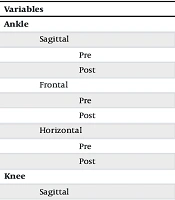
Table 2 presents the pre- and post-intervention kinematic variables for both groups. In the healthy group, the ankle sagittal-plane angle changed from 38.35 ± 7.27° at baseline to 38.02 ± 5.61° post-intervention, whereas in the concurrent deformities group, it changed from 39.01 ± 6.87° to 39.85 ± 5.04°. Similarly small pre-to-post changes were observed for ankle frontal- and transverse-plane kinematics, as well as for knee and hip joint kinematics, in both groups. After adjustment for baseline values using ANCOVA, no significant between-group differences were observed for any kinematic variable (all P > 0.05).
Table 2.
Kinematics of the Ankle, Knee, and Hip Joints a
| Variables | Healthy | Concurrent Deformities | F | Effect Size | P-Value |
|---|---|---|---|---|---|
| Ankle | |||||
| Sagittal | 0.638 | 0.041 | 0.437 | ||
| Pre | -38.35 ± 7.27 | -39.01 ± 6.87 | |||
| Post | -38.02 ± 5.61 | -39.85 ± 5.04 | |||
| Frontal | 10.259 | 0.077 | 0.280 | ||
| Pre | -7.02 ± 8.01 | -5.42 ± 9.40 | |||
| Post | -2.49 ± 12.46 | -5.11 ± 9.85 | |||
| Horizontal | 10.487 | 0.090 | 0.242 | ||
| Pre | -15.96 ± 12.83 | -11.05 ± 7.45 | |||
| Post | -10.14 ± 14.98 | -11.45 ± 9.59 | |||
| Knee | |||||
| Sagittal | 0.009 | 0.001 | 0.926 | ||
| Pre | -91.01 ± 13.67 | -96.45 ± 10.91 | |||
| Post | -91.22 ± 8.34 | -94.47 ± 11.21 | |||
| Frontal | 0.479 | 0.031 | 0.499 | ||
| Pre | 11.26 ± 4.68 | 19.35 ± 8.67 | |||
| Post | 10.35 ± 5.52 | 17.94 ± 6.98 | |||
| Horizontal | 0.447 | 0.029 | 0.514 | ||
| Pre | 2.79 ± 8.29 | 5.24 ± 8.48 | |||
| Post | .10 ± 8.28 | 3.57 ± 8.24 | |||
| Hip | |||||
| Sagittal | 0.234 | 0.015 | 0.635 | ||
| Pre | -99.89 ± 11.62 | -95.57 ± 13.09 | |||
| Post | -100.57 ± 9.95 | -95.55 ± 13.65 | |||
| Frontal | 0.356 | 0.023 | 0.560 | ||
| Pre | 7.73 ± 5.03 | 6.26 ± 4.06 | |||
| Post | 6.54 ± 4.93 | 6.03 ± 4.10 | |||
| Horizontal | 0.027 | 0.002 | 0.871 | ||
| Pre | 2.42 ± 11.10 | 7.44 ± 7.86 | |||
| Post | 1.05 ± 7.65 | 4.61 ± 8.92 |
a (-) dorsiflexion, eversion, flexion, adduction, and internal rotation; (+) plantarflexion, inversion, extension, abduction, and external rotation.
As shown in Table 3, both groups demonstrated only small pre-to-post changes in ankle, knee, and hip joint kinetics. For example, the ankle sagittal-plane moment changed from 0.0037 ± 0.0073 to 0.0039 ± 0.0057 in the healthy group and from 0.0033 ± 0.0024 to 0.0035 ± 0.0019 in the concurrent deformities group. Similar patterns were observed for the frontal- and transverse-plane moments of the ankle, knee, and hip joints. ANCOVA revealed no significant between-group differences after adjusting for baseline values for any kinetic variable (all P > 0.05).
Table 3.
Kinetics of the Ankle, Knee, and Hip Joints a
| Variables | Healthy | Concurrent deformities | F | Effect size | P-value |
|---|---|---|---|---|---|
| Ankle | |||||
| Sagittal | 0.409 | 0.027 | 0.532 | ||
| Pre | 0.0037 ± 0.0073 | 0.0033 ± 0.0024 | |||
| Post | 0.0039 ± 0.0005 | 0.0035 ± 0.0019 | |||
| Frontal | 0.832 | 0.053 | 0.376 | ||
| Pre | -0.0034 ± 0.0009 | -0.0032 ± 0.0013 | |||
| Post | -0.0033 ± 0.0008 | -0.0033 ± 0.0012 | |||
| Horizontal | 0.307 | 0.020 | 0.588 | ||
| Pre | -0.0016 ± 0.0005 | -0.0029 ± 0.0038 | |||
| Post | -0.0014 ± 0.0004 | -0.0016 ± 0.0007 | |||
| Knee | |||||
| Sagittal | 0.083 | 0.006 | 0.777 | ||
| Pre | -0.0779 ± 0.0087 | -0.0828 ± 0.0269 | |||
| Post | -0.0773 ± 0.0065 | -0.0784 ± 0.0490 | |||
| Frontal | 0.644 | 0.041 | 0.435 | ||
| Pre | 0.0145 ± 0.0081 | 0.0227 ± 0.0103 | |||
| Post | 0.0112 ± 0.0093 | 0.0193 ± 0.0109 | |||
| Horizontal | 0.485 | 0.031 | 0.497 | ||
| Pre | -0.0128 ± 0.0131 | -0.0167 ± 0.0202 | |||
| Post | -0.0077 ± 0.0042 | -0.01119 ± 0.0134 | |||
| Hip | |||||
| Sagittal | 0.056 | 0.004 | 0.816 | ||
| Pre | 0.1807 ± 0.0346 | 0.1914 ± 0.0786 | |||
| Post | 0.2016 ± 0.0361 | 0.2065 ± 0.0803 | |||
| Frontal | 0.569 | 0.037 | 0.462 | ||
| Pre | 0.0278 ± 0.1174 | 0.0900 ± 0.0924 | |||
| Post | 0.0434 ± 0.0494 | 0.0242 ± 0.0773 | |||
| Horizontal | 0.002 | 0.001 | 0.963 | ||
| Pre | -0.0068 ± 0.0461 | 0.0254 ± 0.0424 | |||
| Post | 0.0157 ± 0.0697 | 0.0288 ± 0.0959 |
a (-) dorsiflexion, eversion, flexion, adduction, and internal rotation; (+) plantarflexion, inversion, extension, abduction, and external rotation.
5. Discussion
The present study found no significant between-group differences in any of the examined ankle, knee, or hip kinetic and kinematic variables following the fatigue protocol. Although small pre-to-post changes were observed descriptively in both groups, the ANCOVA analyses did not detect evidence that athletes with concurrent flexible flatfoot and genu varum responded differently to the fatigue protocol than healthy athletes. These findings should not be interpreted as demonstrating equivalent biomechanical responses between groups, but rather as indicating that no statistically significant differences were detected under the conditions of the present study.
The absence of statistically significant between-group differences may indicate that the fatigue protocol, the SLS task, or both were insufficiently sensitive to reveal measurable biomechanical differences between groups. Alternatively, the magnitude of any true differences may have been too small to be detected with the present sample size. Regarding kinematics, these findings partially align with the results of Weeks et al. (39). Their study was the first investigation of SLS biomechanics pre- and post-fatiguing intervention in healthy participants. They reported that fatiguing exercise increases trunk motion in all cardinal planes, as well as pelvic rotation, tilt, and obliquity. They did not report any changes at the knee and hip (except in the sagittal plane). Preferential proximal movement of the pelvis and trunk has been reported as a strategy to preserve upright stability, thereby reducing the force contribution of the thigh muscles and the moment demand on the knee. The trunk and pelvis stabilize the ankle, knee, and hip joints (40). In a recent systematic review, it was found that trunk movement in the sagittal plane increases after lower-extremity muscle fatigue. This proximal strategy helps unload fatigued lower-extremity structures (41). In our study, we did not investigate trunk or pelvic kinematics, which limited our ability to explore these compensatory mechanisms.
Our results are inconsistent with those reported in previous studies evaluating single-leg hop landing (42), single-leg landing (43, 44), and cutting. These inconsistencies are likely attributable to the fact that the SLS is a relatively slow, stable, and controlled task compared with more dynamic activities such as landing, hopping, and cutting, which place greater demands on the neuromusculoskeletal system. Previous research has shown that even mild fatigue can have a pronounced effect during high-impact tasks, in which neuromuscular demands and loading rates are significantly higher (45). Therefore, the SLS may not have been sufficiently challenging to reveal fatigue-induced biomechanical differences, even in individuals with concurrent flexible flatfoot and genu varum deformities.
An alternative explanation for the present findings is that the fatigue protocol itself may not have elicited sufficient neuromuscular fatigue to produce measurable alterations in lower-limb biomechanics during the SLS. Although fatigue was confirmed using predefined subjective and objective criteria, these measures do not necessarily reflect the extent of biomechanical impairment or ensure that fatigue was induced to the same magnitude across all participants. Furthermore, different fatigue protocols target different physiological mechanisms, and task-specific biomechanical adaptations may depend on both the type and severity of fatigue induced (46). It is therefore possible that a more demanding or sport-specific fatigue protocol, or one producing greater reductions in neuromuscular performance, would have resulted in detectable biomechanical changes.
Our results indicated no statistically significant differences between athletes with and without concurrent flexible flatfoot and genu varum deformities in any of the examined kinetic parameters across the sagittal, frontal, and transverse planes following the fatigue protocol. No previous research has investigated the effects of lower-extremity fatiguing exercise on lower-limb kinetics during SLS. However, several studies have investigated the effects of fatigue during other tasks such as running (47), single-leg landing (48), and drop-jump landing (49). A systematic review found that lower-extremity kinetics in all cardinal planes during overground running do not change with distance-running fatigue (47). One possible explanation for this lack of significant kinetic change is that the torque and force demands during SLS remain below the joint torque and force that fatigued muscles can produce (50). SLS requires a moderate level of muscle activation; during SLS, the activity of the gluteus maximus, gluteus medius, and rectus femoris is reported to be 35%, 30%, and 26% of their maximum voluntary isometric contraction (51). Further evidence suggests that participants can compensate for fatigue by activating unaffected muscles during tasks (41). A study by Turpin et al. (52) investigating fatigue-related adaptations in muscle coordination during cyclic exercise showed increased activation in certain muscle groups to compensate for the declining performance of fatigued muscles, highlighting the body's ability to adapt neuromuscular control strategies in response to fatigue. Another study investigating the effects of two fatigue protocols on lower-limb joint mechanics, stiffness, and energy absorption during drop landings found that after fatigue, there was a significant increase in energy absorption by the hip extensors and a corresponding decrease in the plantar flexors. This suggests a compensatory shift toward greater reliance on proximal muscles to absorb impact when distal muscles are fatigued (53). In addition, Webster et al. (54), who examined lateral hop performance in individuals with and without chronic ankle instability (CAI), showed that those with CAI demonstrate elevated activation of the gluteus maximus muscle both pre- and post-fatigue. This indicates a compensatory strategy involving increased proximal muscle activation to stabilize the ankle joint when fatigued. Another important consideration is the possibility that the present study was underpowered to detect small between-group differences. Although no significant effects were identified, the effect sizes for all kinematic and kinetic variables were generally small, suggesting that any true differences, if present, were likely modest. Given the relatively small sample size and the substantial inter-individual variability observed in several outcome measures, the study may not have had sufficient statistical power to detect subtle fatigue-related biomechanical adaptations. Consequently, the absence of significant between-group differences should be interpreted cautiously and should not be considered definitive evidence that meaningful differences do not exist. Although statistical significance and clinical significance are distinct concepts, the observed changes in joint angles and moments were generally small in magnitude and are unlikely to represent meaningful alterations in movement patterns during this specific task. Nevertheless, caution is warranted when interpreting these findings because the study was not designed to establish clinical equivalence, and subtle biomechanical changes that may become clinically relevant during more demanding functional or sport-specific activities cannot be excluded.
This study has several limitations. First, the generalizability of the results may be limited because of the small sample size. Second, only male collegiate athletes with and without concurrent flexible flatfoot and genu varum were included, which limits the applicability of the results to non-athletic populations with similar deformities. Third, while the study examined kinematics and kinetics at the lower-extremity joints, trunk and pelvic kinematics—important for understanding lower-extremity joint stability mechanisms under fatigue—were not analyzed. Fourth, the fatigue protocol was applied only to male participants, which limits the applicability of these findings to female athletes. Fifth, generalizability was limited to the specific fatigue protocol used in this study. Another limitation of this study is that multiple kinematic and kinetic variables were analyzed simultaneously without applying a formal correction for multiple comparisons. Given the exploratory nature of this investigation and the biomechanically interdependent variables assessed across the lower-limb kinetic chain, a conservative correction was not applied to avoid inflating the risk of a type II error. However, this approach increases the potential risk of type I error, and the reported findings should therefore be interpreted with caution. Although fatigue was confirmed using combined subjective and objective criteria, group-level data confirming that a statistically comparable magnitude of fatigue was achieved in both the deformity and control groups were not reported. It is therefore possible that between-group differences in the degree of induced fatigue may have partly influenced the post-fatigue kinetic and kinematic outcomes. A further limitation of this study is the absence of investigator blinding during marker placement, data processing, and statistical analysis. Because flexible flatfoot and genu varum are visually identifiable deformities, blinding the investigator responsible for marker placement was not feasible, and data processing and statistical analyses were likewise not performed by a blinded assessor. Finally, the study focused solely on the SLS, which is relatively controlled and less dynamic than other tasks such as cutting or landing.
5.1. Conclusions
The results indicated no significant differences between athletes with and without concurrent flexible flatfoot and genu varum deformities across all examined angles and moments of the ankle, knee, and hip joints. While dynamic tasks such as single-leg hopping, landing, and cutting show significant kinematic and kinetic changes post-fatigue, more stable and controlled movements such as SLS may show only minor changes, even in athletes with concurrent flexible flatfoot and genu varum deformities.