This investigation examined the neuroprotective impacts of HES in a rat PD paradigm following repeated RES administration. The results demonstrated that HES prevents RES-elicited cataleptic behavior, and improves passive avoidance and working memory. In fact, the amelioration of RES-induced cognitive and motor deficits occurs through the modulation of the hippocampal activity of CAT, SOD and GPx enzymes, the reduction of lipid peroxidation and AChE activity in the hippocampus. In addition, HES prevents the reduction of neuronal density in CA1 and CA3 areas of hippocampus by modulating the antioxidant capacity and cholinergic activity of hippocampus.
Reserpine inhibits vesicular monoamine transporter 1 (VMAT-1) and vesicular monoamine transporter 2 (VMAT-2), leading to a reduction in vesicular monoamine storage and a noteworthy decrease in monoamine concentrations within the brain (
24). This effect results in both motor and non-motor symptoms, neurochemical alterations, and heightened oxidative stress in the brains of humans and animals, resembling the changes noted in individuals with PD (
24).
Reserpine induces cognitive impairments in laboratory animals, such as attention deficits, learning disorders, and episodic and executive memory dysfunctions (
25).
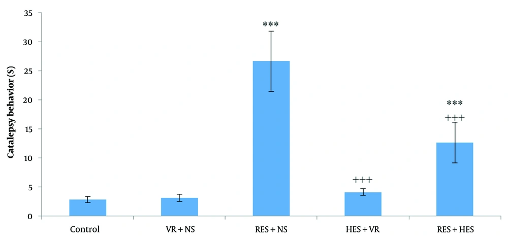
The current investigation demonstrated a notable enhancement in cataleptic behavior in the RES + NS group, consistent with prior research (
26). Conversely, treatment with HES prevented RES-elicited cataleptic behavior (
Figure 1). Cataleptic behavior is characterized by an animal's inability to adjust to an externally imposed posture, analogous to the challenges individuals with PD face when initiating movements (
27). This behavioral change is associated with dysfunction in specific brain regions, including the striatum and globus pallidus, and alterations in the nigrostriatal system's dopamine levels (
27). Consequently, the catalepsy test is a valuable tool for assessing motor impairments related to PD and can be elicited by dopamine receptor antagonists (
28). It is proposed that dopamine receptor agonists may mitigate this behavior. Therefore, HES might activate dopaminergic receptors, providing protective effects during the catalepsy test (
29).
Reserpine causes disturbances in the storage of synaptic vesicular monoamines. The depletion of monoamines at nerve terminals and the increase in intracellular monoamine levels result in the auto-oxidation of dopamine and its oxidative metabolism by monoamine oxidase (MAO), leading to oxidative stress (
30). Since PD motor impairments are associated with oxidative stress, they are mitigated by antioxidant compounds. As demonstrated in
Table 1, HES likely reduced RES-elicited cataleptic behavior by enhancing the antioxidant defense system.
Regarding non-motor changes, cognitive deficits are widespread in PD, often preceding motor symptoms. Progressive parkinsonism elicited by repeated doses of RES is a valuable tool for evaluating non-motor disorders, including cognitive deficits in rodents (
18). This investigation noted cognitive impairment in the passive avoidance and working memory tests. As mentioned, pathways other than the nigrostriatal pathway play an essential role in manifesting PD's non-motor symptoms (
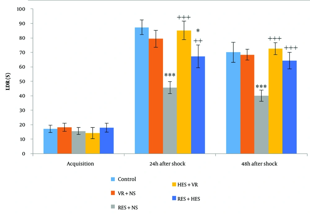
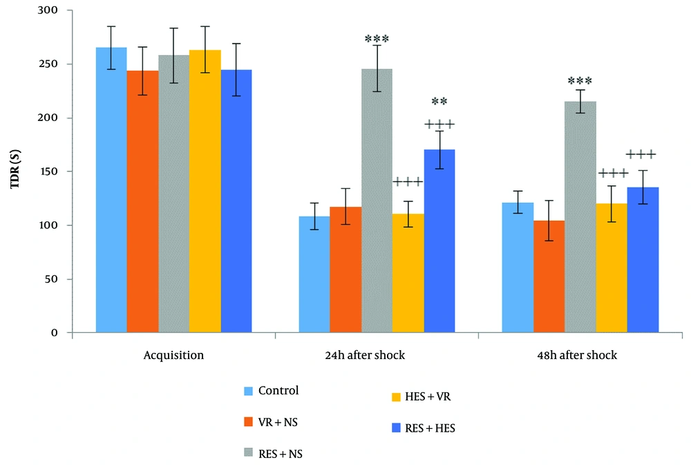
27). The current investigation demonstrated impaired passive avoidance memory by employing the shuttle box test 24 hours after the final dose of RES.
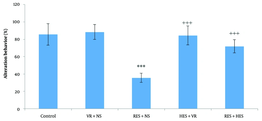
Additionally, the percentage of alternation behavior in the Y-maze, indicative of spatial working memory or short-term memory performance, was noteworthyly reduced in rats receiving RES. Despite this, HES noteworthyly improved spatial working memory by increasing the percentage of alternation behavior. Cognition-related brain areas, such as the hippocampus and medial frontal cortex, are innervated by serotonergic and dopaminergic projections, which stem from the raphe complex and the mesocorticolimbic pathway, respectively (
31).
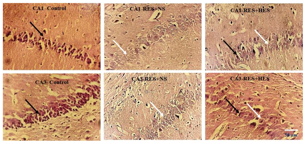
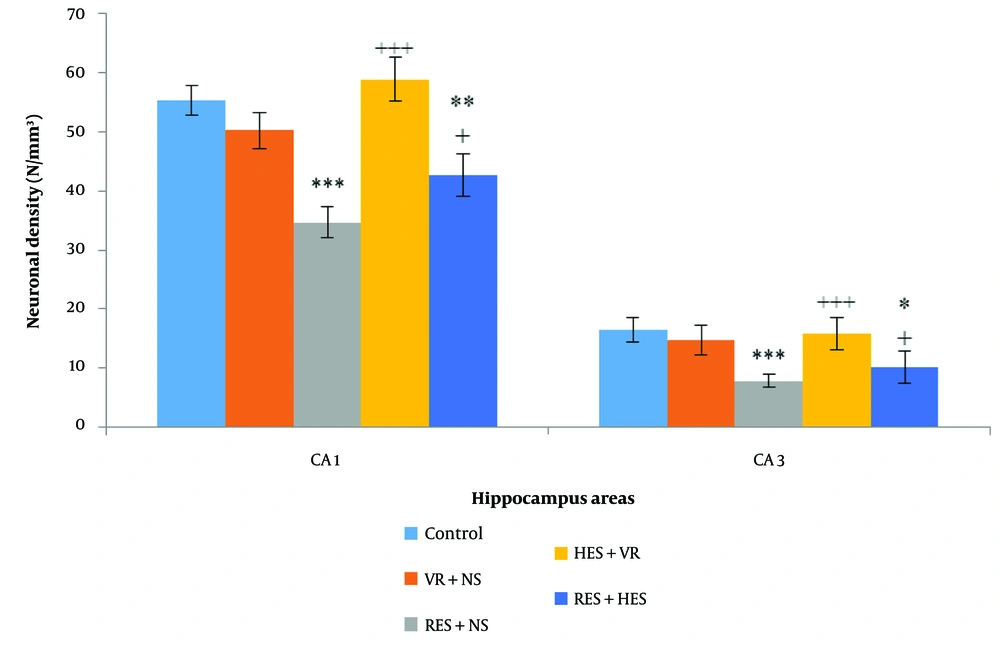
The memory impairment elicited by RES in this investigation was likely due to neurochemical changes in the hippocampus, which damaged pyramidal neurons in the CA1 and CA3 regions.
Reserpine, as an inducer of oxidative stress, can reduce CAT, GPx, SOD, reduced glutathione, and adenosine triphosphate (ATP) enzymes (
32). Increased lipid peroxidation (MDA) and NO have also been noted in multiple brain regions, including the midbrain and striatum, in a rat paradigm of PD. Increased lipid peroxidation results from free radical attacks on the cell membrane, leading to phospholipid oxidation and cell death (
32). However, some research has reported contradictory results, possibly due to different RES doses, administration methods, and brain regions studied. For example, repeated administration of low doses of RES (0.1 mg/kg) has cumulative impacts on lipid peroxidation in the striatum. Simultaneously, these alterations have not been observed in rats' hippocampus. Catalase function is generally reduced in all brain regions except the striatum (
7). However, in the current investigation, employing a dose of 0.2 mg/kg body weight RES, the enzyme levels of CAT, SOD, and GPx in the hippocampus of the RES + NS group demonstrated a noteworthy decrease in relative assessment with the control group at the end of the treatment duration. It seems that oxidative stress in the hippocampus following RES administration is sufficient for neuronal degeneration and reduced neuronal density in the hippocampus. Oxidative stress can weaken the brain's antioxidant system by producing free radicals, leading to irreversible brain damage (
33). Oxidative stress and chronic inflammation caused by PD result in neuronal cell damage by altering the levels of miRNAs and their target proteins. However, according to prior research, antioxidants can protect neuronal cells by modulating the levels of miRNAs and their intended proteins (
34).
Flavonoids, widely used in multiple fruits and vegetables, have numerous biological and pharmacological impacts. Flavonoid-rich diets are effective in regulating brain function. Hesperidin has potential antioxidant impacts in vivo and may act as a neuroprotective agent in the brain. This compound stabilizes biological membranes as an antioxidant, thus preventing cell membrane damage in neurological diseases such as Alzheimer's, Parkinson's, and Huntington's (
35). In this investigation, HES increased the function levels of the enzymes CAT, SOD, and GPx and decreased MDA levels, consistent with prior research (
14). Hesperidin increases the activity of antioxidant enzymes through upregulation of SIRT1 gene and inhibition of NADPH oxidase 4 (NOX4) enzyme (
36). Also, studies have shown that HES decreases interleukin 1 beta (IL-1β), tumor necrosis factor (TNF-α) and MDA and increases hippocampal brain derived neurotrophic factor (BDNF) levels in the depression model. In addition, oxidative stress parameters, including SOD, CAT, nuclear factor erythroid 2-related factor 2 (Nrf2) and heme oxygenase-1 (HO-1), were significantly increased following HES treatment in this model (
37). By activating the Nrf2-antioxidant response element (Nrf2/ARE) pathway, HES activates the main antioxidant systems, and by stimulating the BDNF/protein kinase A (PKA)/ cAMP-response element binding protein (CREB) signaling pathway in the amygdala and hippocampus, it shows anxiolytic effects (
38).
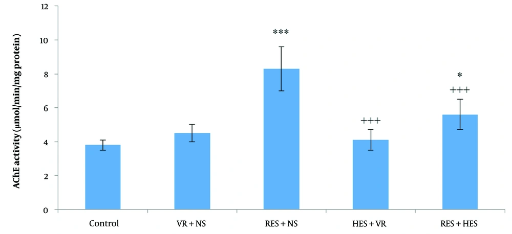
Although decreased AChE function in the midbrain of the RES paradigm of PD has been noted, which may be due to damage to cholinergic neurons (
39), and noteworthy inhibition of AChE function has been recorded in the thalamus of PD patients (
40), in the current investigation, AChE function in the hippocampus of rats in the PD paradigm of increased following RES treatment, which could explain cognitive impairment and memory and learning deficits in this paradigm of. Prior research has demonstrated that AChE function levels occur before neurodegeneration in Alzheimer's disease. Acetylcholinesterase hydrolyzes acetylcholine in the synaptic cleft, and changes in AChE function are considered an early indicator of cognitive disorders (
41). In this investigation, HES improved learning and memory deficits caused by RES in the PD paradigm of rats by reducing AChE expression. Additionally, the noteworthy reduction in hippocampal AChE function after HES treatment may be due to its AChE-inhibiting impact.
Prior studies have demonstrated that HES can inhibit apoptosis and cell cycle arrest, thereby preventing cell death in various brain regions (
38). In this investigation, the administration of HES led to an increase in neuronal density in the CA1 and CA3 regions of the hippocampus. This increase was associated with enhanced working memory, passive avoidance, and a reduction in cataleptic behavior in the RES + HES group in contrast to the vehicle-treated group (VR + NS). Hesperidin reduces oxidative stress in PD by regulating Nrf2 nuclear factor kappa-light-chain-enhancer of activated B cells (NF-κB) and reducing apoptotic cells (
42). Chronic administration of HSP (HES precursor for five weeks) exhibited neuroprotective impacts against oxidative stress in the brains of mice. These impacts were associated with decreased lipid peroxidation levels and activation of endogenous antioxidant defense mechanisms, including CAT, SOD, and glutathione (GSH)-related enzymes (
43). By scavenging high ROS levels and boosting antioxidant defense mechanisms—primarily by upregulating the expression of Nrf2 and HO-1—HES has demonstrated impressive antioxidant effects. By suppressing apoptotic cell death, lowering ROS and MDA generation, raising SOD and GSH expression, and triggering Kelch-like ECH-associated protein 1 (KEAP-1) and Nrf2/HO-1 signaling, HES considerably guards against severe oxidative stress (
44).
5.1. Conclusions
The current investigation demonstrated that HES exhibits neuroprotective impacts, improves both passive avoidance and working memory, enhances cholinergic function, and prevents the reduction of neuronal density in multiple subareas of the hippocampus in the RES paradigm of PD. However, the limitations of this investigation should be considered, as molecular methods for evaluating the mechanisms underlying the protective impacts of HES were not conducted. Some of the limitations of the study can be mentioned: (1) The study lacks molecular insights or signaling pathway into HES's protective mechanisms; (2) findings are based solely on the RES model, which may not fully represent human PD; (3) the focus was on motor and cognitive functions, with limited exploration of other behavioral domains; (4) the sample size or possibility of potential biases. Further research is essential to enhance the understanding of PD prevention and therapy. This research should investigate the impact of HES on motor and non-motor modifications within the RES paradigm of PD. At the end of this research and for future studies, we recommended the investigation of molecular mechanisms underlying HES's neuroprotective effects, exploration of the long-term effects of HES on motor and non-motor symptoms, evaluation of the potential synergistic effects of combining HES with other treatments, additional behavioral assessments for non-motor symptoms such as anxiety and depression in the RES model of PD and conduction of the clinical trials to evaluate the efficacy and safety of HES for PD treatment.