1. Background
Diabetic foot ulcers (DFUs) are among the most severe and debilitating complications of diabetes mellitus, affecting millions of individuals worldwide (1). Characterized by chronic, non-healing wounds, DFUs are strongly associated with high morbidity, reduced quality of life, and a markedly elevated risk of lower-extremity amputation (2). A major contributor to this poor prognosis is the susceptibility of DFUs to bacterial infection, particularly infection with multidrug-resistant organisms. Infection not only delays wound healing by prolonging the inflammatory phase but also exacerbates tissue destruction, creating a vicious cycle that complicates clinical management and imposes a substantial economic burden on healthcare systems (3).
Standard wound care for DFUs typically includes debridement, offloading, and the administration of systemic antibiotics for infected ulcers (4). However, the increasing prevalence of antimicrobial resistance and the limited penetration of systemic antibiotics into ischemic wound beds have compromised the effectiveness of conventional treatments. Consequently, there is an urgent need for alternative, adjunctive topical therapies capable of eradicating local pathogens while promoting tissue regeneration (5). In recent years, medical ozone therapy has emerged as a promising modality in wound care. Ozone exhibits potent antimicrobial activity against bacteria, fungi, and viruses, while also enhancing local tissue oxygenation, promoting angiogenesis, and modulating immune responses (6). When formulated as ozonated olive oil, ozone provides a stable, lipid-soluble delivery system that continuously releases reactive oxygen species (ROS) and lipid ozonation products directly into the wound bed, thereby fostering an optimal microenvironment for healing without inducing systemic side effects (7).
Beyond the physiological aspects of wound healing, effective DFU management requires a robust patient-centered approach. Chronic wounds often lead to psychological distress, diminished self-efficacy, and treatment fatigue, underscoring the need for patient empowerment (8). Empowered patients are more likely to engage in proactive health behaviors, particularly foot self-care. Adherence to foot self-care practices, such as daily inspection, proper hygiene, and appropriate footwear, is essential not only for preventing ulcer recurrence but also for supporting the healing of existing wounds (9). However, the interplay among advanced topical treatments, clinical symptom relief, and psychosocial outcomes, such as patient empowerment, remains largely unexplored. It is plausible that an effective, easy-to-apply topical intervention such as ozonated olive oil could reduce the physical burden of infection, thereby improving patients' confidence and subsequent adherence to self-care routines.
2. Objectives
Although preliminary evidence supports the antimicrobial efficacy of ozonated oils in various dermatological conditions (10, 11), there is a paucity of rigorous randomized controlled trials evaluating their specific impact on DFU infections, particularly in conjunction with patient-reported outcomes. Bridging the gap between clinical efficacy and patient empowerment is essential for holistic diabetic foot management. Therefore, this randomized controlled trial aimed to investigate the efficacy of topical ozonated olive oil in diabetic foot infection resolution, patient empowerment, and foot self-care behaviors in individuals with DFUs. By addressing both the biological and behavioral aspects of DFU management, this study seeks to provide comprehensive evidence to support the integration of ozonated olive oil into standard diabetic foot care protocols.
3. Methods
3.1. Study Design and Setting
A two-arm, parallel-group randomized controlled trial was conducted to investigate the effects of adjunctive topical ozonated olive oil on diabetic foot infection, patient empowerment, and foot self-care among individuals with diabetic foot ulcers. The study was conducted between December 2025 and June 2026 in two outpatient wound-care clinics in Bandar Abbas, southern Iran. The study protocol was developed in accordance with the Declaration of Helsinki and is reported in accordance with the Consolidated Standards of Reporting Trials (CONSORT) statement.
3.2. Participants
Adults with type 2 diabetes mellitus and a clinically diagnosed diabetic foot ulcer were screened for eligibility at the participating outpatient clinics. Because clinical resolution of infection was defined as the primary endpoint, eligibility required the presence of a clinically infected diabetic foot ulcer at baseline, according to the International Working Group on the Diabetic Foot/Infectious Diseases Society of America (IWGDF/IDSA) classification (12).
Participants were eligible if they were aged ≥ 18 years, had a confirmed diagnosis of type 2 diabetes mellitus, had a diabetic foot ulcer with clinical evidence of infection, were able to communicate in Persian and complete the study questionnaires, and provided written informed consent.
Patients were excluded if they had a severe systemic infection requiring immediate hospitalization, critical limb ischemia requiring urgent vascular intervention, osteomyelitis requiring immediate surgical management, an indication for urgent major amputation, extensive tissue necrosis incompatible with conservative wound management, a known allergy to the study preparation, or concurrent participation in another interventional wound-care study. These exclusion criteria were necessary for ethical and safety reasons, as such patients require urgent specialized care that cannot be delayed for study participation. However, these criteria limit the generalizability of the findings to less severely ill populations. Participants were withdrawn if they discontinued the assigned treatment, underwent major amputation, required a treatment strategy incompatible with the study protocol, or were unavailable for follow-up assessment.
3.3. Sample Size
The sample size was calculated to detect a clinically meaningful difference in the primary outcome, clinical resolution of diabetic foot infection, at the prespecified primary endpoint (two-month follow-up). Based on published data from similar wound-care trials in diabetic foot populations (12, 13) and local clinical audit data from the participating centers, we assumed a control-group clinical resolution rate of 35% at two months. An absolute improvement of 25 percentage points (to 60% in the intervention group) was considered clinically meaningful. Using a two-sided χ2 test (superiority hypothesis) with α = 0.05 and power = 80%, the required sample size was 39 participants per group. Accounting for an anticipated attrition rate of 15% (based on previous wound-care trials in our center), the inflated sample size was 45 participants per arm, resulting in a total planned sample of 90 participants. The calculation was performed using G*Power software, version 3.1.9.7 (effect size w = 0.50 corresponding to proportions of 0.35 vs. 0.60).
Accordingly, 90 eligible participants were enrolled and randomly allocated in a 1:1 ratio to the intervention and control groups, with 45 participants in each group. During the study period, three participants in the intervention group and two participants in the control group were lost to follow-up. Thus, 42 participants in the intervention group and 43 participants in the control group completed the study and were included in the final per-protocol analysis.
3.4. Randomization and Allocation
After confirmation of eligibility and completion of baseline assessments, participants were randomly assigned to the intervention or control group using a computer-generated randomization sequence with a 1:1 allocation ratio. To minimize potential selection bias, allocation was concealed using sequentially numbered, opaque, sealed envelopes prepared independently of participant recruitment.
A researcher not involved in the clinical assessment generated the allocation sequence. After enrollment, the designated coordinator opened the next envelope in sequence and implemented the assigned treatment. Because the intervention involved application of a topical preparation with a recognizable procedure, blinding of participants and treatment providers was not feasible. Outcome assessments were performed by two independent wound-care specialists (designated as blinded assessors) who were not involved in treatment allocation, application, or routine clinical care. However, true blinding could not be guaranteed. Topical oil application may leave residual odor, visible residue, or dressing characteristics that could potentially reveal treatment allocation to assessors. Therefore, blinding of the outcome assessors should be considered partial at best, and the primary outcome remains susceptible to detection bias. To minimize the risk of unblinding, treatment application occurred in separate clinic areas after outcome assessments were completed, and participants were instructed not to discuss their treatment with the assessing clinicians. Despite these measures, the subjective nature of clinical infection-resolution assessment means that residual detection bias cannot be fully excluded. This represents a fundamental limitation of the study design. Statistical analysis was conducted by a researcher who was not involved in treatment allocation.
3.5. Intervention: Topical Ozonated Olive Oil
Participants allocated to the intervention arm received topical ozonated olive oil as an adjunct to standard diabetic foot-ulcer care for four consecutive weeks. The preparation was formulated using medical-grade ozone and olive oil under controlled production conditions. The ozonated olive oil (commercial name: Heal Ozone) was supplied by Nasim Ozone Company with the following product identifiers: Unique Identifier (UID: 29486042328361021251), Global Trade Item Number (GTIN: 06260001510593), and Lot No. D1404 - 2.
The ozonated olive oil preparation was produced by bubbling medical-grade ozone (60 μg/mL) through pharmaceutical-grade olive oil for 24 hours at 25 °C under light-protected conditions until a target peroxide value of 600 - 700 mEq O2/kg was achieved. The final product had the following physicochemical characteristics: peroxide value of 658 ± 24 mEq O2/kg; acidity value of 0.8 ± 0.1% (expressed as oleic acid); iodine value of 82 ± 3 g I2/100 g; and viscosity of 85 ± 5 mPa·s at 25 °C. The oxidation profile, characterized by Fourier-transform infrared spectroscopy, demonstrated the presence of both ozonides (C–O–O stretching at 1105 cm-1) and peroxides (O–O stretching at 870 cm-1), confirming the formation of the expected bioactive ozonation products. The product was packaged in amber glass bottles, stored at 15 - 25 °C protected from light, and had an in-use stability of 30 days after opening. A single production type was used for all participants, and consistency was verified by peroxide-value determinations at release and at study completion (658 ± 24 vs. 641 ± 19 mEq O2/kg; P = 0.41).
At each treatment session, the ulcer was first inspected and cleansed with sterile normal saline. After gentle removal of visible exudate and debris, approximately 6 mL of ozonated olive oil was applied topically to the affected ulcer area once daily for 28 consecutive days. The preparation was distributed evenly over the wound surface and then covered with the routine dressing used in the participating clinics.
Participants and/or their caregivers received standardized instructions regarding the application procedure, dressing care, and avoidance of other non-prescribed topical preparations. Adherence was monitored throughout the intervention using treatment records and participant reports. Any treatment interruption, local intolerance, or adverse event was documented.
3.6. Control Group
Participants allocated to the control group received standard diabetic foot-ulcer care according to the routine protocol of the participating outpatient wound-care clinics. The minimum standardized wound-care protocol applied to both groups included: 1) wound cleansing with sterile normal saline at each clinic visit; 2) sharp debridement performed by trained wound-care nurses when necrotic or nonviable tissue was present; 3) moisture-retentive dressing selection based on wound exudate characteristics (foam dressings for moderate exudate, hydrocolloid for low exudate, alginate for highly exudative wounds); 4) offloading using total-contact casts or removable cast walkers for plantar ulcers, or appropriate footwear modifications for non-plantar ulcers; 5) glycemic management targeting HbA1c < 8.0% with endocrinology consultation when needed; and 6) systemic antibiotic therapy according to IWGDF/IDSA guidelines when clinically indicated (12). Antibiotic selection was based on wound culture and sensitivity when available, or on empirical therapy targeting common pathogens (Gram-positive cocci as first-line).
Systemic antibiotic treatment, offloading, glycemic management, debridement, or referral for specialist intervention was provided whenever clinically indicated and according to the treating physician’s assessment. The same standard-care principles were applied to participants in both groups. The only planned difference between groups was the addition of topical ozonated olive oil in the intervention arm.
3.7. Outcome Measures
The primary outcome was clinical resolution of diabetic foot infection. The secondary outcomes were patient empowerment and diabetic foot self-care.
All outcomes were assessed at three predefined time points: baseline (T0), immediately after completion of the 4-week intervention (T1), and two months after completion of the intervention (T2). The primary outcome was clinical resolution of diabetic foot infection at the two-month follow-up (T2), as prespecified in the trial protocol and registry. The immediate post-intervention assessment (T1) was prespecified as a secondary time point to characterize the trajectory of the treatment response. This designation was made before data collection and was not modified after observing the results.
3.8. Primary Outcome: Diabetic Foot Infection
Diabetic foot infection was assessed using the IWGDF/IDSA clinical classification of diabetes-related foot infection (12). Current IWGDF/IDSA guidance recommends diagnosing diabetes-related soft-tissue infection clinically based on local or systemic manifestations of inflammation and grading infection severity using the IWGDF/IDSA classification system.
At baseline, participants were categorized according to infection severity as uninfected, mildly infected, moderately infected, or severely infected. Clinical assessment considered the presence and extent of erythema, local swelling or induration, tenderness or pain, increased local temperature, purulent discharge, depth of tissue involvement, and systemic manifestations.
Because all participants were required to have clinical evidence of infection at enrollment, the primary endpoint was defined as clinical resolution of diabetic foot infection, operationalized as a transition from an infected classification at baseline to the uninfected category following treatment. In addition, changes in infection severity across the IWGDF/IDSA categories were recorded as a secondary clinical indicator of treatment response.
Infection status at each time point was assessed by the two independent, blinded wound-care specialists who had completed standardized training on IWGDF/IDSA classification. Before study initiation, both assessors completed a calibration exercise using 20 standardized case vignettes; inter-rater agreement was excellent (weighted κ = 0.87; 95% CI, 0.78 - 0.96). During the study, the assessors independently classified each participant, and any disagreements were resolved by consensus discussion.
The infection assessment was performed at baseline, immediately after the 4-week treatment period, and at the 2-month follow-up. Additional clinical evaluations were performed when deterioration or other clinically relevant signs required reassessment.
It is important to note that this trial’s primary outcome was clinical infection resolution, not complete wound healing (epithelialization) or reduction in ulcer area. The term 'wound healing' should not be inferred from these findings. Furthermore, the primary outcome was based on clinical signs and symptoms of infection according to IWGDF/IDSA criteria; microbiological eradication was not assessed. Therefore, the term 'infection resolution' in this manuscript refers exclusively to clinical resolution, not microbiological clearance.
3.9. Secondary Outcome 1: Patient Empowerment
Patient empowerment was measured using the Iranian Diabetes Empowerment Scale (IR-DES-28) (14). The IR-DES-28 is a 28-item Persian-language instrument specifically validated for Iranian patients with type 2 diabetes. Its psychometric properties were evaluated in a sample of 100 Iranian patients with type 2 diabetes using construct, criterion, and concurrent validity procedures, as well as internal consistency, test–retest reliability, and split-half reliability. The total scale demonstrated a Cronbach's alpha of 0.89, while test–retest reliability was 0.74 (P < 0.01) (14). The validation study identified three principal domains related to psychological self-management, goal achievement, and goal setting.
The IR-DES-28 contains 28 items scored on a 5-point Likert scale (1 = strongly disagree to 5 = strongly agree), yielding a total score range of 28 - 140, with higher scores indicating greater empowerment. The validated Persian version has established a minimally important difference (MID) of 5 points based on anchor-based methods (14). Each item is scored according to the original response format of the instrument, with higher overall scores indicating greater diabetes-related empowerment. The questionnaire was completed at T0, T1, and T2.
3.10. Secondary Outcome 2: Diabetic Foot Self-Care
Foot self-care was evaluated using the Persian version of the Diabetic Foot Self-Care Questionnaire of the University of Málaga (DFSQ-UMA) (15). The Persian adaptation was psychometrically evaluated in 407 Iranian patients with diabetes using exploratory and confirmatory factor analyses. The validated Persian version contains 15 items and demonstrated a three-factor structure explaining 56.74% of the total variance, with a Cronbach's alpha of 0.865 for the total instrument. The authors concluded that the Persian DFSQ-UMA has satisfactory validity and reliability for assessing diabetic foot self-care among Iranian patients with diabetes (16).
The DFSQ-UMA contains 15 items scored on a 5-point Likert scale (1 = never to 5 = always), yielding a total score range of 15 - 75, with higher scores indicating better foot self-care. No established MID is available for the Persian DFSQ-UMA; therefore, changes were interpreted descriptively and benchmarked against distribution-based estimates (0.5 SD = 3.7 points). The instrument evaluates behaviors related to foot examination and care, footwear and socks, and other diabetes-specific foot self-care practices. Higher scores indicate more appropriate foot self-care behaviors according to the validated Persian scoring system. The questionnaire was administered at T0, T1, and T2.
3.11. Clinical and Sociodemographic Variables
A structured data-collection form was used to record participants' demographic and clinical characteristics, including age, sex, educational level, marital status, duration of diabetes, diabetes treatment, history of previous diabetic foot ulcer, ulcer duration, ulcer location, and baseline infection severity. Relevant clinical characteristics were obtained through patient interviews, physical examination, and review of medical records.
Baseline characteristics were used to assess comparability between the randomized groups and to identify clinically relevant covariates for subsequent statistical analyses.
3.12. Intervention Adherence and Safety
Adherence to the topical intervention was monitored throughout the 4-week treatment period. Participants were asked to report missed applications, treatment interruptions, local discomfort, or other unexpected symptoms. Adverse events (AEs) were defined as any untoward medical occurrence in a participant, regardless of causal relationship to the study intervention. Serious adverse events (SAEs) were defined as events resulting in death, life-threatening conditions, hospitalization, persistent or significant disability, or requiring intervention to prevent permanent impairment. AEs were actively collected at each study visit through structured interviews and physical examination, supplemented by participant self-reports between visits via a dedicated telephone line. Severity was graded using the Common Terminology Criteria for Adverse Events (CTCAE) version 5.0 (grades 1 - 5) (13). Causality assessment was performed by the treating physician using the World Health Organization-Uppsala Monitoring Centre criteria (certain, probable, possible, unlikely, unrelated). AEs were classified as related to treatment if rated as certain, probable, or possible. Adverse events were documented and evaluated by the clinical team.
Particular attention was given to local reactions, including erythema, burning, itching, irritation, increased pain, allergic manifestations, and ulcer deterioration. Serious clinical events, including progression of infection, hospitalization, surgical intervention, or amputation, were also recorded.
3.13. Data Collection Procedure
Baseline assessments were completed before initiation of the intervention. Participants were subsequently followed for four weeks during the treatment phase. Immediately after completion of the 4-week intervention, the primary and secondary outcomes were reassessed. A further assessment was conducted two months after completion of treatment to determine whether changes in infection status, patient empowerment, and foot self-care were maintained over time.
To enhance consistency, clinical assessments were conducted using standardized procedures and predefined criteria. The same instruments and assessment procedures were used across all study time points.
3.14. Statistical Analysis
Statistical analyses were performed using IBM SPSS Statistics, version 27.0 (IBM Corp., Armonk, NY, USA). The distribution of continuous variables was evaluated using graphical methods and the Shapiro–Wilk test. Continuous variables were summarized as mean ± standard deviation or median and interquartile range, as appropriate, whereas categorical variables were presented as frequencies and percentages.
Baseline comparability between the intervention and control groups was evaluated using the independent-samples t-test or Mann–Whitney U test for continuous variables and the chi-square test or Fisher's exact test for categorical variables. Baseline significance testing was not used to assess randomization success but rather to identify potential covariates for adjusted analyses.
The primary efficacy analysis was conducted in the intention-to-treat (ITT) population, defined as all randomized participants analyzed according to their assigned treatment group. The per-protocol (PP) population, consisting of participants who completed the study without major protocol deviations, was used as a secondary analysis. For the primary binary outcome, missing data were handled using multiple approaches to assess robustness: 1) primary analysis: multiple imputation by chained equations (MICE) with 50 imputed datasets using logistic regression models including baseline infection severity, ulcer duration, age, and treatment group as predictors; 2) sensitivity analysis 1: baseline observation carried forward (BOCF, assuming no resolution in dropouts); 3) sensitivity analysis 2: best-case/worst-case bounds (assuming all intervention dropouts resolved and all control dropouts unresolved; then the reverse).
For the primary outcome, the proportion of participants achieving clinical resolution of diabetic foot infection was compared between groups. Relative risks (RRs), absolute risk differences, 95% confidence intervals (CIs), and corresponding P values were reported where appropriate. Changes in IWGDF/IDSA infection severity were also examined using appropriate categorical-data analyses.
The principal longitudinal analyses for patient-reported outcomes were conducted in the ITT population using linear mixed-effects models with all available observations from all 90 randomized participants, analyzed according to randomized allocation under the missing-at-random assumption. The models included fixed effects for treatment group, time, and the group × time interaction, with participant-level random intercepts and an unstructured covariance matrix to account for within-subject correlation. Restricted maximum likelihood (REML) estimation was used. Model diagnostics included examination of residual normality, homoscedasticity, and influence statistics; no material violations were identified.
The magnitude of between-group effects was reported using appropriate effect-size estimates with 95% CIs. To distinguish confirmatory from exploratory findings, all analyses at the primary endpoint (T2) are presented as the principal results. The post-intervention (T1) analyses are reported as secondary or exploratory. No multiplicity adjustment was applied because the primary hypothesis was tested at a single prespecified time point. Secondary outcomes (patient empowerment and foot self-care) were analyzed using a prespecified hierarchical approach, with patient empowerment considered before foot self-care. All statistical tests were two-sided, and P < 0.05 was considered statistically significant.
Sensitivity analyses using baseline-adjusted models were conducted, with adjustment for age, duration of diabetes, BMI, HbA1c, and baseline infection severity. The full adjusted-model estimates are presented in Table S1 in the Supplementary File.
4. Results
4.1. Participant Flow and Baseline Characteristics
A total of 147 patients were assessed for eligibility. Of these, 57 were excluded: 31 did not meet the inclusion criteria (18 without clinical infection, 7 with critical limb ischemia, 4 with severe systemic infection, and 2 with osteomyelitis requiring surgery), 19 declined to participate, and 7 were participating in another interventional study. The remaining 90 eligible participants with clinically infected diabetic foot ulcers were enrolled and randomly allocated in a 1:1 ratio to the ozonated olive oil group (n = 45) or the standard-care control group (n = 45). During follow-up, three participants in the intervention group and two in the control group were lost to follow-up. Reasons for loss to follow-up in the intervention group were as follows: 1 participant withdrew consent (moved to another city), 1 was unable to attend follow-up visits (work obligations), and 1 was withdrawn due to a protocol deviation (use of a non-study topical antimicrobial). In the control group, 1 participant withdrew consent (personal reasons), and 1 was lost to follow-up due to hospitalization for an unrelated condition (acute cholecystitis). No loss to follow-up was attributed to adverse events or treatment dissatisfaction. Therefore, 42 participants in the intervention group and 43 in the control group completed the study and were included in the per-protocol analysis (overall retention rate: 94.4%). An intention-to-treat analysis was also conducted, including all 90 randomized participants. For the primary binary outcome, the primary ITT analysis used multiple imputation for missing data. Sensitivity analyses using BOCF and best-case/worst-case bounds were also conducted. ITT and PP findings for the primary outcome are both reported in Figure 1.
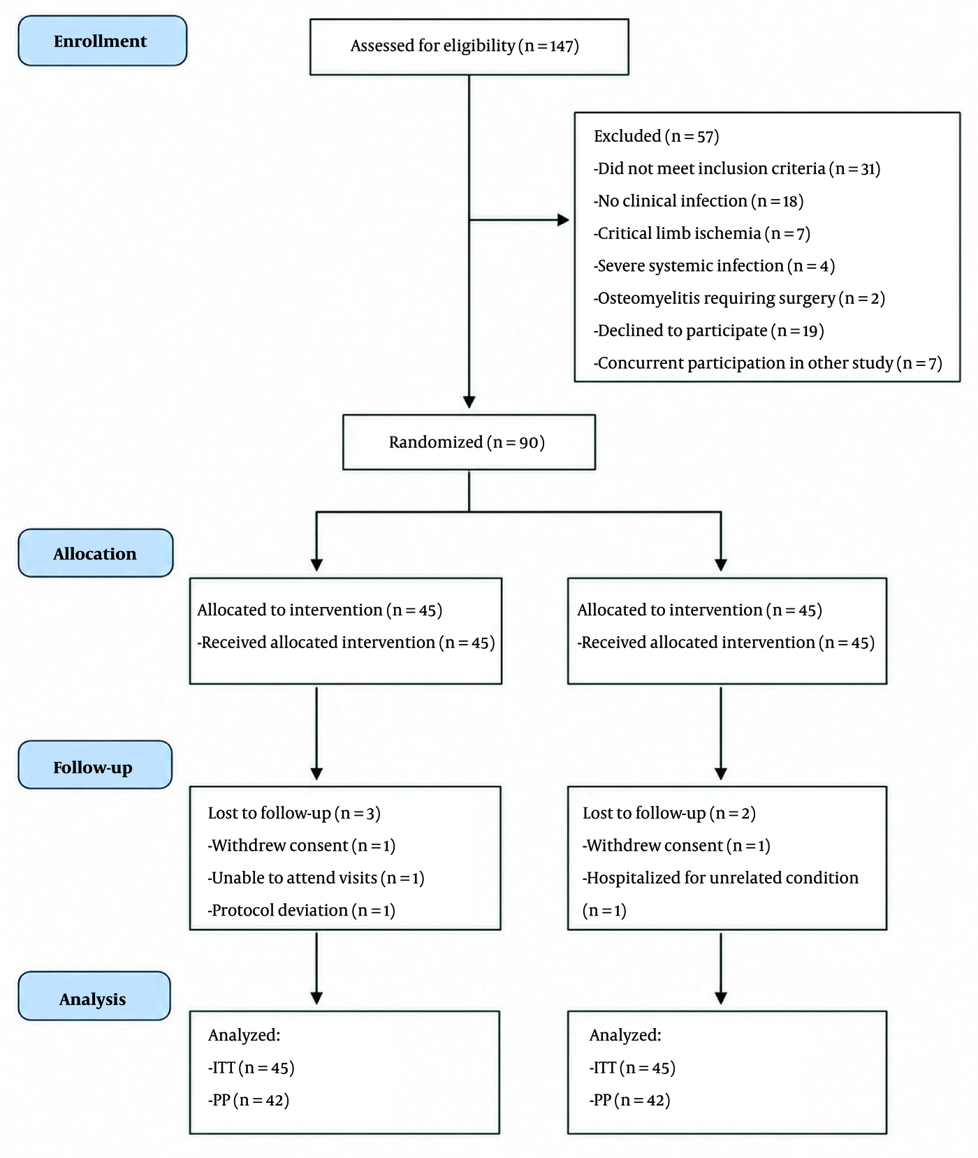
Figure 1.
CONSORT flow diagram
Baseline demographic and clinical characteristics of the intention-to-treat population (N = 90) are summarized in Table 1. Baseline characteristics were well balanced between groups. The mean age was 57.1 ± 8.0 years in the intervention group and 55.9 ± 8.5 years in the control group (P = 0.49). Men accounted for 62.2% and 57.8% of the intervention and control groups, respectively (P = 0.67). Mean body mass index (BMI) was 28.2 ± 4.5 kg/m2 in the intervention group and 28.0 ± 4.2 kg/m2 in the control group (P = 0.83). The mean duration of diabetes was comparable (10.4 ± 4.0 vs. 10.2 ± 4.1 years; P = 0.82). Mean HbA1c was 8.1 ± 1.4% and 8.0 ± 1.3%, respectively (P = 0.73). The distribution of educational attainment was also similar between groups (P = 0.79).
Table 1.
Baseline Demographic and Clinical Characteristics of the Intention-to-Treat Population (N = 90) a
| Characteristics | Ozonated Olive Oil (n = 45) | Standard Care (n = 45) | P-Value |
|---|---|---|---|
| Age, y | 57.1 ± 8.0 | 55.9 ± 8.5 | 0.49 |
| Gender (male) | 28 (62.2) | 26 (57.8) | 0.67 |
| BMI, kg/m2 | 28.2 ± 4.5 | 28.0 ± 4.2 | 0.83 |
| Educational level | 0.79 | ||
| Primary or less | 15 (33.3) | 17 (37.8) | |
| Secondary | 20 (44.4) | 20 (44.4) | |
| Tertiary or above | 10 (22.2) | 8 (17.8) | |
| Duration of diabetes, y | 10.4 ± 4.0 | 10.2 ± 4.1 | 0.82 |
| HbA1c, % | 8.1 ± 1.4 | 8.0 ± 1.3 | 0.73 |
| Ulcer duration, d | 22 (14 - 36) | 23 (14 - 33) | 0.91 |
| Ulcer area, cm2 | 3.7 ± 2.0 | 3.6 ± 1.9 | 0.80 |
| Ulcer location | 0.88 | ||
| Plantar surface | 20 (44.4) | 19 (42.2) | |
| Toes | 15 (33.3) | 16 (35.6) | |
| Dorsal surface | 6 (13.3) | 6 (13.3) | |
| Other | 4 (8.9) | 4 (8.9) | |
| Peripheral neuropathy | 35 (77.8) | 33 (73.3) | 0.62 |
| Peripheral arterial disease | 10 (22.2) | 11 (24.4) | 0.80 |
a Values are presented as mean ± SD, median (IQR), or No. (%). Percentages are based on the intention-to-treat denominators (45 per group). Abbreviations: BMI, Body Mass Index; HbA1c, glycated hemoglobin; IQR, interquartile range.
All participants had clinically infected diabetic foot ulcers at baseline. Mild infection was present in 28 participants (62.2%) in the intervention group and 27 (60.0%) in the control group, whereas moderate infection was present in 17 (37.8%) and 18 (40.0%), respectively (P = 0.83). The median ulcer duration before enrollment was 22 days (IQR, 14 - 36) in the intervention group and 23 days (IQR, 14 - 33) in the control group (P = 0.91). Ulcers were predominantly located on the plantar surface (44.4% vs. 42.2%) and toes (33.3% vs. 35.6%) (P = 0.88). The mean baseline ulcer area was 3.7 ± 2.0 cm2 in the intervention group and 3.6 ± 1.9 cm2 in the control group (P = 0.80). Peripheral neuropathy was present in 77.8% and 73.3% of participants, respectively (P = 0.62), and peripheral arterial disease in 22.2% and 24.4% (P = 0.80). Baseline patient-reported outcomes were also similar, with mean IR-DES-28 scores of 71.9 ± 8.2 vs. 71.3 ± 8.7 (P = 0.74) and DFSQ-UMA scores of 51.0 ± 7.0 vs. 50.9 ± 7.5 (P = 0.95).
For reference, baseline characteristics of the per-protocol population (n = 85) are presented in Table S2 in the Supplementary File.
4.2. Clinical Resolution and Infection Severity
Clinical resolution of diabetic foot infection was the prespecified primary outcome. Because all participants had infected ulcers at baseline, none met the criterion for clinical resolution at T0. During follow-up, infection severity improved in both groups. Participants with moderate infection could transition to mild infection and subsequently to the uninfected category, whereas participants with mild infection could achieve clinical resolution directly.
Immediately after the 4-week intervention, clinical resolution was observed in 21 of 42 participants (50.0%) in the intervention group compared with 14 of 43 (32.6%) in the control group. The absolute between-group difference was 17.4 percentage points and was not statistically significant (χ2 = 2.67, P = 0.102). Based on these frequencies, the relative risk for clinical resolution post-intervention was 1.54 (95% CI, 0.92 to 2.57). The corresponding risk difference was 17.4 percentage points (95% CI, −3.4 to 38.2). In the ITT sensitivity analysis using multiple imputation, clinical resolution was observed in 22.7 of 45 (50.4%) in the intervention group versus 14.9 of 45 (33.1%) in the control group (RR = 1.52, 95% CI, 0.90 to 2.56; P = 0.118). Using BOCF, clinical resolution was observed in 21 of 45 (46.7%) versus 14 of 45 (31.1%) (χ2 = 2.29, P = 0.130; RR = 1.50, 95% CI, 0.89 to 2.52).
At the prespecified primary endpoint (two-month follow-up), 25 participants (59.5%) in the intervention group and 16 (37.2%) in the control group achieved clinical resolution in the per-protocol analysis. The absolute difference was 22.3 percentage points, and the categorical comparison was statistically significant (χ2 = 4.24, P = 0.040). The RR for clinical resolution at two months was 1.60 (95% CI, 1.01 to 2.54), and the risk difference was 22.3 percentage points (95% CI, 1.5 to 43.1). In the primary ITT analysis using multiple imputation, the estimated clinical resolution rates were 56.8% (95% CI, 42.1% to 71.5%) in the intervention group versus 36.4% (95% CI, 22.9% to 49.9%) in the control group (RR, 1.56; 95% CI, 1.01 to 2.41; P = 0.044). However, the BOCF sensitivity analysis did not reach statistical significance: 25 of 45 (55.6%) versus 16 of 45 (35.6%) (χ2 = 3.66, P = 0.056; RR = 1.56, 95% CI, 0.98 to 2.49). Best-case/worst-case bounds yielded RRs ranging from 1.28 to 1.89, with corresponding P values from 0.032 to 0.210. The sensitivity of the primary result to the choice of missing-data method indicates that the treatment effect estimate is fragile and should be interpreted with appropriate caution. The wide 95% confidence intervals for the risk difference in both analyses suggest imprecision in the effect estimates, and a larger trial would be required to estimate the treatment effect with greater certainty. The observed pattern suggested progressive improvement rather than an abrupt treatment response; between T1 and T2, four additional participants in the intervention group and two in the control group achieved clinical resolution.
The distribution of infection status across follow-up is summarized in Table 2.
Table 2.
Distribution of Infection Status and Clinical Resolution Across Follow-Up (Per-Protocol Population) a
| Time Points and Infection Status | Ozonated Olive Oil | Standard Care | Absolute Difference (pp) | P-Value |
|---|---|---|---|---|
| Baseline | ||||
| Uninfected | 0 (0.0) | 0 (0.0) | — | — |
| Mild infection | 26 (61.9) | 26 (60.5) | — | 0.90 |
| Moderate infection | 16 (38.1) | 17 (39.5) | — | 0.90 |
| Post-intervention | ||||
| Clinical Resolution (Uninfected) | 21 (50.0) | 14 (32.6) | 17.4 | 0.102 |
| Still infected | 21 (50.0) | 29 (67.4) | — | — |
| Two-month follow-up | ||||
| Clinical Resolution (Uninfected) | 25 (59.5) | 16 (37.2) | 22.3 | 0.040 |
| Still infected | 17 (40.5) | 27 (62.8) | — | — |
a Values are expressed as No. (%). The per-protocol population consists of 42 participants in the intervention group and 43 in the control group who completed the study with no major protocol deviations.
Effect estimates for the primary outcome across both time points and analytical approaches are summarized in Table 3.
Table 3.
Effect Estimates for the Primary Outcome at Each Follow-Up Time Point a
| Time Points and Analysis Populations | Missing-Data Method | Intervention | Control | RR | Risk Difference, pp | P-Value |
|---|---|---|---|---|---|---|
| Post-intervention (T1) | ||||||
| PP | Complete case | 21/42 (50.0) | 14/43 (32.6) | 1.54 (0.92 to 2.57) | 17.4 (−3.4 to 38.2) | 0.102 |
| ITT | Multiple imputation | 22.7/45 (50.4) | 14.9/45 (33.1) | 1.52 (0.90 to 2.56) | 17.3 (−4.6 to 39.2) | 0.118 |
| ITT | BOCF | 21/45 (46.7) | 14/45 (31.1) | 1.50 (0.89 to 2.52) | 15.6 (−5.0 to 36.1) | 0.130 |
| ITT | Best-case/worst-case | — | — | 1.28 to 1.89 | 10.2 to 27.8 | 0.032 to 0.210 |
| Two-month follow-up (T2, primary endpoint) | ||||||
| PP | Complete case | 25/42 (59.5) | 16/43 (37.2) | 1.60 (1.01 to 2.54) | 22.3 (1.5 to 43.1) | 0.040 |
| ITT (primary analysis) | Multiple imputation | 25.6/45 (56.8) | 16.4/45 (36.4) | 1.56 (1.01 to 2.41) | 20.4 (0.4 to 40.4) | 0.044 |
| ITT | BOCF | 25/45 (55.6) | 16/45 (35.6) | 1.56 (0.98 to 2.49) | 20.0 (−0.5 to 40.5) | 0.056 |
| ITT | Best-case/worst-case | — | — | 1.28 to 1.89 | 12.4 to 31.6 | 0.032 to 0.210 |
a Values are expressed as n/N (%) or (95% CI). The primary analysis is highlighted in bold. For multiple imputation, values represent pooled estimates across 50 imputed datasets. Best-case/worst-case bounds present the range of RRs and risk differences under extreme assumptions about missing outcomes (best-case: all intervention dropouts resolved and all control dropouts unresolved; worst-case: the reverse). Abbreviations: PP, per-protocol (intervention n = 42, control n = 43); ITT, intention-to-treat (intervention n = 45, control n = 45); BOCF, baseline observation carried forward; RR, relative risk; CI, confidence interval; pp, percentage points; T1, immediate post-intervention assessment (secondary time point); T2, two-month follow-up (prespecified primary endpoint).
4.3. Patient Empowerment and Foot Self-Care
Patient empowerment (IR-DES-28) and foot self-care (DFSQ-UMA) both demonstrated progressive improvement over time. However, treatment-specific effects differed between the two outcomes.
For patient empowerment, the mean change from baseline post-intervention was +9.1 points in the intervention group versus +3.2 points in the control group. At the two-month follow-up, the changes were +11.0 and +4.3 points, respectively. Linear mixed-effects modeling confirmed a significant treatment-related improvement that persisted after the intervention. The between-group differences of 4.24 points post-intervention and 6.47 points at two months approach or exceed the established MID of 5 points for the IR-DES-28. This suggests that the observed improvement in patient empowerment is not only statistically significant but may be clinically meaningful, particularly at the two-month follow-up, when the difference exceeded the MID threshold. However, given the unblinded design and the self-reported nature of patient empowerment, these findings should be interpreted as exploratory and hypothesis-generating rather than confirmatory. The possibility of performance bias, expectancy effects, and the Hawthorne effect cannot be excluded.
For foot self-care, both groups improved substantially from baseline (mean changes of +4.5 vs. +4.4 post-intervention, and +7.1 vs. +6.3 at follow-up). However, the model demonstrated a significant overall effect of time (P < 0.001) but no statistically significant group × time interaction, indicating no additional treatment-specific effect associated with topical ozonated olive oil. The improvement in both groups likely reflects the structured foot-care education provided as part of standard care, enhanced awareness resulting from study participation (Hawthorne effect), and the supportive effect of regular clinical contact. Descriptive and longitudinal analytic results for both patient-reported outcomes are summarized in Table 4.
Table 4.
Longitudinal Changes and Mixed-Effects Analysis of Patient-Reported Outcomes a
| Outcome / Time Point | Ozonated Olive Oil (n = 42) | Standard care (n = 43) | Mean Change (Intervention) | Mean Change (Control) | Between-Group Difference (β) | 95% CI | P-Value |
|---|---|---|---|---|---|---|---|
| IR-DES-28 | |||||||
| Baseline | 72.4 ± 8.3 | 71.0 ± 8.8 | — | — | — | — | — |
| Post-intervention | 81.5 ± 8.1 | 74.2 ± 8.7 | +9.1 | +3.2 | 4.24 | 1.63 to 6.86 | 0.001 |
| Two-month follow-up | 83.4 ± 8.0 | 75.3 ± 8.9 | +11.0 | +4.3 | 6.47 | 3.85 to 9.09 | < 0.001 |
| DFSQ-UMA | |||||||
| Baseline | 51.2 ± 7.1 | 50.8 ± 7.6 | — | — | — | — | — |
| Post-intervention | 55.7 ± 7.3 | 55.2 ± 7.5 | +4.5 | +4.4 | 0.13 | −2.30 to 2.57 | 0.914 |
| Two-month follow-up | 58.3 ± 7.1 | 57.1 ± 7.4 | +7.1 | +6.3 | 0.87 | −1.57 to 3.31 | 0.484 |
a Values are expressed as mean ± SD. β represents the estimated between-group difference in change from baseline derived from linear mixed-effects models. Sample sizes: intervention n = 42, control n = 43. Patient-reported outcomes (IR-DES-28 and DFSQ-UMA) should be interpreted as exploratory due to the unblinded design and inherent susceptibility to performance and expectancy biases.
Figure 2A-C shows changes in clinical resolution and mean scores over time.
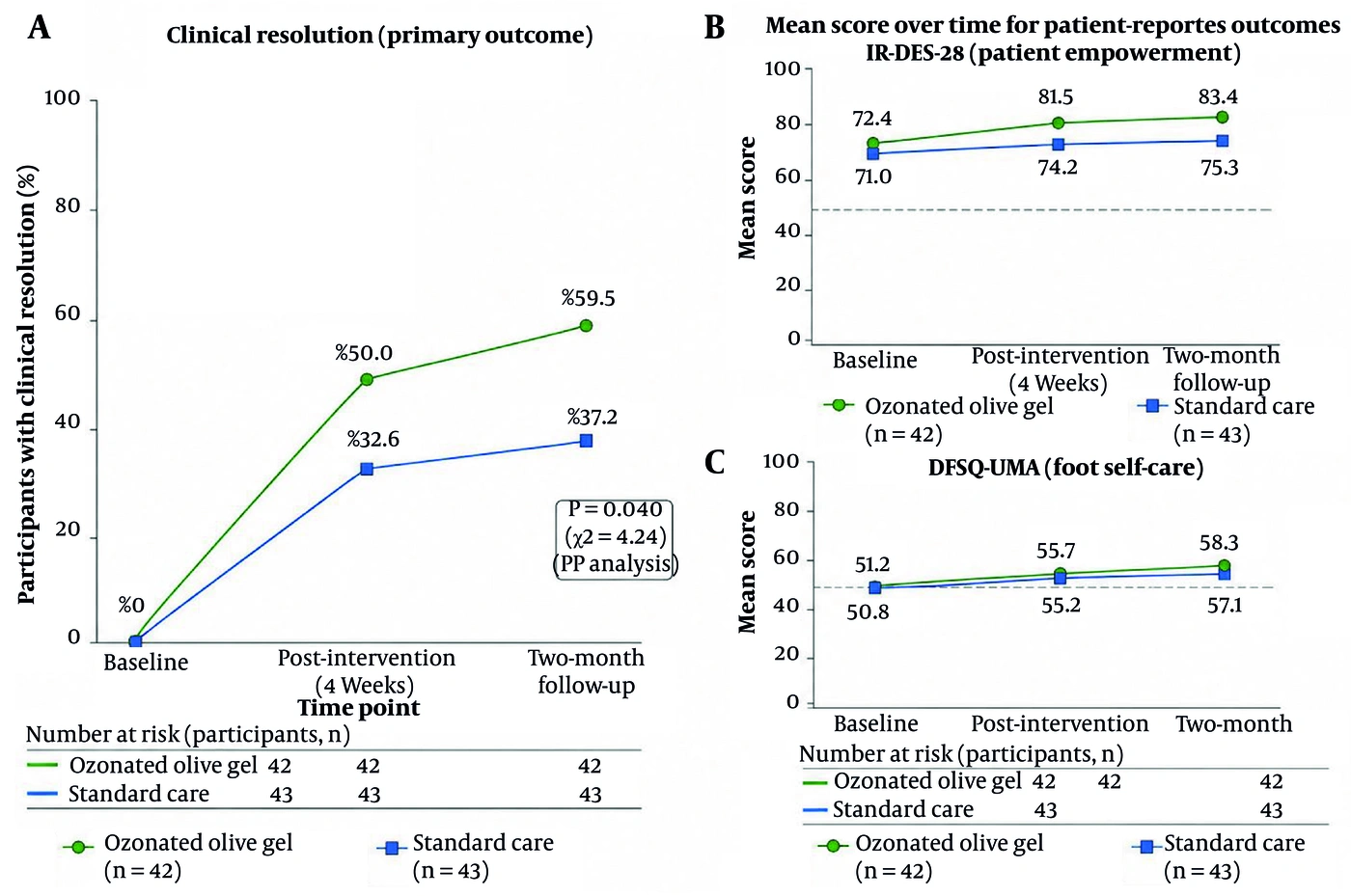
Figure 2.
Changes in clinical resolution and mean scores over time. Panel A shows the proportion of participants achieving clinical resolution of infection at each time point (per-protocol population). Panel B shows mean IR-DES-28 scores (range 28 - 140) over time. Panel C shows mean DFSQ-UMA scores (range 15 - 75) over time. Error bars represent 95% confidence intervals. PP = per-protocol population.
4.4. Mixed-Effects Model Estimates for Patient-Reported Outcomes
To further quantify the longitudinal treatment effects observed in the descriptive analysis, linear mixed-effects models were fitted for both patient-reported outcomes in the ITT population (N = 90) using all available observations under the missing-at-random assumption. The models included fixed effects for group, time, and the group × time interaction, with participant-level random intercepts and an unstructured covariance matrix. The key parameters of interest were the group × time interaction terms, which estimate the differential change from baseline between the intervention and control groups at each follow-up time point. The full model estimates are presented in Table 5. Figure 3 presents the between-group differences from the mixed-effects models.
Table 5.
Linear Mixed-Effects Model Estimates for Patient-Reported Outcomes a
| Outcomes and Fixed Effects | β | SE | 95% CI | df | P-Value |
|---|---|---|---|---|---|
| IR-DES-28 | |||||
| Intercept | 71.02 | 1.29 | 68.48 to 73.56 | 166 | < 0.001 |
| Group (Intervention vs. Control) | 1.38 | 1.82 | −2.21 to 4.97 | 83 | 0.450 |
| Time (Post-intervention vs. Baseline) | 3.18 | 1.17 | 0.88 to 5.48 | 166 | 0.007 |
| Time (Two-month follow-up vs. Baseline) | 4.27 | 1.16 | 1.98 to 6.56 | 166 | < 0.001 |
| Group × Time (Post-intervention) | 4.24 | 1.33 | 1.63 to 6.86 | 166 | 0.001 |
| Group × Time (Two-month follow-up) | 6.47 | 1.34 | 3.85 to 9.09 | 166 | < 0.001 |
| DFSQ-UMA | |||||
| Intercept | 50.81 | 1.15 | 48.54 to 53.08 | 166 | < 0.001 |
| Group (Intervention vs. Control) | 0.42 | 1.62 | −2.77 to 3.61 | 83 | 0.796 |
| Time (Post-intervention vs. Baseline) | 4.42 | 1.09 | 2.27 to 6.57 | 166 | < 0.001 |
| Time (Two-month follow-up vs. Baseline) | 6.28 | 1.10 | 4.12 to 8.44 | 166 | < 0.001 |
| Group × Time (Post-intervention) | 0.13 | 1.24 | −2.30 to 2.57 | 166 | 0.914 |
| Group × Time (Two-month follow-up) | 0.87 | 1.25 | −1.57 to 3.31 | 166 | 0.484 |
a The model includes 85 participants (intervention n = 42, control n = 43) contributing up to 3 observations each. Denominator degrees of freedom (df = 166) are approximated using the Satterthwaite method. The intercept represents the estimated mean baseline score for the control group. The Group effect represents the estimated baseline difference between groups. Time effects represent the mean change from baseline in the control group. Group × Time interactions represent the additional change in the intervention group relative to the control group at each follow-up time point.
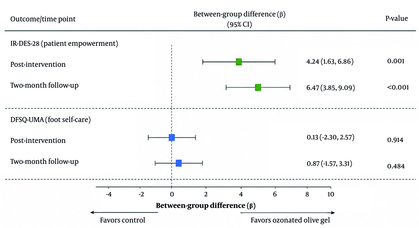
Figure 3.
Forest plot of between-group differences (mixed-effects model estimates)
4.5. Sensitivity and Adjusted Analyses
Sensitivity analyses using baseline-adjusted models yielded findings consistent with the primary longitudinal analyses. Adjustment for age, duration of diabetes, BMI, HbA1c, and baseline infection severity did not materially change the direction, magnitude, or significance of the observed treatment effects. For patient empowerment, the treatment-by-time interaction remained statistically significant after adjustment. In contrast, the treatment-by-time interaction for foot self-care remained nonsignificant. These findings support the robustness of the principal longitudinal conclusions. The full adjusted-model estimates are presented in Table S1 in the Supplementary File.
4.6. Concomitant Treatments
Systemic antibiotic therapy was administered to all participants in both groups according to the study protocol, as all patients had clinically infected diabetic foot ulcers at enrollment. The prescribed regimen consisted of oral ciprofloxacin 500 mg twice daily (BID) and oral clindamycin 300 mg three times daily (TID), administered based on the treating physician's prescription and in accordance with IWGDF/IDSA guideline recommendations for empirical therapy of diabetic foot infections (12). This regimen provides appropriate antimicrobial coverage against common pathogens implicated in diabetic foot infections (including staphylococci, streptococci, and anaerobic organisms) and is consistent with standard clinical practice. The mean duration of antibiotic treatment was 12.4 ± 4.2 days in the intervention group and 13.1 ± 4.8 days in the control group (P = 0.48). Antibiotic adherence was monitored at each clinic visit and was satisfactory in both groups (mean self-reported adherence > 95%). No participants required escalation to intravenous antibiotics or modification of the prescribed antibiotic regimen due to treatment failure or adverse reactions. Debridement was performed in 35 participants (83.3%) in the intervention group and 37 (86.0%) in the control group (P = 0.73), with mean frequencies of 2.8 ± 1.1 and 3.0 ± 1.2 sessions during the 4-week treatment period (P = 0.43). Dressing types were similarly distributed between groups: foam dressings (38.1% vs. 41.9%), hydrocolloid (26.2% vs. 23.3%), alginate (21.4% vs. 18.6%), and gauze (14.3% vs. 16.3%) (P = 0.68). Appropriate offloading was used in 27 participants (64.3%) in the intervention group and 26 (60.5%) in the control group (P = 0.72). No revascularization procedures were performed in either group during the study period. Glycemic medication adjustments were required in 10 participants (23.8%) in the intervention group and 9 (20.9%) in the control group (P = 0.75) (Table 6).
Table 6.
Concomitant Treatments During the Study Period a
| Treatments | Ozonated Olive oil (n = 42) | Standard Care (n = 43) | P-Value |
|---|---|---|---|
| Systemic antibiotics | 42 (100.0) | 43 (100.0) | 1.00 |
| Antibiotic regimen | Ciprofloxacin 500 mg BID + Clindamycin 300 mg TID | Ciprofloxacin 500 mg BID + Clindamycin 300 mg TID | — |
| Antibiotic duration, days | 12.4 ± 4.2 | 13.1 ± 4.8 | 0.48 |
| Antibiotic adherence, % (self-reported) | 96.2 ± 3.1 | 95.8 ± 3.5 | 0.58 |
| Debridement | 35 (83.3) | 37 (86.0) | 0.73 |
| Debridement sessions | 2.8 ± 1.1 | 3.0 ± 1.2 | 0.43 |
| Offloading | 27 (64.3) | 26 (60.5) | 0.72 |
| Glycemic medication adjustment | 10 (23.8) | 9 (20.9) | 0.75 |
| Revascularization procedures | 0 (0.0) | 0 (0.0) | 1.00 |
| Intravenous antibiotic escalation | 0 (0.0) | 0 (0.0) | 1.00 |
a Values are expressed as No. (%) or mean ± SD.
4.7. Adherence and Safety
Adherence to the prescribed once-daily application regimen was satisfactory among participants in the intervention group. A total of six mild adverse events were reported during the study period. In the intervention group, one participant experienced mild transient erythema at the application site (CTCAE grade 1), which resolved spontaneously within 48 - 72 hours without treatment modification or discontinuation. In the control group, three participants experienced mild transient erythema (CTCAE grade 1), and two participants reported mild local pruritus (CTCAE grade 1); all events resolved spontaneously within 48 - 72 hours without pharmacological intervention or treatment modification. All six events were assessed by the treating physician using WHO-UMC causality criteria. The single case of erythema in the intervention group was rated as possibly related to the study intervention. The events in the control group were rated as unlikely related or unrelated to the study intervention, as no topical study product was applied in that group. No local intolerance requiring treatment discontinuation, no systemic adverse events, and no serious adverse events were recorded in either group throughout the follow-up period (through the two-month assessment). No participant required withdrawal because of an adverse reaction. Serious clinical events, including infection deterioration, hospitalization, major surgical intervention, and amputation, were not observed during the study, and no major safety signal was identified. The explicit denominators for adverse-event assessment were all 42 participants in the intervention group and all 43 participants in the control group who completed the study, as well as the 5 participants lost to follow-up (3 intervention, 2 control), for whom no adverse events were reported before withdrawal. The complete adverse-event profile is summarized in Table 7.
Table 7.
Adverse Events and Safety Outcomes During the Complete Study Period (Through Two-Month Follow-Up) a
| Adverse Events | Ozonated olive oil (n = 42) b | Standard care (n = 43) b | Total (n = 85) b | CTCAE Grade | WHO-UMC Causality |
|---|---|---|---|---|---|
| Local events | |||||
| Erythema at application site | 1 (2.4) | 3 (7.0) | 4 (4.7) | Grade 1 | Intervention: Possible; Control: Unlikely/Unrelated |
| Pruritus at application site | 0 (0.0) | 2 (4.7) | 2 (2.4) | Grade 1 | Unlikely/Unrelated |
| Burning sensation | 0 (0.0) | 0 (0.0) | 0 (0.0) | — | — |
| Allergic reaction | 0 (0.0) | 0 (0.0) | 0 (0.0) | — | — |
| Wound deterioration | 0 (0.0) | 0 (0.0) | 0 (0.0) | — | — |
| Systemic events | |||||
| Any systemic adverse event | 0 (0.0) | 0 (0.0) | 0 (0.0) | — | — |
| Serious adverse events (SAEs) | |||||
| Hospitalization | 0 (0.0) | 0 (0.0) | 0 (0.0) | — | — |
| Amputation | 0 (0.0) | 0 (0.0) | 0 (0.0) | — | — |
| Death | 0 (0.0) | 0 (0.0) | 0 (0.0) | — | — |
| Withdrawal due to adverse event | 0 (0.0) | 0 (0.0) | 0 (0.0) | — | — |
a Values are expressed as No. (%). Adverse events were actively collected at each study visit and via telephone follow-up between visits. Severity was graded using CTCAE version 5.0 (grades 1 - 5). Causality assessment was performed using WHO-UMC criteria (certain, probable, possible, unlikely, unrelated). All six reported events were mild (Grade 1), self-limiting, and resolved without pharmacological intervention or treatment discontinuation. No Grade ≥ 2 adverse events were reported.
b Denotes participants who completed the study. The 5 participants lost to follow-up (3 in the intervention group, 2 in the control group) had no adverse events reported prior to withdrawal; therefore, the safety assessment denominators for the pre-withdrawal period were 45 per group.
5. Discussion
This randomized controlled trial investigated the efficacy of topical ozonated olive oil on clinical infection resolution, patient empowerment, and foot self-care behaviors in individuals with DFUs. The findings should be interpreted in the context of substantial methodological limitations, including statistical fragility of the primary outcome, susceptibility to detection bias due to partial blinding, absence of microbiological validation, and inadequate statistical power for the observed effect size. Given these limitations, this study should be considered hypothesis-generating rather than providing evidence of treatment efficacy.
The primary efficacy finding is statistically fragile. Although the addition of ozonated olive oil to standard care was associated with a higher clinical resolution rate at the prespecified two-month primary endpoint in the multiple imputation ITT analysis, this result did not reach statistical significance under the conservative BOCF sensitivity analysis. This dependence on missing-data assumptions indicates that the treatment effect estimate is not robust and should not be interpreted as evidence of efficacy. The best-case/worst-case bounds further illustrate this fragility, with P values ranging from 0.032 to 0.210 depending on assumptions about missing outcomes. The observed effect size (approximately 20 - 22 percentage points) was also smaller than the 25-percentage-point difference assumed in the sample size calculation, indicating that the trial was underpowered to detect clinically meaningful differences. The wide confidence intervals reflect this imprecision. This finding is consistent with the broader literature on wound-care interventions, in which initial single-center trials often report larger effect sizes than subsequent multicenter confirmatory studies (17).
Critically, this study did not include any microbiological endpoints. Wound cultures, bacterial burden quantification, and microbiological eradication were not assessed. Clinical resolution of infection, as defined by the IWGDF/IDSA classification, reflects the resolution of clinical signs and symptoms of inflammation but does not directly demonstrate antimicrobial efficacy. This distinction is well established in the diabetic foot literature, in which clinical improvement frequently occurs without complete microbiological eradication, and vice versa (18). The proposed antimicrobial mechanism of action of ozonated olive oil therefore remains unverified in this study population. Improvements in clinical infection status could have resulted entirely from standard care components, including systemic antibiotics (administered to all participants), debridement, offloading, and glycemic management, without any specific contribution from topical ozonated olive oil. Similar concerns about confounding by concomitant standard care have been raised in other trials of adjunctive topical therapies for diabetic foot infections (19). Any inference of antimicrobial efficacy from these findings would be inappropriate.
The unblinded design represents a fundamental methodological weakness. Although outcome assessors were formally masked and instructed not to inquire about treatment assignment, true blinding could not be guaranteed. Topical oil application may leave residual odor, visible residue, or dressing characteristics that could potentially reveal treatment allocation. Given the subjective nature of clinical infection resolution assessment, this introduces a substantial risk of detection bias that cannot be fully mitigated through statistical methods (20). The primary outcome results should therefore be interpreted with considerable caution, and the possibility that the observed difference reflects detection bias rather than a true treatment effect cannot be excluded. Future trials should consider blinded, centralized photographic assessment by independent experts as the standard for rigorous wound-care research.
A difference in patient empowerment scores was observed between groups, with greater improvement in the intervention group. However, this observation must be interpreted with extreme caution and should not be construed as evidence of a treatment effect. The unblinded design makes patient-reported outcomes particularly susceptible to performance bias, expectancy effects, and the Hawthorne effect. Participants who knew they were receiving an active topical treatment may have reported greater empowerment owing to additional attention, perceived treatment efficacy, or an expectation of benefit, rather than any specific pharmacological effect. No causal inference is warranted from these data. Whether adjunctive topical therapies can influence psychological empowerment in diabetic foot care remains an open question requiring evaluation in rigorously blinded trials with appropriate attention controls.
Foot self-care behaviors (DFSQ-UMA) improved substantially and equally in both groups, with no treatment-specific effect. This lack of a treatment-by-time interaction is consistent with studies suggesting that behavioral habits, such as daily foot inspection and footwear modification, are strongly influenced by the structured education and routine clinical contact inherent in standard DFU management (21). The significant improvement observed in both groups likely reflects the structured foot-care education provided as part of standard care, increased awareness resulting from study participation (Hawthorne effect), and the supportive effect of regular clinical contact (22). The absence of a treatment-specific effect indicates that the addition of ozonated olive oil did not confer a specific advantage in behavioral outcomes beyond what was achieved through standard care and study participation.
The safety profile observed in this trial was favorable, with no serious adverse events and only mild, self-limiting local reactions. However, the small sample size and short follow-up period limit the ability to detect rare or delayed adverse events. Longer-term safety data would be required before any clinical recommendations could be considered.
5.1. Study Limitations
Despite the high retention rate (94.4%), this study has several fundamental limitations that preclude definitive conclusions.
First, the primary outcome is statistically fragile. The treatment effect is significant only under the multiple imputation assumption and loses significance under the conservative BOCF analysis. This sensitivity indicates that the primary finding is not robust and should not be presented as evidence of efficacy. Second, the unblinded design represents a major potential source of detection bias. Despite efforts to mitigate the risks of unblinding, the claim of blinded outcome assessment is internally inconsistent with the acknowledgment that residual odor, visible residue, or dressing characteristics could reveal allocation. True blinding could not be guaranteed, and the primary outcome remains susceptible to detection bias. This is particularly concerning given the subjective nature of clinical infection resolution assessment. Third, the absence of microbiological validation is a major limitation. The intervention’s proposed mechanism is antimicrobial activity, but the study did not include wound cultures, bacterial burden quantification, or any microbiological endpoint. Clinical resolution of infection does not necessarily reflect antimicrobial efficacy, as improvements could result from standard care alone (systemic antibiotics, debridement, offloading). Future trials should incorporate serial wound cultures or molecular microbiological assessments to confirm the antimicrobial mechanism of action and to explore potential effects on biofilm formation and antimicrobial resistance. Fourth, the trial was underpowered for the observed effect size. The actual effect (approximately 20 - 22 percentage points) was smaller than the planned effect (25 percentage points) used in the sample size calculation. The wide confidence intervals for the primary outcome (RR: 1.01 - 2.41 in ITT analysis; risk difference: 0.4 - 40.4 percentage points) indicate imprecision. A post hoc power calculation indicates that a future confirmatory trial would require approximately 130 participants per arm (260 total) to detect a 20-percentage-point difference with 80% power.
Fifth, the patient-reported outcomes are particularly susceptible to bias in the unblinded design. The observed differences in patient empowerment cannot be attributed to a specific treatment effect and may reflect nonspecific effects such as additional attention, expectation of benefit, or social desirability. These findings should be considered exploratory observations only. Sixth, the two-month follow-up period may be insufficient to evaluate long-term outcomes such as ulcer recurrence, sustained behavioral change, or late complications. Longer follow-up periods (at least 6 - 12 months) are recommended in future trials. Seventh, the exclusion of patients with severe systemic infection, critical limb ischemia, and osteomyelitis requiring immediate surgical management limits the generalizability of the findings to more severely ill populations. These exclusions were necessary for ethical and safety reasons. Eighth, the study was conducted in a specific geographic region (Bandar Abbas, southern Iran) with particular demographic, cultural, and healthcare system characteristics. Multicenter trials across diverse geographic and cultural settings are needed to assess the generalizability of these findings.
5.2. Conclusions
In this randomized controlled trial of 90 adults with infected diabetic foot ulcers, adjunctive topical ozonated olive oil was associated with a higher rate of clinical infection resolution at the two-month follow-up in the multiple imputation ITT analysis. However, this finding was not statistically significant under conservative missing-data assumptions (BOCF, P = 0.056) and is therefore fragile and dependent on analytical choices. The trial was underpowered for the observed effect size, the outcome assessment was susceptible to detection bias due to partial blinding, and no microbiological validation was performed. Patient empowerment showed greater improvement in the intervention group, but this observation is highly susceptible to performance and expectancy biases and should be considered exploratory only. Foot self-care improved similarly in both groups without a treatment-specific effect.
These preliminary findings do not support clinical implementation and should be considered hypothesis-generating only. Larger, adequately powered, multicenter trials with rigorous blinding, active placebo controls, and microbiological endpoints are required before any clinical recommendations can be made.