


1. Background
Following the emergence of COVID-19 in late December 2019 in Wuhan, China, with a cluster of unexplained pneumonia cases, the World Health Organization (WHO) declared it a worldwide pandemic in March 2020. The infection caused by severe acute respiratory syndrome coronavirus 2 (SARS-CoV-2) primarily affects the respiratory system, leading to various symptoms that range from mild to life-threatening conditions such as acute respiratory distress syndrome (ARDS) or even death (1-3).
Despite extensive efforts to control the pandemic, the disease persists, and its progression remains unpredictable. Numerous studies have investigated the prognostic factors of severe COVID-19, including demographic characteristics (age, male sex, habitual profile, and preexisting comorbidities), physical characteristics (respiratory failure, hypotension, hypoxemia, tachycardia), laboratory factors (elevated serum procalcitonin, myocardial injury markers, white blood cell count (WBC), erythrocyte sedimentation rate (ESR), C-reactive protein (CRP), lactate, creatinine, D-dimer, abnormal liver function tests, and decreased lymphocyte count and albumin), and radiological factors such as consolidative infiltrate and pleural effusion, along with a high sequential organ failure assessment (SOFA) score (4).
Chest computed tomography (CT) is a non-invasive diagnostic procedure with high sensitivity for the early diagnosis of patients with SARS-CoV-2. It has been recognized that chest CT scans have higher sensitivity than real-time reverse transcription-polymerase chain reaction (RT-PCR) tests for COVID-19 diagnosis, although some symptomatic patients may have normal chest CT results. In addition, chest CT is considered an easily accessible imaging modality for diagnosing pneumonia in many healthcare centers (5, 6).
2. Objectives
Given the rapid and easy access to chest CT and the promising evidence supporting its role in determining the prognosis of COVID-19, we aimed to evaluate the association between the severity of COVID-19 and the pattern of pulmonary involvement observed in chest CT scans.
3. Methods
3.1. Study Designs
This cross-sectional study was conducted at Imam Hossein Hospital in Tehran, Iran, which served as a regional tertiary COVID-19 center during the pandemic. The study was carried out over two months, from March 6, 2020, to May 6, 2020. The ethics committee at Shahid Beheshti University of Medical Sciences and Health approved the study (ethics code: IR.SBMU.RETECH.REC.1399.107). Additionally, all patients provided informed consent prior to participating in the study. If a patient was unable to provide consent, their family was responsible for giving consent on their behalf.
3.2. Study Population
Patients were recruited from the COVID-19 ICU at Imam Hossein Hospital. The inclusion criteria for selecting eligible participants were patients over 18 years of age with a confirmed diagnosis of severe to critical COVID-19 during the study period, based on the polymerase chain reaction (PCR) test. Patients were excluded if they did not undergo chest CT, had a poorly qualified chest CT scan, had a previous history of respiratory disease, were heavy smokers, or did not consent to participate.
3.3. Severity of COVID-19
The WHO has classified the severity of COVID-19 into four categories: Mild, moderate, severe, and critical. Severe COVID-19 is characterized by the presence of dyspnea, a respiratory rate of 30 or more breaths per minute, blood oxygen saturation of 93% or less, a partial pressure of arterial oxygen to fraction of inspired oxygen (PaO2/FiO2) ratio of less than 300, and/or lung infiltrates greater than 50% within 24 - 48 hours. In contrast, critical COVID-19 is defined by respiratory failure, septic shock, and/or multiple organ dysfunctions, according to WHO recommendations (7).
3.4. Interventions and Data Collection
Patients' electronic medical records were reviewed to collect information about their characteristics, including age, gender, habitual profile, comorbidities, and clinical presentation. This information was obtained using a questionnaire. All patients then underwent high-resolution CT (HRCT) of the lungs, with subsequent chest CT scans performed using the low-dose method.
3.5. Chest Computed Tomography Findings
An expert radiologist reported the CT findings using the international standard method of the Fleischner Society and a review of the viral pneumonia literature. A semi-quantitative scoring system was designed to analyze morphologic lung involvement. The five lobes of the lungs—right upper lobe (RUL), right middle lobe (RML), right lower lobe (RLL), left upper lobe (LUL), and left lower lobe (LLL)—were visually reviewed for ground-glass opacity (GGO) and consolidation twice and independently. Each pattern was then scored from 0 to 5 based on the percentage of involvement: (0) indicates no involvement, (1) indicates involvement of 5% or less, (2) indicates 5 - 25%, (3) indicates 26 - 49%, (4) indicates 50 - 75%, and (5) indicates involvement of 76% or more. The scores for each lobe were added together to determine the total scores for GGO and consolidation. The PI score was calculated by summing either the total GGO scores and total consolidation scores or the GGO and consolidation scores for all five lobes (8).
3.6. Statistical Analysis
Statistical analysis was performed using SPSS version 26 (IBM, NY, USA). All continuous variables were presented as mean and median, while all categorical variables were expressed as percentages and numbers. Pearson's chi-square test was used to compare the CT findings of patients. A P-value of less than 0.05 was considered statistically significant, and the confidence interval was set at 95%.
4. Results
4.1. Study Population and Clinical Data
All patients hospitalized in the intensive care unit (ICU) were analyzed retrospectively between March and May 2020. We included 47 patients in our study who met the inclusion criteria. Of these, 27.6% were female, and 72.4% were male, with a mean age of 62 ± 14 years. At the time of the CT scan, three out of 47 patients were intubated, and after the CT examination, 34 patients required invasive mechanical ventilation. However, ten patients were managed through non-invasive methods.
Thirty-nine patients (82.9%) had comorbidities, with hypertension being the most common. In 44 patients (93%), laboratory blood tests showed elevated levels of CRP greater than 5.9 mg/L, with a mean CRP level of 76 mg/L, and lymphocytopenia (lymphocyte count < 1.1 × 10³/mm3) was observed in 31 patients (66%). Table 1 provides a summary of the baseline characteristics of the patients.
Table 1.Baseline Clinical and Laboratory Characteristics of Patients with Severe to Critical COVID-19 Patients
| Variables | Values |
|---|---|
| Patient demographics | |
| Total patients | 47 (100) |
| Male | 34 (72.4) |
| Female | 13 (27.6) |
| Age (y); range | 25 - 87 |
| Age (y); mean ± SD | 62 ± 14 |
| Laboratory test | |
| C-reactive protein (mg/L; normal range 0.00 - 5.90) | |
| Increased | 44 (93) |
| Normal | 1 (2) |
| CRP mean level | 76 |
| White blood cell (× 10³/mm³; normal range 4.4 - 11) | |
| Increased | 14 (30) |
| Decreased | 5 (10) |
| Normal | 28 (60) |
| Lymphocyte (× 10³/mm³ normal range 1.5 - 3.0) | |
| Increased | 4 (9) |
| Decreased | 31 (66) |
| Normal | 12 (25) |
| Neutrophil (× 10³/mm³, normal range 1.5 - 8.0) | |
| Increased | 30 (64) |
| Decreased | 0 (0) |
| Normal | 17 (36) |
Abbreviation: CRP, C-reactive protein.
4.2. Chest Computed Tomography Scan Findings
COVID-19 CT features and their distribution are shown in Tables 2. and 3. All patients had bilateral lung involvement. As shown in Table 2 and Figure 1, the lower lobes had a higher injury score compared to the upper and middle lobes (2.9 vs. 2.68 and 2.31, respectively, with P = 0.001 and P = 0.009). The semi-quantitative score for the LLL was 2.9, compared to 2.45 for the LUL (P = 0.001). The highest score was observed in the RLL compared to the middle and upper lobes. Right lower lobe injury with a score of 3 was found in 25 cases (53.2%), while LLL involvement with a score of 3 was found in 20 cases (42.5%). Involvement of all five lobes was observed in 42 patients (89%). The minimum score was 3.25, and the maximum score was 21.25 (CI: 12 - 15; mean, 13.25).
Table 2.The Semi-Quantitative Score of Each Lung Lobe in Patients with Severe to Critically Ill COVID-19 Patients
| Variables | Score 1 | Score 2 | Score 3 | Score 4 | Score 5 |
|---|---|---|---|---|---|
| RUL | 4 (1 - 8) | 10 (6 - 15) | 18(12 - 24) | 12 (8 - 16) | 0 |
| RML | 6 (3 - 9) | 15 (10-20) | 14 (9 - 20) | 4 (1 - 8) | 3 (1 - 6) |
| RLL | 2 (0 - 5) | 9 (5 - 14) | 25 (19 - 30) | 6 (3 - 10) | 4 (2 - 6) |
| LUL | 7 (4 - 10) | 14 (9 - 19) | 12 (7 - 18) | 10 (6 - 14) | 1 (0 - 4) |
| LLL | 5 (2 - 8) | 10 (6 - 15) | 20 (14 - 26) | 9 (5 - 13) | 3 (1 - 6) |
Abbreviations: RUL, right upper lobe; RML, right middle lobe; RLL, right lower lobe; LUL, left upper lobe; LLL, left lower lobe.
Table 3.The Computed Tomography Features of Patients with Severe to Critical COVID-19
| Features | No. (%) | 95% CI |
|---|---|---|
| CT appearance | ||
| GGO | 26 (55) | (24 - 26) |
| Consolidation | 14 (30) | (9 - 19) |
| Equal GGO/consolidation | 9 (19) | (5 - 14) |
| Reverse halo | 1 (2) | (0 - 4) |
| Cavity | 0 | 0 |
| Nodule | 0 | 0 |
| Pleural effusion | 10 (21) | (6 - 15) |
| Pericardial effusion | 3 (6) | (1 - 7) |
| Distribution | ||
| Peripheral | 35 (74) | (32 - 36) |
| Perihilar | 1 (2) | 2 (0 - 4) |
| Peribronchovascular | 36 (76) | (34 - 36) |
Abbreviations: CT, computed tomography; GGO, ground-glass opacity; RUL, right upper lobe; RML, right middle lobe; RLL, right lower lobe; LUL, left upper lobe; LLL, left lower lobe.
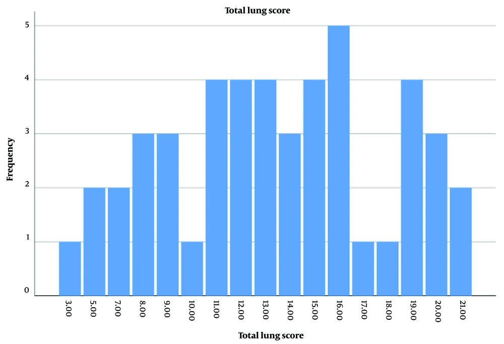
Figure 1.
The semi-quantitative score of each lung lobe in patients with severe to critically ill COVID-19 patients
In addition, the most frequently observed features were GGO in 26 patients (55.31%), consolidation in 14 patients (30%), and pleural effusion in 10 patients (21%). Among those with pleural effusion, seven patients had mild pleural effusion, three had moderate pleural effusion (with only one case of bilateral pleural effusion), and pericardial effusion was seen in 3 cases (6.4%) (Table 3). Furthermore, peripheral distribution, peribronchovascular distribution, round opacity, crazy-paving pattern, linear opacity, and non-specific pattern were observed in 74%, 76%, 23%, 8%, 8%, and 34% of the patients, respectively (Table 4).
Table 4.The Computed Tomography Features of Patients with Severe to Critical COVID-19
| CT Features Sub-analysis | Patients | |
|---|---|---|
| Number of involved lobes | Values | 95% CI |
| 0 | 0 | NA |
| 1 | 0 | NA |
| 2 | 2 (4.3) | (0 - 6) |
| 3 | 1 (2.1) | (0 - 4) |
| 4 | 4 (8.5) | (1 - 9) |
| 5 | 40 (85) | (38 - 40) |
| Frequency of lobe involvement | ||
| RUL | 44 (94) | (39 - 46) |
| RML | 42 (90) | (37 - 45) |
| RLL | 46 (98) | (43 - 47) |
| LUL | 44 (94) | (39 - 46) |
| LLL | 47 (10) | (45 - 47) |
| GGO pattern | ||
| Crazy paving | 4 (8) | (1 - 8) |
| Round opacities | 11 (23) | (7 -14) |
| Linear opacities | 4 (8) | (1 - 8) |
| Nonspecific | 16 (34) | (14 - 16) |
Abbreviations: CT, computed tomography; GGO, ground-glass opacity; RUL, right upper lobe; RML, right middle lobe; RLL, right lower lobe; LUL, left upper lobe; LLL, left lower lobe.
5. Discussion
The COVID-19 pandemic is considered a global concern. Despite numerous efforts to manage the pandemic, including efforts to achieve herd immunity through vaccination, the pandemic remains ongoing as the virus continues to mutate. While COVID-19 primarily presents with mild respiratory symptoms, SARS-CoV-2 can affect any organ system, leading to a wide range of clinical presentations. Additionally, patients may experience rapid progression to acute lung injury and ARDS when accompanied by septic shock. Severe multi-systemic involvement in COVID-19 is associated with a poor prognosis, leading to significant morbidity and mortality if not promptly diagnosed. Therefore, timely diagnosis and treatment of COVID-19 are crucial (1-3). In this context, chest CT can contribute to the early diagnosis of COVID-19. Moreover, certain radiological features might have prognostic value and play a significant role in effectively managing critically ill patients.
Despite the abundance of literature on chest CT findings in COVID-19 patients, few studies have evaluated the relationship between the extent of lung disease and the severity of COVID-19 using a semi-quantitative scoring system. The present study demonstrated an association between the extent of changes observed in chest CT scans of COVID-19 patients and the severity of the disease. In cases of COVID-19 with symptoms and signs of respiratory distress, the most common chest CT findings were GGO (55%) and consolidation (30%). Bilateral involvement was observed in all patients, with the most frequently affected areas being the peribronchovascular distribution (76%) and peripheral distribution (74%). Most patients had pulmonary involvement in all five lobes (minimum score: 3.25; maximum score: 21.25; CI: 12-15; mean: 13.25).
Supporting our findings, Andrea Leonardi et al. identified a mixed pattern of GGO and consolidation as the most frequently observed pattern in critically ill patients. They established a cut-off of 23.0% lung involvement as a marker of critical COVID-19, with 96% sensitivity and 96% specificity (9). Similarly, a report on 25 critical COVID-19 patients showed a significantly higher prevalence of chest CT scan scores, consolidation, and number of involved lobes in critically ill patients (10). Likewise, a study on 60 COVID-19 patients found sub-pleural ground-glass opacities and consolidation to be the most common radiologic findings. Furthermore, there was a significant difference in semi-quantitative scores between the early, progressive, and severe stages of the disease (11). A recent study on 739 COVID-19 patients, including 72 admitted to the ICU, suggested that a total pulmonary involvement (PI) score or PI density score of ≥ 8 (15) could serve as a marker for ICU admission and severe COVID-19 (12).
In terms of laboratory findings, our results showed that 93% of patients had elevated CRP levels, and lymphocytopenia was evident in 66% of the patients, consistent with the existing literature (6, 13, 14).
Taking all considerations into account, a semi-quantitative analysis of chest CT findings could serve as a valuable technique to facilitate the timely diagnosis of severe COVID-19. However, our study has several limitations. Firstly, we analyzed only a small number of severe to critical COVID-19 patients without including a control group of mild to moderate COVID-19 cases. As a result, it is not possible to draw a firm conclusion. Secondly, we primarily focused on radiological parameters and did not measure key laboratory parameters that reflect impaired respiratory physiology. Additionally, we did not analyze the association between laboratory and radiologic findings and severe COVID-19. Thirdly, we did not consider the potential role of confounding factors, such as comorbidities, age, and sex, on the severity of COVID-19. More extensive studies that include all COVID-19 patients and consider all prognostic factors are needed to better understand the utility of the semi-quantitative scoring system as a prognostic marker for COVID-19.
5.1. Conclusions
The present study revealed that patients with bilateral and diffuse lung involvement, who obtained higher scores on their initial chest CT scans, were more likely to experience severe respiratory disease. Our results suggest that chest CT can play a crucial role in identifying patients at risk of respiratory failure in the early stages of the disease, facilitating timely and preemptive treatment.


