1. Background
Drawing diagrams is not restricted to learning anatomy and is an integral part of various arenas of medicine (1). Visual explanation of concepts influences learning, retention, and recall (2, 3). Therefore, medical undergraduates are encouraged to draw diagrams to explain both simple and complicated concepts (4, 5). However, first-year medical students are not trained and sufficiently skilled in drawing diagrams. No formal curricular elements introduces medical students to drawing diagrams. In learning human anatomy, students are compelled to draw diagrams, which is rather confusing and incomprehensible. Starting from the first courses of human anatomy, medical students are compelled to draw diagrams, (6) and their summative and formative assessments often rely on their skills in drawing diagrams.
Although drawing diagrams is considered the mainstream learning method of anatomy, curricular evaluations should be focused on the competency of drawing skills in medical students without considering their initial artistic and drawing abilities. Consequently, these students often resort to unscientific methods of drawing, including the outline tracing of diagrams and asking for the help of artists in drawing. Although such practices are rampant in numerous medical colleges, no efforts are made to quantify the problem or take meaningful steps toward addressing the issue; this leads to the perpetuation of drawing incompetency. Therefore, efforts must be dedicated to simplifying the drawing process, encouraging students to draw scientifically accurate diagrams, and imbibing practices of drawing diagrams to explain various concepts (7). The basic principles upon which such efforts may be implemented are as follows:
- Internalization of the concepts leading to reflective diagrams
- Sequencing the drawing process
- Following the fundamentals of drawing diagrams (e.g., central positioning, symmetry, and use of proper colors) to highlight the core theme of the illustration
In anatomy learning, drawing well-defined two-dimensional depictions of proper anatomical structures is spatially associated with proper labelling (4) The drawing method of choice could improve the overall comprehension of musculoskeletal anatomy (8). Sequence graphics could be used to address the lacunae of drawing skill development in medical undergraduates (9). In a sequence graphics illustration, the components of diagrams appear in the form of a sequence, and students build the diagram sequentially (10). In this technique, emphasis is placed on the core component of each diagram, and other non-core or accessory components are of lesser or secondary importance. Sequence graphics properly orients the learner toward the core components of a diagram, as well as the sequential addition of the components, thereby improving overall learning, retention, and recall (10).
Exclusive life drawing sessions focusing on artistic theories, practical applications, and associations of anatomical concepts have delivered a positive and reinforcing environment among poor learners (11). The observe-reflect-draw-edit-repeat sequence has been reported to enhance the anatomy knowledge of first-year undergraduates in a medical school in the United Kingdom (12). Moreover, lower visual processing and poor conceptual basis are known to be associated with errors in drawing (13).
2. Objectives
The present study aimed to use the method of sequence graphics to evaluate medical undergraduates in terms of drawing moderately complex diagrams.
3. Methods
This quasi-experimental pilot study was conducted on six medical students, and four moderately complex diagrams were evaluated regarding the usefulness of sequence graphics.
3.1. Study Setting
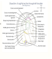
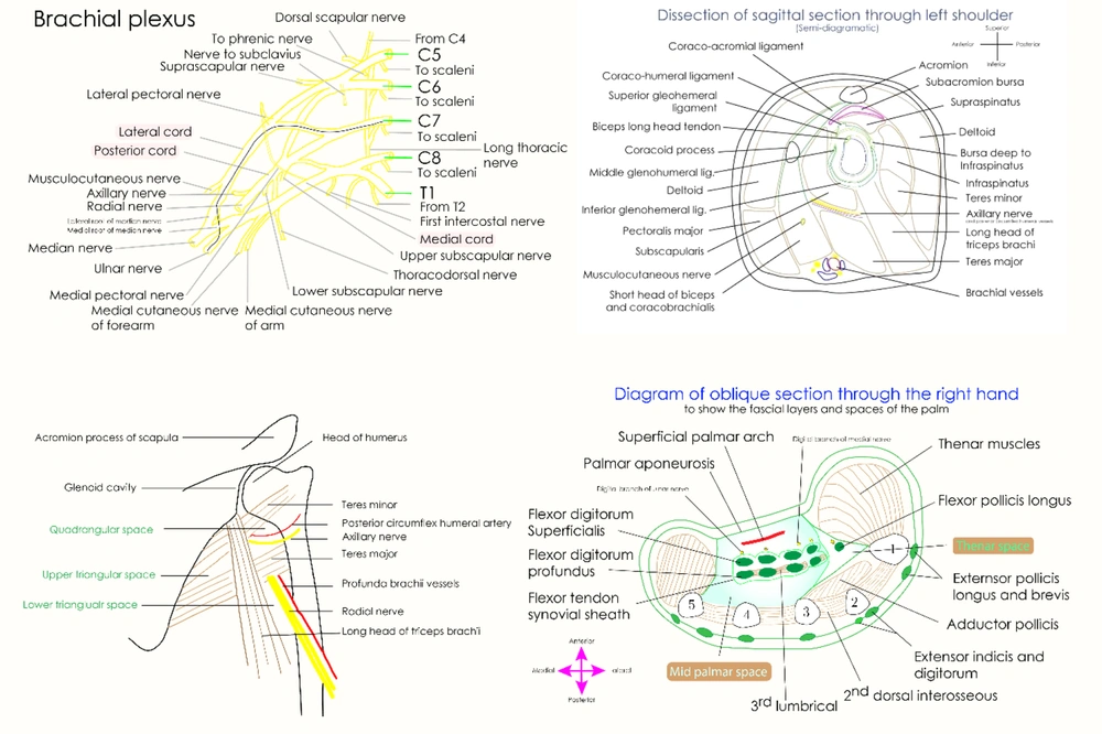
In total, six students were asked to draw four moderately complex diagrams in gross anatomy. The selection of the diagrams was validated by two subject experts (Figure 1). The students were given A4-sized papers to draw the diagrams and were free to choose the colors. Relevant concepts to the diagrams were incorporated into routine interactive lecture sessions and cadaveric dissection. The proposal was approved by the Institutional Ethics Committee (letter number: AEC/Rev/2016/14 dated 30-11-2016) before starting the study.
3.2. Diagram Selection
The four selected diagrams were schematic representations of the brachial plexus, sagittal section of the shoulder joint showing the relations, spaces around the scapula, and section of the hand showing the thenar and midpalmar space (Figure 1). Core and accessory components were identified before asking the students to draw the diagram (Table 1). The diagrams were selected given their distinct core and accessory components. As the diagrams had more than four components to be represented by the students, they were designated as moderately complex diagrams. Two experienced faculty members of the department validated the selected diagrams before the drawing exercise. The selected diagrams were useful for understanding the anatomical basis of specific clinical conditions. Each diagram had two core component and a minimum of two accessory components. Drawing the diagrams required a fundamental understanding of relevant anatomical core concepts, 3D orientation, and clinical applicative knowledge prior to drawing.
| No. | Diagram | Core Component | Accessory Components |
|---|---|---|---|
| 1 | Brachial plexus (schematic representation) | Roots, trunks, divisions, and cords, branches from cords | Branches from roots and trunks, differential shading of anterior and posterior divisions, showing C7 component in ulnar nerve |
| 2 | Sagittal section of shoulder joint showing relations | Glenoid labrum in center surrounded by four rotator cuff muscles, relation of neurovascular structures below joint | Coracoacromial arch, subdeltoid bursa, deltoid muscle, and pectoralis major muscle |
| 3 | Spaces surrounding scapula | Highlighting boundaries of three named spaces, contents of these triangles | Orientation of fibers of teres major and triceps with each other, thickness of radial nerve and axillary nerve |
| 4 | Section of hand showing thenar and midpalmar space | Boundaries and contents of thenar and midpalmar space | Superficial and deep tendon relations, position of adductor pollicis, and relative sizes of thenar and hypothenar eminences |
3.3. Conventional Drawing
The students were asked to draw four diagrams consecutively during the dissection hour, and three routinely used textbooks were used as references (14-16). Following that, the students were asked to trace the diagrams in the textbooks after studying each. Notably, the students were free to draw the diagram by whichever method they were comfortable with, and the exercise was labelled as conventional drawing.
3.4. Sequence Graphics
On the next day, the students drew the same diagrams using the sequence graphics method. The intended diagrams were created using the Inkscape software (open-source vector graphic editor) (17) and exported as scalable vector graphics (SVGs). The SVG images were animated to appear consecutively using CSS and JavaScript, (18) and the final animated sequence was exported as a video file. At the next stage, the video was projected on a large screen, and the students were asked to follow the video to draw the diagrams. The video would be paused and replayed if necessary. The students synchronously traced the diagrams along with the video in order to achieve the same end results simultaneously. Several parameters were noted for each diagram throughout the drawing process to evaluate the diagrams. To evaluate these parameters, the drawing process was video-recorded by the six student volunteers, and the videos and final images were carefully assessed in terms of the following parameters by two evaluators. Notably, the mean value of the findings was considered for the final analysis.
The parameters considered for evaluating the drawn diagrams were as follows:
- Time taken to draw the diagram
- Whether the students labelled the diagram before drawing the next diagram
- Whether the students added navigation marks to each diagram
- The first component drawn and its added sequence
- Drawing centered and symmetrical diagrams
- Representation of the core component
- Use of proper colors (black for the bones and outlines, red for arteries, blue for veins, yellow for the nerves, brown for the muscles, and green for the tendons)
Data obtained from the mentioned parameters were compiled and compared with the continuous variables and expressed as mean and standard deviation. In addition, immediate feedback was obtained from the students based on a five-point scale using a form consisting of six simple questions.
4. Results
4.1. Conventional Drawing
The students initially decided to draw the diagrams by themselves based on three provided references. Out of 24 instances, 22 were the same for all the students, and only the sagittal section of the shoulder (student 4) and hand spaces (student 3) differed among the subjects. Table 2 shows the pre-defined parameters during and after drawing the diagrams using the conventional method. While using the conventional drawing method, the students took significantly more time to complete the diagram, the outcomes were not uniform, and several missing core and accessory components were detected (Table 3).
| Parameter | Student 1 | Student 2 | Student 3 | Student 4 | Student 5 | Student 6 |
|---|---|---|---|---|---|---|
| 1. Brachial Plexus (Schematic Representation) | ||||||
| Time (second) | 580 | 490 | 660 | 720 | 920 | 840 |
| Labelling | No | Yes | No | Yes | Yes | No |
| Navigation | No | No | No | No | Yes | No |
| First | Roots | Roots | Lateral cord | Lateral cord | Roots | Roots |
| Next | Trunk | Trunk | Division | Divisions | Trunk | Branches |
| Last | Branches | Branches | Branches | Branches | Branches | Branches |
| 2. Sagittal Section of Shoulder Showing Relations | ||||||
| Time (second) | 920 | 940 | 840 | 920 | 880 | 900 |
| Labelling | No | No | Yes | Yes | Yes | Yes |
| Navigation | No | No | No | No | Yes | No |
| First | Skin | Coracoid process | Skin | Joint line | Glenoid cavity | Skin |
| Next | Deltoid | Rotator cuff | Deltoid | Rotator cuff | Rotator cuff | Coracoid process |
| Last | Rotator cuff | Non-rotator cuff | Rotator cuff | Non-rotator cuff | Non-rotator cuff | Rotator cuff |
| 3. Spaces Surrounding Scapula | ||||||
| Time (second) | 660 | 540 | 600 | 630 | 720 | 680 |
| Labelling | No | No | Yes | Yes | Yes | No |
| Navigation | No | No | No | No | Yes | No |
| First | Skin | Skin | Bones | Thenar muscles | Bones | Thenar muscles |
| Next | Thenar muscles | Thenar muscles | Superficial arch | Bones | Thenar muscles | Hypothenar |
| Last | Spaces shading | Long tendons | Long tendons | Skin | Skin | Bones |
| 4. Section of Hand Showing Thenar and Midpalmar Spaces | ||||||
| Time (second) | 740 | 560 | 620 | 820 | 700 | 760 |
| Labelling | No | Yes | No | Yes | Yes | Yes |
| Navigation | No | No | No | Yes | No | No |
| First | Metacarpal bones | Thenar eminence | Thenar eminence | Palmar skin | Dorsal skin | Palmar skin |
| Next | Interossie muscles | Palmar skin | Hypothenar eminence | Doral skin | Palmar skin | Dorsal skin |
| Last | Skin and outline | Tendons | Tendons | Space boundaries | Bones | Tendons |
a Sequence and complete labelling; depiction of navigation marks to indicate direction and provide orientation; first, next, and last components or parts of drawn diagram.
| Diagram | Core Component | Accessory Components | Centeredness | Symmetry |
|---|---|---|---|---|
| Brachial plexus | 3/6 | 2/6 | 3/6 | NA |
| Sagittal section of shoulder joint | 2/6 | 2/6 | 2/6 | 2/6 |
| Spaces surrounding scapula | 3/6 | 3/6 | 3/6 | NA |
| Spaces of hand | 4/6 | 2/6 | 2/6 | 2/6 |
Abbreviations: NA, not applicable.
4.2. Diagram Evaluation
Out of 24 diagrams (4 diagrams by each of the 6 students), 20 were drawn with proper colors. Among four diagrams, two had unconventional colors to show the tendons in the section of the hand and sagittal section of the shoulder joint diagram. The other two diagrams had a different shade of yellow and other colors to represent various components of the brachial plexus. Table 2 shows a compilation of other parameters.
4.3. Specific Comments on Each Diagram
Regarding the brachial plexus, two students failed to show the posterior cord behind the other cords, and four students did not depict a larger radial nerve, median nerve, and ulnar nerves compared to the other nerves. Out of six students who were closely observed, two students started their diagrams from the lateral cord rather than the roots. Three students made minor mistakes in labelling the branches of the cords, while improper labelling was not considered in the evaluation of the diagrams as it was not previously defined in the methodology of the research.
In terms of the sagittal section of the shoulder joint, four students failed to show the proper relation of the rotator cuff to the glenoid cavity. In addition, two students failed to draw the coracoacromial arch above the glenoid cavity with a subacromial bursa, and one student illustrated the pectoralis major anteriorly and deltoid posteriorly while in reality, this deltoid section is appreciated both anteriorly and posteriorly. Two students also depicted larger posterior muscles than anterior structures, and one student drew smaller branchial vessels below the joint.
Regarding the spaces surrounding the scapula, two students drew the teres major above the long head of the triceps. Moreover, the spatial relations of three triangles were not as per the anatomical relations in the diagrams of two students. Three students illustrated the radial nerves as simple lines, and two students drew the entire scapula through coracoid and acromion processes to show the spaces related to its lateral border. The researchers only considered the lateral border and the humeral shaft to represent the spaces in question.
In terms of the spaces of the hand, two students failed to represent the midpalmar and thenar spaces in their diagrams. In addition, three students failed to illustrate the flexor pollicis longus tendon, and four students failed to draw the median nerve branches. As these structures are of particular clinical significance, they are often noted separately. Notably, three students depicted equal heights in drawing the thenar and hypothenar eminences.
4.4. Sequence Graphics
All the students traced the diagrams in tandem with the recorded sequence graphics videos. The videos would be paused and replayed an average of six times each, and the mean duration of the videos was 95 seconds. All the students started and ended drawing at the same time. Specifically regarding the brachial plexus diagram, the time taken was 330 seconds. The students started the diagram at the left corner of the page with the representation of roots C5-T1 and continued by forming the trunks, divisions, and cords. The branches of the cords were drawn later and sequentially labelled. All the diagrams uniformly had radial nerve as the largest and posterior cord behind the other structures.
The time taken for drawing the sagittal section of the shoulder joint was 610 seconds. The rotator cuff muscles were uniformly shown around the glenoid labrum, and there was a proper gap between the coracoacromial arch and glenoid accommodating the subacromial bursa. Overall, symmetry and centeredness were maintained in all the diagrams.
The students took 340 seconds to draw the diagram for the spaces surrounding the scapula. All the diagrams properly illustrated the special relation of the teres major, humerus shaft, radial nerve, and contents of these triangles. On the other hand, none of the students traced the entire scapula and shaded the bones, muscles or neurovascular structures entirely.
The time taken for drawing the spaces of the hand was estimated at 420 seconds. All the students uniformly depicted the core and accessory components in the section of the hand, with branches of the median nerve and tendon of the flexor pollicis longus shown in appropriate places. The students started the diagram from the sections of the metacarpal bones, respecting the fact that the drawing diagrams must be started with the skeletal elements forming the skeletal system of the body. Furthermore, a clear distinction was observed between the sizes of the thenar and hypothenar eminences. Overall, sequence graphics resulted in uniform, centered, labelled, large diagrams with defined core and accessory components, which were drawn in lesser time compared to conventional drawing.
4.5. Immediate Feedback of the Drawing Exercise
All the students agreed that sequence graphics imparted better drawing skills (mean score: 4.8/5) and felt that it was easier to draw the diagrams (score: 5/5). Moreover, all the students stated that sequence graphics could lead to better diagram drawing in formative and summative assessments (score: 4.5/5). The students liked to have sequence graphics for diagram drawing of gross anatomy in the future (score: 4.6/5). They also opined that sequence graphics saved time (score: 5/5) and believed that sequencing the diagrams made the concepts clear (score: 4.5/5).
5. Discussion
Sequence learning is inherent to human cognitive psychology, and a smooth and simple transition is taking place between conscious and unconscious behavioral tendencies (19). The concept of sequential learning, which is also referred to as the ‘learning arc’, has been extensively used in various educational areas by properly determining the expectations of students in terms of learning. (20). Sequence graphics presents an orderly fashion for the construction of diagrams, thereby helping medical students appreciate and represent the core components associated with the high-risk relations of anatomical structures.
Medical undergraduate learning is based on the performance of students in cognitive-based tests (21). However, the assessment and encouragement of the innate artistic aptitude of these students have a small scope. When students start formal education with human anatomy, their ability to draw diagrams and illustrate various concepts in a pictorial or schematic manner is extensively tested (22). Unfortunately, outdated or updated curricula are not inclined toward enhancing these essential skills in future clinicians. Physicians with better communication skills (both verbal and schematic) are highly successful in their career across the globe (23). Although drawing improves the visual, kinesthetic, and auditory learning styles of medical students and is a preferred method of learning, (8) efforts have rarely been dedicated to imparting drawing skills in medical undergraduates. Reports in this regard confirm the positive impact of drawing on the overall wellbeing of medical students, (11) as well as the concept retention (8) and clinical application of basic medical sciences (24). Notably, these studies are mainly focused on the perceptions of the participants. In the present study, we attempted to objectively evaluate the process of drawing and its outcomes. Automated diagram evaluation using UML (Unified Modeling Language) classes based on the similarities to the reference diagrams are in use among higher education technologies (25). However, such automation may not be applicable to medical illustrations.
Anatomy drawing skills may be viewed as stepwise progression from imitation, precision, articulation, and naturalization (26). Using sequence graphics, we intended to create a significant construct of drawing skills in the medical students. Sequence graphics is aimed at imparting this essential and rarely addressed skill in medical undergraduates. The fundamental principle of sequence graphics is the time-tested method of initially constructing concepts with skeletal elements, followed by adding other details to the anatomical images. As a result, the concept is anatomically and diagrammatically comprehensive given the meaningful sequential additions of the details to the central element. Notably, efforts are made to sequence clinically relevant details over essential anatomical structures, and a video format adds to visual memory and long-term retention.
In the present study, the primary and secondary output measures clearly indicated that the sequence graphics of drawing resulted in better diagrams of moderately complex anatomical illustrations. Therefore, the researchers intend to replicate the study with more participants. Due to logistic issues, the parameters noted in this study (especially the time taken to draw the diagrams and representation of the first, second, and last elements of the diagrams) may not be replicated in large groups of participants. Nevertheless, the objective evaluation of the drawn diagrams is possible.
A similar study conducted by Viveka and Sudha demonstrated that sequence graphics with a pre-defined key component contributed to the drawing skills of participants (9). In the present study, a similar principle was considered to create the sequence graphics videos. As videos generally have more visual impact on the long-term memory, this method seems to translate to better drawing skills.
5.1. Limitations of the Study
One of the limitations of our study was that robust methods of the objective evaluation of medical illustrations were not available in the literature. In addition, simple extrapolation of the principles of art and drawing to anatomical images may not be completely justifiable. In this pilot study, indigenous and primitive evaluation methods were employed to assess diagram drawing, and more refined methods may add an objective angle to diagram evaluation.
5.2. Conclusion
According to the results, sequence graphics resulted in uniform, centered, labelled, and large diagrams with defined core and accessory components, which were drawn in lesser time compared to conventional drawing.

