Reagents and Abs
Zymosan isolated from Saccharomyces cerevisiae (Dectin-1 agonist), was purchased from Sigma-Aldrich. Specific Abs against p38 and phospho-(Thr180/Tyr182)-p38 were purchased from Cell Signalling Technology (Beverly, MA). Anti–phospho-(Ser345)-p47phox, Ab p47phox were purchased from Santa Cruz. Biotechnology Dimethyl sulfoxide (DMSO) was purchased from Sigma. ELISA kits for IL-6, IL-10, TNF-α, and IL-12p40 were purchased from BD Pharmingen (Franklin Lakes, NJ).Also, standard wedelolactone was purchased from Sigma Aldrich. Additionally, Chloroform, methanol, toluene, acetone, formic acid, ethyl acetate, and acetonitrile were purchased from Merk.
Cell culture
Primary bone marrow derived-macrophages (BMDMs) were isolated from Swiss mice. The BMDMs were differentiated for 5–7 days in the medium containing Dulbecco’s modified Eagle’s medium (DMEM, Gibco-BRL, Gaithersburg, MD) with 10% L929 cell-conditioned medium (as a source of M-CSF), 10% heat- inactivated fetal
bovine serum (FBS) (Gibco-BRL), 1mM sodium pyruvate, 50 U/mL penicillin, 50 µg/mL streptomycin and 5x10⁵ M ß-mercaptoethanol, sodium pyruvate, non-essential amino acids, penicillin G (100 IU/mL), and streptomycin (100 µg/mL).
Plant material
The Eclipta prostrata (L). L. was collected in Thai Binh provinces, Vietnam and identified (TTC01, TTC02, TTC03) by the Institute of Ecology and Biological Resources Vietnam Academy of Science and Technology (VAST), Hanoi.
Isolation of wedelolactone
The leaves of
Eclipta prostrata (L) L. were extracted with methanol by Soxhlet, as previously described (
12). The solvent was removed and the residues were suspended in water separately and heated on steam bath below 80 °C. After filtrating, the aqueous phase was partitioned with ethyl acetate. The organic phase was filtered and the solvent was evaporated to reach light brown powder. The powder was subjected to fractionation by column chromatography on silica gel (glass column (1×80) cm), and the final silica length was 50 cm. The mobile phase was prepared from chloroform: methanol (70: 30). The crud and partial purified extract were subjected TLC, the solvent system (toluene: acetone: formic acid: 11: 6: 1). The purified sample and standard wedelolactone were measured by HPLC, as previously described (
13). The purified sample and standard wedelolactone were diluted with methanol 10 mL. The solution was filtered through a 0.45 μm membrane filter before HPLC analysis and the injection volume was 20 µL. HPLC was performed using a Shimadzu LC-20AT pump system equipped with a Shimadzu SPD-M20A Photodiode array detector and the detection wavelength set at 351 nm. The separation was obtained using a reversed-phase column (cosmosil 5C18-AR-II 4.6 mm x 250 mm, 5μm) with the mobile phase, acetonitrile: water (35: 65 % v/v). The pH of mobile phase was adjusted to 3.2. The experiment was performed at room temperature and the flow was fixed at 1.0 mL/min.
Experimental animals
All experiments described in this study were performed using Swiss mice. All animal-related procedures and care were reviewed and approved by the national institute of hygiene and epidemiology (Vietnam). These mice used for the zymosan challenge were 8–10 weeks old (18-22 g). These experimental groups were matched age and sex.
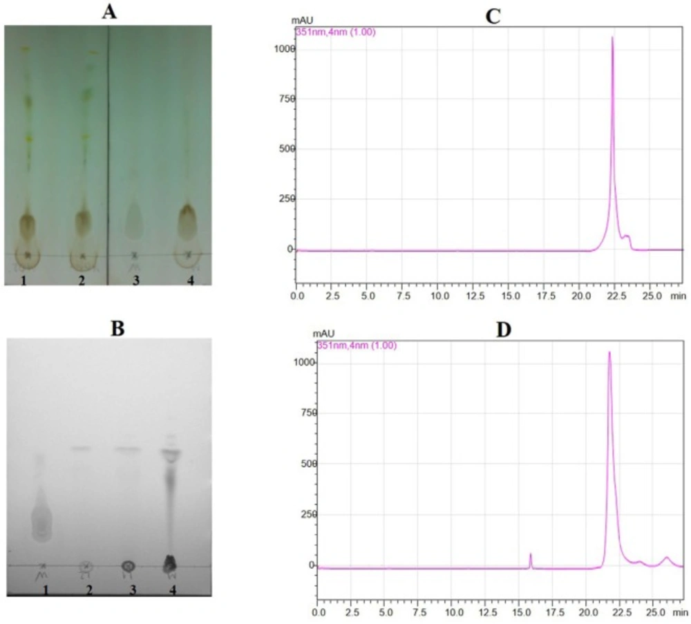
Thin layer chromatography for crude sample and partial purified sample, and HPLC for standard wedelolactone and sample wedelolactone. (A) Transfer crude sample extract in TLC (1,2,4: cude sample; 3 for standard wedelolactone). (B) Transfer partial purified sample in TLC (1: standard wedelolactone; 2, 3, 4: partial purified sample). (C) HPLC figure for standard wedelolactone. (D) HPLC Figure for sample wedelolactone
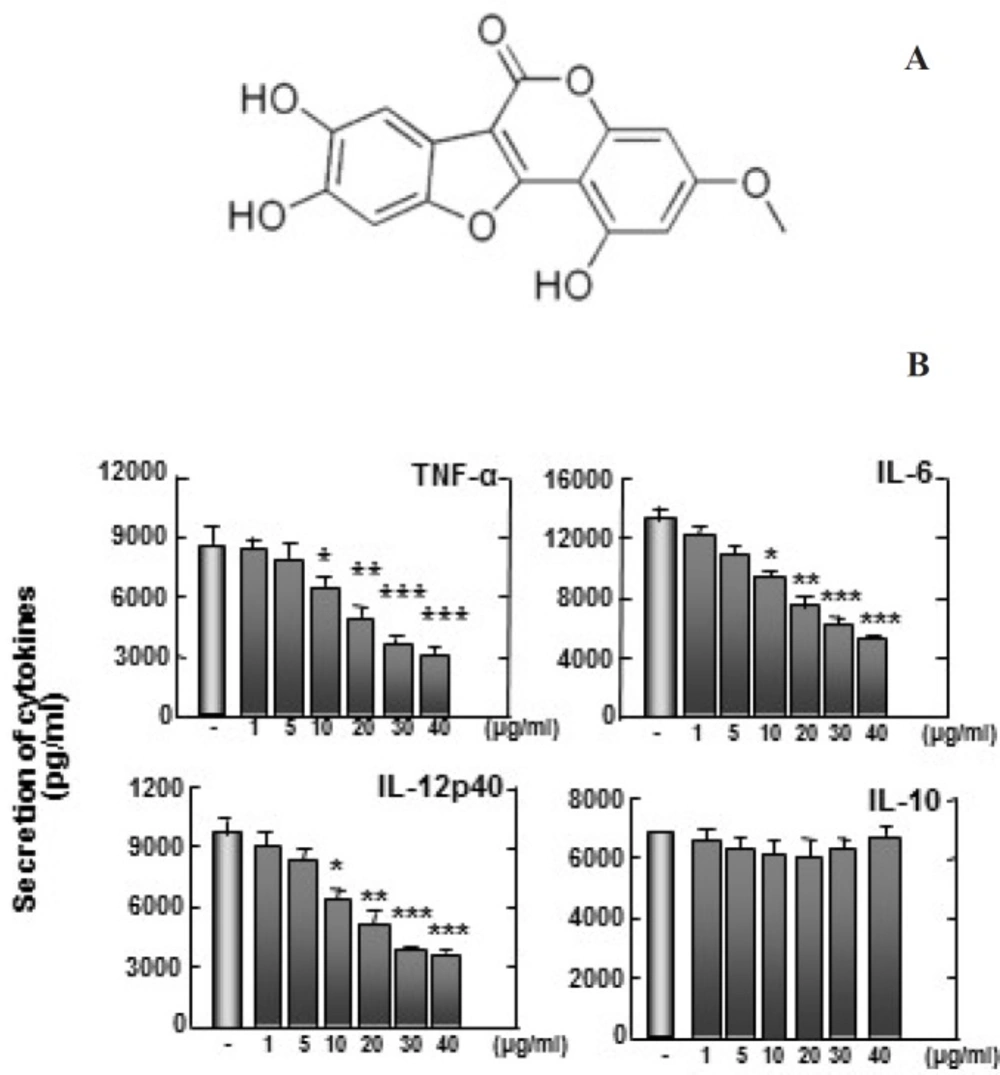
(A) Structure of wedelolactone. (B) Zymosan-induced TNF-α, IL-6, and IL-12p40 production are inhibited by wedelolactone, but not IL-10. Bone marrow-derived macrophages (BMDMs) from mice were treated with wedelolactone at concentrations of 0, 1, 5, 10, 20, 30, 40 µg/mL in DMSO 0.1% in 45 min before stimulation with zymosan. Supernatants were harvested 18 h after stimulation with 100 µg/mL zymosan to induce inflammation. Concentrations of IL-10, IL-12p40, IL-6, and TNF-α in the culture supernatants were determined by ELISA. The results are expressed the mean ± SD of five experiments. Significant differences (**P < 0.01; ***P < 0.001) compared with cultures without wedelolactone are indicated
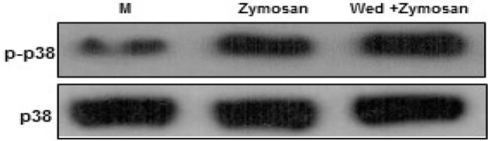
Wedelolactone negatively regulates zymosan-induced phosphorylation of p38 MAPK activation in BMDMs. The cells were pretreated solvent control (0.1% DMSO) or wedelolactone (30 µg/mL) for 45 min. Then the cells were stimulated with zymozan (100 µg/mL) for 30 min. The cells were harvested after 1 hour lysed with ice-cold lysis buffer, and subjected to western blot analysis to detect the activation of mitogen-activated protein kinases (MAPKs) (p38). M, media control; Wed, wedelolactone. The results are representative of three experiments
Wedelolactone inhibits the zymosan-induced production of ROS in BMDMs
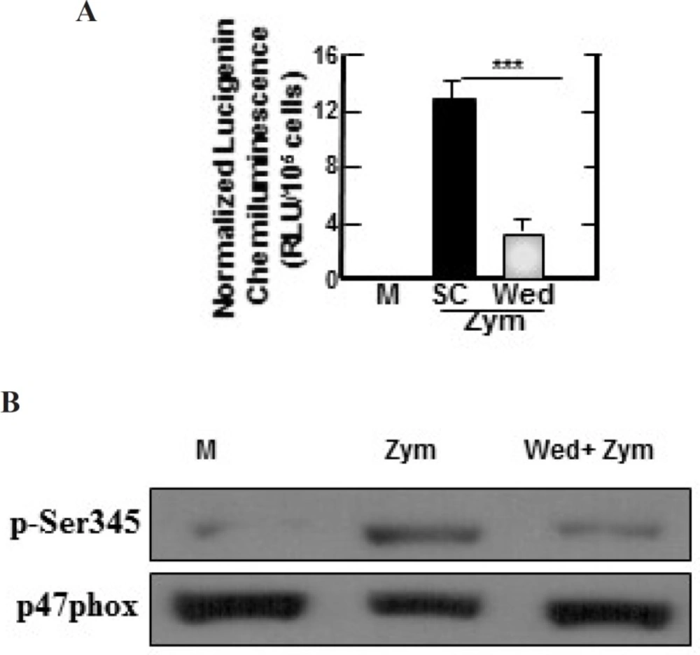
Zymosan-induced ROS-generating NADPH oxidase activities, phosphorylation of p47phox in BMDMs are inhibited by wedelolactone. (A) BMDMs were pretreated with solvent control (0.1% DMSO) or wedelolactone (30 µg/mL) for 45 min. Then the cells were stimulated with zymozan (100 µg/mL) for 60 min. NADPH oxidase activity in the cells were measured as described the materials and methods. M, media control; SC, solvent control; Wed, wedelolactone, Zym, zymosan. The results are expressed the mean ± SD of three experiments. (***, P < 0.001) compared with cultures without wedelolactone are indicated. (B) The cells were pretreated with solvent control or wedelolactone (30 µg/mL) for 45 min. Then the cells were stimulated with zymozan (100 µg/mL) for 15 min. The cells were lysed and analyzed by SDS-PAGE and immunoblotting with anti–phospho-Ser345–p47phox Ab (p-Ser345) or anti-p47phox Ab (p47phox). M, media control; Wed, wedelolactone; Zym, zymosan. The results are representative of three experiments
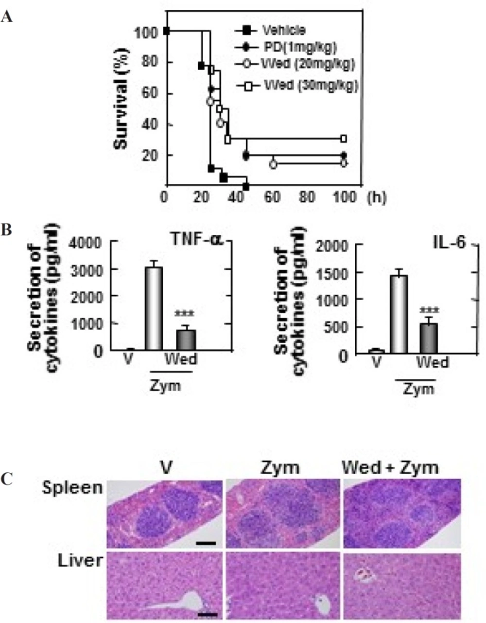
Wedelolactone protects mice against the zymosan-induced inflammatory responses. (A) Wild-type (WT) mice (n = 25 per group) were given wedelolactone (20 mg/kg) or (30 mg/kg) or vehicle or prednisone (1 mg/kg) orally for 24 h before intraperitoneal injection by zymosan (2 mg/kg). Viability was assessed every 5 h for the first 40 h and every 10 h thereafter. Wed, wedelolactone, PD, prednisone. (B) Serum levels of TNF-α, IL-6 were measured in mice that had or had not been given wedelolactone (30 mg/kg) or vehicle by using ELISA at 18 h after zymosan (2 mg/kg) injection. V, vehicle; Zym, zymosan,; Wed, wedelolactone. The data are expressed the mean ± SD of three experiments. Statistical differences (***P < 0.001) compared to the mice were not given wedelolactone are indicated. (C) Sections of spleen and liver from mice were injected with zymosan (2mg/kg) for 24 h after giving administration of wedelolactone (30 mg/kg) or vehicle. H&E staining was performed (scale bars: upper, 100 µm). V, vehicle; Zym, zymosan; Wed, wedelolactone. The images are representative of sections from five mice per group
Enzyme-linked immunosorbent assay
BMDMs were treated as indicated and processed for analysis by sandwich ELISA, as previously described (
14). The levels of cytokines secreted by cell culture and serum were analyzed by ELISA reagent (BD Pharmingen). All assays were performed as recommended by the manufacturers.
Western blotting
BMDMs were treated as indicated and processed for analysis by western blotting, as previously described (
15). The membranes were developed by a chemiluminescence assay (ECL; Amersham-Pharmacia).
Measurement of intracellular ROS
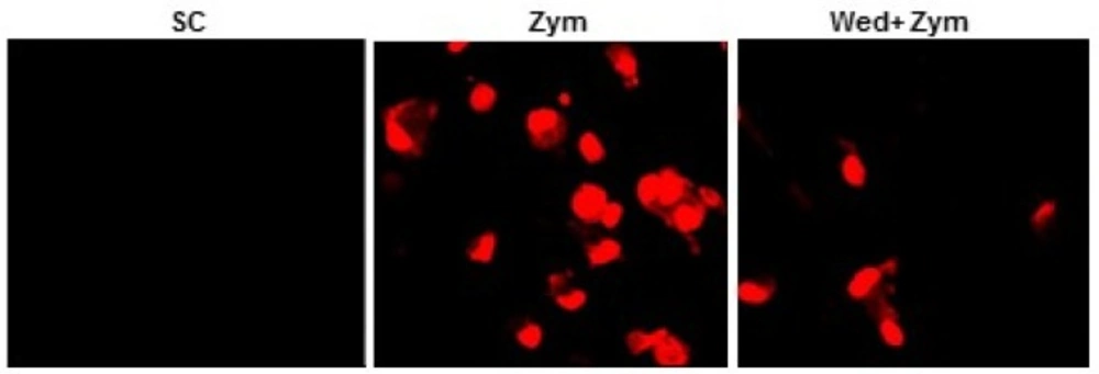
Intracellular superoxide levels were measured as previously described (
15). Briefly, BMDMs were treated (60 min) with zymosan after incubating with or without wedelolactone for 45 min. Then, the cells were incubated with either 2 µM DHE (Calbiochem) for 15 min at 37°C in 5% CO
2. The cells were examined by laser-scanning confocal microscopy (LSM 510).
Determination of NADPH oxidase activity
NADPH oxidase activities were evaluated by lucigenin chemiluminescence assay (5x10
-6 mol/L lucigenin, Sigma) in the presence of its substrate NADPH (10
-4 mol/L, Sigma) as described previously (
15).
Experimental animals and zymosan-induced sepsis model
Mice were injected intraperitoneal (i.p) by zymosan (2 mg/g of body weight; Sigma) was diluted in sterile phosphate-buffered saline (PBS). Wedelolactone was dissolved in a mixture of dimethyl sulfoxide (DMSO; Sigma): polyethylene glycol (PEG; Sigma) 400: distilled water (DW) (1: 4: 5). The mice were orally administered wedelolactone (20 mg/kg, 30 mg/kg) or prednisolone (1 mg/kg). The same amount of DMSO: PEG400: DW (1: 4: 5) mixture were orally administered to control group (vehicle). The mice were monitored for 5 days post-injection. The survival of mice was assessed by very 5 h for the first 40 h and every 10 h thereafter as previously described (
15).
Histological analysis
Histological analysis was used haematoxylin and eosin as described previously (
15)
Statistical analysis
For statistical analysis, data obtained from independent experiments are presented as mean ± SD and were analyzed by the Student’s t-test with a Bonferroni adjustment or ANOVA for multiple comparisons. The differences were considered significant at P < 0.05.