Repeated Administration of Mercury Intensifies Brain Damage in Multiple Sclerosis through Mitochondrial Dysfunction

Author(s):
Farzad KahriziFarzad Kahrizia, Ahmad SalimiAhmad Salimia, b, c, Farshid NoorbakhshFarshid Noorbakhshd, Mehrdad FaiziMehrdad Faizia, Freshteh MehriFreshteh Mehria, Parvaneh NaserzadehParvaneh Naserzadeha, c, Nima NaderiNima Naderia,*, Jalal PourahmadJalal Pourahmada,*
aDepartment of Pharmacology and Toxicology, Faculty of Pharmacy, Shahid Beheshti University of Medical Sciences, Tehran, Iran.
bDepartment of Pharmacology and Toxicology, School of Pharmacy, Ardabil University of Medical Science, Ardabil, Iran.
cStudents research committee, School of Pharmacy, Shahid Beheshti University of Medical Sciences, Tehran, Iran
dDepartment of Immunology, Faculty of medicine, Tehran University of Medical Sciences, Tehran, Iran.
Corresponding Authors:
*

IJ Pharmaceutical Research:Vol. 15, issue 4; 834-841
Published online:Oct 31, 2016
Article type:Original Article
Received:Jan 31, 2016
Accepted:May 31, 2016
How to Cite:Kahrizi F, Salimi A, Noorbakhsh F, Faizi M, Mehri F, et al. Repeated Administration of Mercury Intensifies Brain Damage in Multiple Sclerosis through Mitochondrial Dysfunction. Iran J Pharm Res. 2016;15(4):e125234. doi: https://doi.org/10.22037/ijpr.2016.1931

Abstract

In this study we investigated the additive effect of mercury on the brain mitochondrial dysfunction in experimental autoimmune encephalomyelitis (EAE) model. Experimental animals (female C57BL/6 mice) are divided into four groups (n = 8); control, Hg, EAE, EAE with Hg. EAE model of MS induced by injecting myelin oligodendrocyte glycoprotein (MOG). Neurobehavioral alterations are recorded and then mice were sacrificed at day 28 and brain mitochondria were isolated and mitochondrial toxicity parameters including mitochondrial swelling, reactive oxygen species (ROS) formation, collapse of mitochondrial membrane potential (MMP) and cytochrome c release were measured. Our results showed that repeated treatment of mercury following induction of EAE in mice significantly increased the neurobehavioral scores, as well as mitochondrial toxicity through ROS formation, mitochondrial swelling, collapse of MMP and cytochrome c release. Our findings proved that repeated exposure with mercury accelerates progression of MS through mitochondrial damage related to oxidative stress and finally apoptosis.

Introduction

Mercury is a heavy metal that has known as a toxicant in living organisms and exposure to mercury compounds continue to be prevalent and significant health risks to human (1). Approximately one million tons of metallic Hg has been extracted from ores during the past five centuries (2). Original routes of human exposure to Hg are environmental contaminant, industrial usage and health care. Multiple sclerosis is a chronic and progressive autoimmune disease in the central nervous system (CNS) of young adults, with over 2.5 million individuals affected worldwide (3). MS has almost unknown etiology and no effective cure at the time being. it`s treatment is supportive and symptomatic (4). Mitochondria extensive dysfunction and concomitant oxidative stress can cause many neurodegenerative diseases such as multiple sclerosis (5). There are many study that show relation between mercury exposure and MS for example mercury-containing amalgam may exert an important risk for patients with autoimmune diseases (6). Mercury concentrations in the cerebrospinal fluid and blood serum of MS patients is higher than healthy individuals (7) .It has already been demonstrated that inhibition of myelin production can be resulted from toxic effect of mercury on human oligodendrocytes (8).
In this study for evaluation of relationship progression of MS and repeated exposure of mercury we used EAE model to induce MS in C57BL/6 mice, then neurobehavioral symptoms observed and recorded. Finally We sacrificed mice at day 28 following EAE induction and then brain mitochondria were isolated to examine mitochondrial damage parameters such as mitochondrial swelling, ROS formation, collapse of MMP and cytochrome c release.

Material and Methods

Animals
All experiments were performed on female C57BL/6 mice (Razi Institute, Karaj, Iran) weighing 20-25 g (12 weeks).The animals were kept under a 12 h light/dark cycle in a temperature controlled (22. ± 2 °C) environment with free access to food and water ad libitum. All studies and animal care procedures were accomplished in accordance with the local ethics committee for animal experimentation and in compliance with guidelines of Shahid Beheshti University of Medical Sciences.
Induction of EAE and experimental design
EAE model for MS was induced in mice with MOG35–55 peptide emulsified in complete Freund’s adjuvant (CFA) using Hooke kits (Hooke Laboratories, Lawrence, MA, USA) according to the manufacturer’s instruction. All drugs and their respective vehicles were injected intraperitoneally. The intact (no EAE) group received CFA and pertussis toxin without MOG. Mercury sulfides suspended in carboxymethyl cellulose (CMC) then it exposed to mice oral daily (1 mg/Kg/day) for 7 days from the onset of EAE induction. (9).
The following groups were used in this study: control group received CFA and pertussis toxin without MOG, EAE group, mercury sulfide group, and EAE + mercury sulfide group. The number of animals in each group were 8(n=8).
Neurological assessment
Mice were daily observed to record behavioral and neurological signs until day 28 after immunization. The signs of EAE were scored protocol of Stromnes et al. (10).
Mitochondrial preparation
At 28th day after induction EAE mice were sacrificed and brain and spinal cord mitochondria were isolated using differential centrifugation (11). Mitochondria were prepared freshly for each experiment and used within 4 h of isolation.
Determination of mitochondrial ROS level
The mitochondrial ROS measurement was performed using the fluorescent probe DCFH-DA. The fluorescence intensity of DCF was measured using Shimadzu RF-5000U fluorescence spectrophotometer at an excitation wavelength of 488 nm and emission wavelength of 527 nm (12, 13).
Determination of the MMP
Mitochondrial uptake of the cationic fluorescent dye, rhodamine123, has been used for the estimation of mitochondrial membrane potential. The mitochondrial fractions (0.5 mg protein/mL) were incubated with 10 µM of rhodamine 123 in MMP assay buffer. The fluorescence was monitored using Shimadzu RF-5000U fluorescence spectrophotometer at the excitation and emission wavelength of 490 nm and 535 nm, respectively (14, 15) .
Determination of mitochondrial swelling
Briefly, isolated mitochondria were suspended in swelling buffer (70 mM sucrose, 230 mM mannitol, 3 mM HEPES, 2 mM tris phosphate, 5 mM succinate and 1 µM of rotenone). The absorbance was measured at 549 nm at 15 min time intervals with an ELISA reader (Tecan, Rainbow Thermo and Austria) (13, 16).
Cytochrome c release assay
The concentration of cytochrome c was determined through using the Quantikine Rat/Mouse Cytochrome c Immunoassay kit provided by R&D Systems, Inc. (Minneapolis, Minn.) according to manufacturer’s instruction (15).
Statistical analysis
The results are expressed as mean ± SEM and were analyzed using Graph Pad
Prism (version 5, Graph pad Software Inc.). A one-way or two-way analysis of variance (ANOVA) followed by Bonferroni’s tests were used for multiple comparison. For all statistical analyses, p<0.05 was considered significant.

Results

EAE scoring
As shown in Figure 1 behavioral and neurological signs were increased in EAE induced group which repeatedly received Hg. In the Hg2+treated group, there were also some behavioral and neurological signs.
Measurement of ROS generation
As shown in Figure 2. ROS significantly (P<0.05) increased in the brain mitochondria isolated from (EAE + Hg2+) group compared with EAE group. ROS were also significantly (P<0.05) increased in EAE group compared with control group.
Determination of mitochondrial membrane potential
As shown in Figure 3. MMP significantly decreased in the brain mitochondria isolated from EAE + Hg group compared with EAE group.
Measurement of mitochondrial swelling
As shown in Figure 4. collapse of optical absorbance at 540 nm which is consistent with mitochondrial swelling, was assayed. Our results showed that of repeated administration of mercuric sulfide significantly enhanced swelling in the brain mitochondria isolated from (EAE + Hg).
Cytochrome c release
As shown in Figure 5. there is significant enhancement in the release of cytochrome c from brain mitochondria in the (EAE + Hg) group compared with EAE group.

Discussion

Using EAE scoring test we proved that, mercury causes behavioral, neuromuscular, sensorimotor disturbances in EAE + Hg treated mice compared to Hg group (Figure 1). Our results showed that in Hg accelerated behavioral dysfunction in EAE model. Moreover there are many previous studies that indicate relationship between MS and mercury. Mercury is a heavy metal that exerts acute and chronic toxic effects on the human body including the nervous and immune systems (17). One of the main pathways of mercury toxicity is damage to mitochondrial function (18). Malfunction of mitochondria has been associated to neurodegenerative diseases (19). The functionality of mitochondrial preparations is very useful in the study of the processes underlying pathologies of these diseases. It has already proven that use of isolated mitochondria is a very powerful tool in the study of pathologies of many neurodegenerative (20). Results of ROS measurement in isolated mitochondria showed that ROS formation was significantly increased in EAE + Hg+2 group (Figure 2). Following the rise of reactive oxygen species formation and consecutive oxidative damage to mitochondria, mitochondrial functions are impaired. Our findings regarding mitochondrial membrane potential collapse (Figure 3.) and mitochondrial swelling (Figure 4.) in EAE + Hg group proved this hypothesis. Previous studies with mercury have also shown increased damage through ROS formation. Our results suggest in this animal model, substantial ROS produced by mercury increases and accelerates neural damage compared to EAE group. The increased mitochondrial ROS formation can cause oxidation of a lipid membrane which consequently results in disruption of the mitochondrial membrane and subsequently the collapse of mitochondrial membrane potential (MMP) and cytochrome c release. (21). A number of studies have reported mitochondrial defects in MS. We also showed that in EAE model mitochondrial damages increased which with repeated exposure with mercury these damage significantly increased.
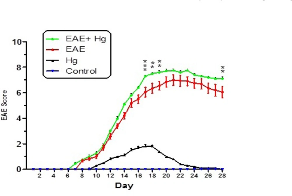
Behavioral and neurological signs in mice experimental groups. Values were presented as score of signs as described in materials and methods (n = 8). ** represents P &lt; 0.01 significant difference, *** represents P &lt; 0.001 significant difference, compared to EAE induced mice group.
Figure 1

Behavioral and neurological signs in mice experimental groups. Values were presented as score of signs as described in materials and methods (n = 8). ** represents P < 0.01 significant difference, *** represents P < 0.001 significant difference, compared to EAE induced mice group.
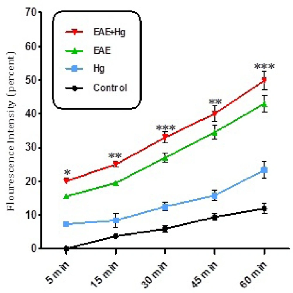
ROS formation. Exposure to mercury (Hg) intensified ROS formation in EAE + Hg group compared to EAE group. Values have been presented as percent ROS formation (n = 8). *** represents significant difference between control and EAE groups, also $$$ represents significant difference (P &lt; 0.001) between EAE and EAE+Hg2+ groups
Figure 2

ROS formation. Exposure to mercury (Hg) intensified ROS formation in EAE + Hg group compared to EAE group. Values have been presented as percent ROS formation (n = 8). *** represents significant difference between control and EAE groups, also $$$ represents significant difference (P < 0.001) between EAE and EAE+Hg2+ groups
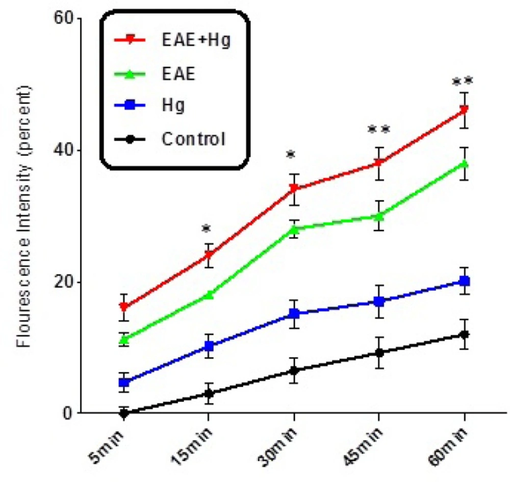
Decline of MMP. Decline of MMP significantly decreased in EAE + Hg group compared to EAE group. Values expressed as percent of MMP collapse (n = 8). *** represents significant difference between control and EAE groups, also $$$ represents significant difference (P &lt; 0.001) between EAE and EAE + Hg groups
Figure 3

Decline of MMP. Decline of MMP significantly decreased in EAE + Hg group compared to EAE group. Values expressed as percent of MMP collapse (n = 8). *** represents significant difference between control and EAE groups, also $$$ represents significant difference (P < 0.001) between EAE and EAE + Hg groups
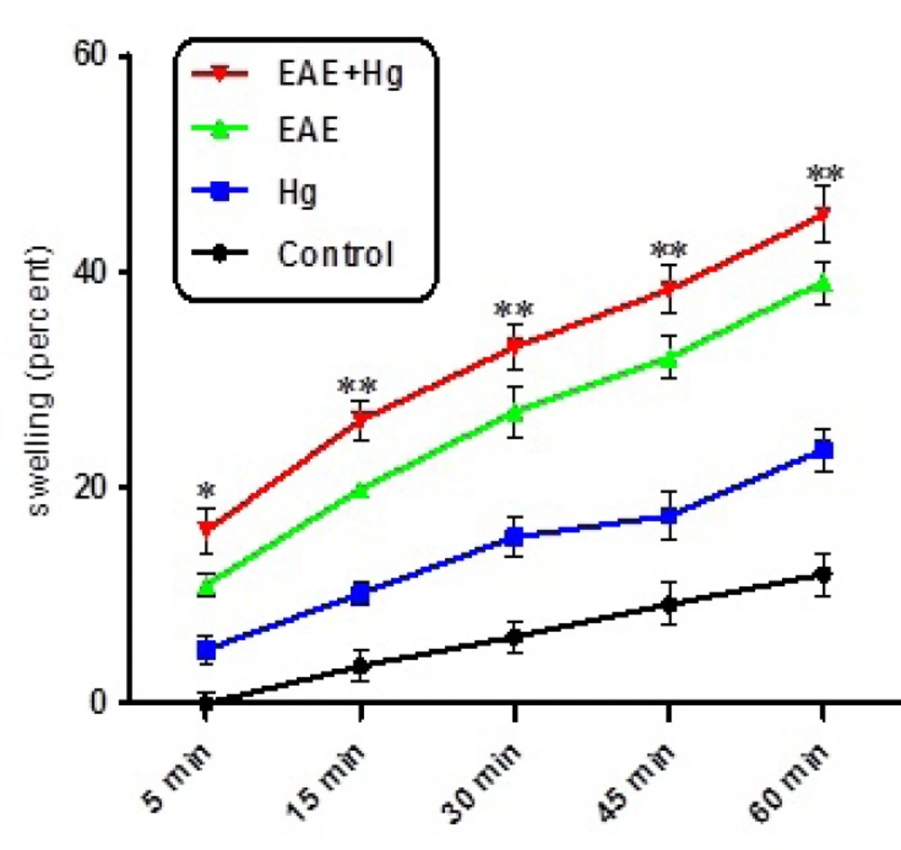
Mitochondrial swelling. Hg enhanced mitochondrial swelling in EAE + Hg group. Values have been presented as decreasing absorbance at 450 nm (n = 8). *** represents significant difference between control and EAE groups, also $$$ represents significant difference (P &lt; 0.001) between EAE and EAE + Hg groups
Figure 4

Mitochondrial swelling. Hg enhanced mitochondrial swelling in EAE + Hg group. Values have been presented as decreasing absorbance at 450 nm (n = 8). *** represents significant difference between control and EAE groups, also $$$ represents significant difference (P < 0.001) between EAE and EAE + Hg groups
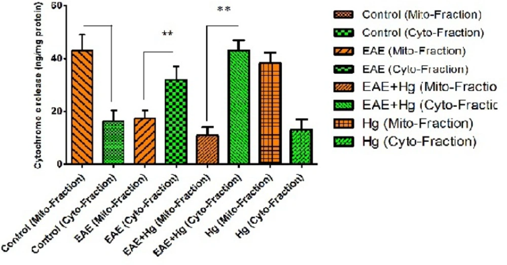
Cytochrome c release. In EAE + Hg and EAE groups cytochrome c released. Values have been presented as mean ± SD (n = 8). *** represents significant difference (P &lt; 0. 001) compared to EAE induced group. Also $$$ represents significant difference (P &lt; 0. 001) between EAE + Hg and EAE groups
Figure 5

Cytochrome c release. In EAE + Hg and EAE groups cytochrome c released. Values have been presented as mean ± SD (n = 8). *** represents significant difference (P < 0. 001) compared to EAE induced group. Also $$$ represents significant difference (P < 0. 001) between EAE + Hg and EAE groups

Our results provided evidence that cumulative mitochondrial damages result in mercury accelerates progression MS through mitochondrial ROS formation that lead to collapse MMP, mitochondrial swelling and finally initiation of apoptosis signaling through mitochondrial pathway. To our knowledge this the first report that provides relationship between mitochondrial events and progression of MS through apoptosis by mercury in brain.
Acknowledgments

References

  • 1.
    Gochfeld M. Cases of mercury exposure, bioavailability, and absorption. Ecotoxicol. Environ. Saf. 2003;56:174-9. [PubMed ID: 12915150].
  • 2.
    Hylander LD, Meili M. 500 years of mercury production: Global annual inventory by region until 2000 and associated emissions. Sci. Total Environ. 2003;304:13-27. [PubMed ID: 12663168].
  • 3.
    Moreau T, Coles A, Wing M, Isaacs J, Hale G, Waldmann H, Compston A. Transient increase in symptoms associated with cytokine release in patients with multiple sclerosis. Brain. 1996;119:225-38. [PubMed ID: 8624684].
  • 4.
    Rietberg MB, Brooks D, Uitdehaag BM, Kwakkel G. Exercise therapy for multiple sclerosis. The Cochrane Library. 2004.
  • 5.
    Beal M. Aging, energy, and oxidative stress in neurodegenerative diseases. Restor. Neurol. Neurosci. 1996;3:180-1.
  • 6.
    Prochazkova J, Sterzl I, Kucerova H, Bartova J, Stejskal VD. The beneficial effect of amalgam replacement on health in patients with autoimmunity. Neuro Endocrinol. Lett. 2004;25:211-8. [PubMed ID: 15349088].
  • 7.
    Attar AM, Kharkhaneh A, Etemadifar M, Keyhanian K, Davoudi V, Saadatnia M. Serum mercury level and multiple sclerosis. Biol. Trace Elem. Res. 2012;146:150-3. [PubMed ID: 22068727].
  • 8.
    Issa Y, Watts D, Duxbury A, Brunton P, Watson M, Waters C. Mercuric chloride: Toxicity and apoptosis in a human oligodendroglial cell line mo3. 13. Biomaterials. 2003;24:981-7. [PubMed ID: 12504520].
  • 9.
    Chuu J-J, Hsu C-J, Lin-Shiau S-Y. Abnormal auditory brainstem responses for mice treated with mercurial compounds: Involvement of excessive nitric oxide. Toxicology. 2001;162:11-22. [PubMed ID: 11311454].
  • 10.
    Stromnes IM, Goverman JM. Active induction of experimental allergic encephalomyelitis. Nat. Protoc. 2006;1:1810-9. [PubMed ID: 17487163].
  • 11.
    Shaki F, Hosseini M-J, Ghazi-Khansari M, Pourahmad J. Depleted uranium induces disruption of energy homeostasis and oxidative stress in isolated rat brain mitochondria. Metallomics. 2013;5:736-44. [PubMed ID: 23629690].
  • 12.
    Babincova M, Bacova Z, Machova E, Kogan G. Antioxidant properties of carboxymethyl glucan: Comparative analysis. J. Med. Food. 2002;5:79-83. [PubMed ID: 12487754].
  • 13.
    Faizi M, Salimi A, Rasoulzadeh M, Naserzadeh P, Pourahmad J. Schizophrenia induces oxidative stress and cytochrome c release in isolated rat brain mitochondria: A possible pathway for induction of apoptosis and neurodegeneration. Iran. J. Pharm. Res. 2014;13:93. [PubMed ID: 24711834].
  • 14.
    Babincová M, Machová E, Kogan G. Carboxymethylated glucan inhibits lipid peroxidation in liposomes. Zeitschrift für Naturforschung C. 1999;54:1084-8.
  • 15.
    Talari M, Seydi E, Salimi A, Mohsenifar Z, Kamalinejad M, Pourahmad J. Dracocephalum: Novel anticancer plant acting on liver cancer cell mitochondria. BioMed Res. Int. 2014;2014.
  • 16.
    Pourahmad J, Faizi M, Abarghuyi S, Salimi A, Nasoohi S. A search for mitochondrial damage in alzheimer’s disease using isolated rat brain mitochondria. Iran. J. Pharm. Res. 2016;15:185-95.
  • 17.
    Taueg C, Sanfilippo D, Rowens B, Szejda J, Hesse J. Acute and chronic poisoning from residential exposures to elemental mercury-michigan, 1989–1990. J. Toxicol. Clin. Toxicol. 1992;30:63-7. [PubMed ID: 1542150].
  • 18.
    Kern JK, Geier DA, Audhya T, King PG, Sykes LK, Geier MR. Evidence of parallels between mercury intoxication and the brain pathology in autism. ‎Acta Neurobiol. Exp. 2011;72:113-53.
  • 19.
    Enríquez JA, Fernández-Sílva P, Montoya J. Autonomous regulation in mammalian mitochondrial DNA transcription. Biol. Chem. 1999;380:737-47. [PubMed ID: 10494823].
  • 20.
    Beal MF. Aging, energy, and oxidative stress in neurodegenerative diseases. Ann. Neurol. 1995;38:357-66. [PubMed ID: 7668820].
  • 21.
    Kowaltowski AJ, Castilho RF, Vercesi AE. Mitochondrial permeability transition and oxidative stress. FEBS Lett. 2001;495:12-5. [PubMed ID: 11322939].

Copyright

© 2016 by School of Pharmacy Shaheed Beheshti University of Medical Sciences and Health Services . This is an Open Access article distributed under the terms of the Creative Commons Attribution License, (http://creativecommons.org/licenses/by/3.0/) which permits unrestricted use, distribution, and reproduction in any medium, provided the original work is properly cited.

Similar Articles

20
Oct
2020
Archives of Neuroscience

Mitochondrial Dysfunction in EAE Mice Brains and Impact of HIF1-α Induction to Compensate Energy Loss

Mahboobeh Mosavimehr,
Seyed Alireza Mesbah-Namin

Mosavimehr M, Mesbah-Namin SA. Mitochondrial Dysfunction in EAE Mice Brains and Impact of HIF1-α Induction to Compensate Energy Loss. Arch Neurosci. 2020;7(4):e104209. doi: https://doi.org/10.5812/ans.104209

31
Oct
2020

Healing Influence of Melilotus Officinalis Herbal Extract on Experimental Autoimmune Encephalomyelitis in C57BL/6 Mice

Mahdieh Hassani,
Maryam Soleimani,
Emran Esmaeilzadeh,
Davood Zare-Abdollahi,
Hamid Reza Khorram Khorshid

Hassani M, Soleimani M, Esmaeilzadeh E, Zare-Abdollahi D, Khorram Khorshid HR. Healing Influence of Melilotus Officinalis Herbal Extract on Experimental Autoimmune Encephalomyelitis in C57BL/6 Mice. Iran J Pharm Res. 2020;19(4):e124663. doi: https://doi.org/10.22037/ijpr.2020.113808.14505

13
Jul
2026
Synthesis and Evaluation of the Effects of Nanoparticles of Atorvastatin and Atorvastatin on Cuprizone-Induced Demyelination: Modulation of the Nrf2/NF-kB Signaling Pathway

Synthesis and Evaluation of the Effects of Nanoparticles of Atorvastatin and Atorvastatin on Cuprizone-Induced Demyelination: Modulation of the Nrf2/NF-kB Signaling Pathway

Samira Shirooie,
Niloofar Heidarizade,
Tayebeh Noori,
Antoni Sureda,
Amin Iranpanah,
Sina Alibeygi

Shirooie S, Heidarizade N, Noori T, Sureda A, Iranpanah A, et al. Synthesis and Evaluation of the Effects of Nanoparticles of Atorvastatin and Atorvastatin on Cuprizone-Induced Demyelination: Modulation of the Nrf2/NF-kB Signaling Pathway. Iran J Pharm Res. 2026;25(1):e170319. doi: https://doi.org/10.5812/ijpr-170319

29
Sep
2025
Jundishapur J Nat Pharm Prod

Combination of Aquatic Exercise with COQ10 Supplementation Improves Oxidative Stress, PTX3 and NF200 Protein Expression in Animal Model of Multiple Sclerosis

Mahsa Porsesh,
Abdolhamid Habibi,
Ali Akbar Alizadeh,
Zohre Ghotbeddin

Porsesh M, Habibi A, Alizadeh AA, Ghotbeddin Z. Combination of Aquatic Exercise with COQ10 Supplementation Improves Oxidative Stress, PTX3 and NF200 Protein Expression in Animal Model of Multiple Sclerosis. Jundishapur J Nat Pharm Prod. 2025;20(4):e164502. doi: https://doi.org/10.5812/jjnpp-164502

21
Sep
2025
Jentashapir J Cell Mol Biol

Aquatic Rehabilitation and CoQ10: Synergistic Effects on Memory Improvement and Myelin Restoration in a Rat Model of Demyelination

Mahsa Porsesh,
Abdolhamid Habibi,
Ali Akbar Alizadeh,
Zohre Ghotbeddin

Porsesh M, Habibi A, Alizadeh AA, Ghotbeddin Z. Aquatic Rehabilitation and CoQ10: Synergistic Effects on Memory Improvement and Myelin Restoration in a Rat Model of Demyelination. Jentashapir J Cell Mol Biol. 2025;16(4):e164888. doi: https://doi.org/10.5812/jjcmb-164888

Download PDF770.85 KB

Crossmark

Crossmark

Checking

Share on
Cited by
Metrics

Ordering Reprints

Articles are published under the Creative Commons license stated on each article. No permission or royalty fee is required for uses permitted by that license. CCC handles optional bulk and customized reprint orders. Any quotation covers production and delivery services only, not copyright permission. > Request Reprints from CCC 

Search Relations

Author(s):

Related Articles