General
All cell lines used in the experiments were obtained from National Cell Bank of Iran (Pasteur Institute, Iran). The chimeric protein coding sequence and the coding sequence for p28 peptide were de novo synthesized by Nedayefan Company (Tehran, IRAN) and supplied in pGE plasmid. Glutathione Resin was purchased from GenScript (MA, USA) and Mouse anti-GST antibody was purchased from Abcam (Abcam, USA). FastDigest TMBamHI, SalI, EcoRI, and XhoI restriction endonucleases and also T4 DNA ligase was purchased from Thermoscientific (USA). The cells were cultivated in RPMI and DMEM 1640 containing 10% FBS and supplemented with 100 U/mL penicillin and 100 mg/mL streptomycin (Sigma, Germany). Amicon filters were obtained from Merck (Merck Millipore, USA).
Construction of recombinant plasmids, expression, and purification of the p28-NRC chimeric protein
The coding sequence of the chimeric protein and the p28 peptide was sub-cloned in the pGEX-5X-1 expression plasmid. This plasmid adds GST tag to the N-terminus of the expressed protein resulting in higher solubility and also aiding in affinity purification of the recombinant protein by glutathione resin. The fidelity of the cloning was confirmed by restriction enzyme digestion and subsequent DNA sequencing. The expression was investigated at various concentrations of IPTG, temperatures, and induction time. Glutathione Resin was packed in an empty column and was used for purification of GST tagged-chimeric protein and the p28 peptide using fast protein liquid chromatography (FPLC). In order to purify the expressed protein, the IPTG-induced bacterial cells were collected by centrifugation and resuspended in phosphate-buffered saline (PBS). Then, the mixtures were sonicated on ice and subsequently centrifuged at 10,000 × g for 15 min. The collected supernatant was used for the purification of GST-tagged chimeric protein and the p28. Elution of GST-tagged chimeric protein and p28 from chromatography medium was performed under mild, non-denaturing conditions using reduced glutathione. Then, the fractions were collected and analyzed by SDS-PAGE and Western blotting. Fractions containing the chimeric protein or p28 were pooled and subjected to dialysis followed by enterokinase cleavage of the GST-tag. When the GST tags removed, protein samples were concentrated using Amicon™ filters. Finally, gel filtration chromatography by Superdex 75 (GE Healthcare, USA) was used to achieve highly pure recombinant protein or p28 peptide suitable for upcoming in-vitro and in-vivo studies.
Cytotoxicity Assay
Cytotoxicity of the chimeric protein was assessed by MTT assay. To measure cytotoxicity, the cells were cultivated in 96 well plates and treated with various concentrations of chimeric protein or p28 peptide for 48 h. Then, the medium was replaced with fresh medium containing 5 mg/mL MTT and further incubated for 4 h. Afterward, the supernatant was replaced with 150 µL Dimethyl sulfoxide (DMSO) to dissolve the formazan crystals and finally, absorbance was read at 450 nm with a microplate reader. Results were calculated as the percent of the absorbance of the treated cells over the absorbance of the untreated control cells. Based on these measurements, the IC50 values of chimeric protein and p28 peptide were calculated.
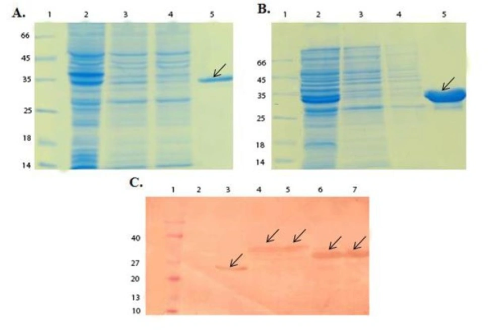
SDS-PAGE and Western blot analysis of GST-chimeric protein and GST-p28 purification. (A) SDS-PAGE analysis of GST- chimeric protein purification. Lane 1; Molecular weight protein marker (26610, Fermentase), Lane 2; Supernatant before applying to the column, Lane 3 and 4; Supernatant after applying to the column, Lane 5; Elution of GST- chimeric protein with 10 mM glutathione. The theoretical MW of the GST-p28 is about 36.5 kDa. Arrow indicated the purified GST-chimeric protein. (B) SDS-PAGE analysis of GST-p28 purification. Lane 1; Molecular weight protein marker26610) , Fermentase), Lane 2; Supernatant before applying to the column Lane 3; Supernatant after applying to the column Lane 4; GST-p28 after elution with the reduced glutathione. The theoretical MW of the GST-p28 is about 32.6 kDa. (C) Western blot analysis of GST, GST-chimeric protein, and GST-p28. Lane 1; Molecular weight protein marker (BM0066, FMC.Bioproducts), Lane 2; Uninduced pGEX, Lane 3; Induced pGEX, Lane 4 and 5; Induced GST- chimeric protein, Lane 6 and 7; Induced GST-p28
SDS-PAGE analysis of enterokinase digestion of the GST-chimeric protein and GST-p28 peptide. (A) SDS-PAGE analysis of enterokinase digestion of the GST-chimeric protein. Lane 1; Molecular weight marker Lane 2; Digested GST- chimeric protein using 1 unit of the enterokinase Lane 3; Undigested GST-chimeric protein Lane 4; Digested GST-chimeric protein using 2 units of the enterokinase. Arrow indicated the chimeric protein. The theoretical MW of the chimeric protein is about 7 kDa. (B) SDS-PAGE analysis of enterokinase digestion of the GST-p28 Lane 1; Molecular weight marker Lane 2; Digested GST-p28 with 1 unit of enterokinase Lane 3; Digested GST-p28 with 2 units of enterokinase Lane 4; Undigested GST-p28. Arrow indicated the p28 peptide. The theoretical MW of the p28 peptide is about 3 kDa
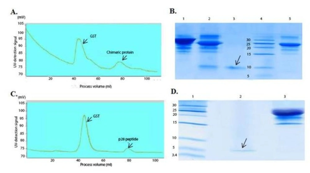
Chromatogram of the chimeric protein and the p28 purification using gel filtration and SDS-PAGE analysis after purification
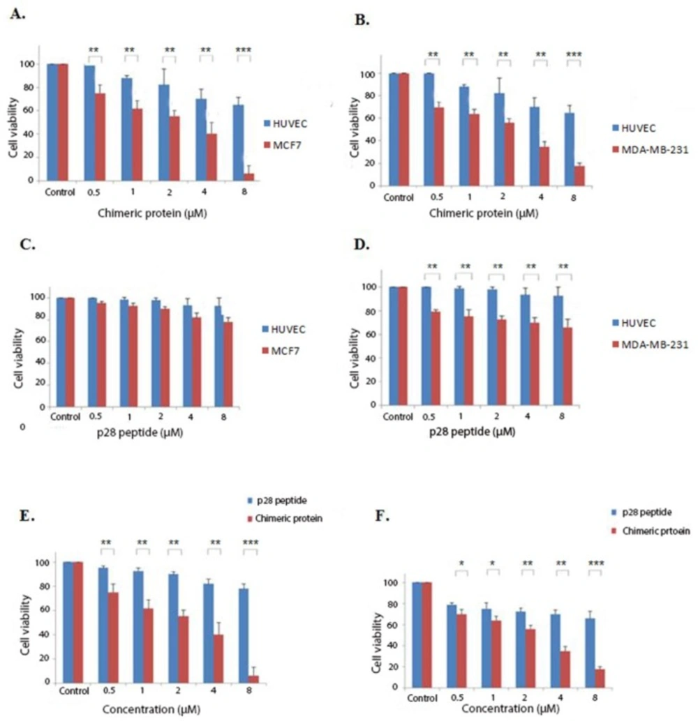
Cytotoxic effects of the chimeric protein and p28 peptide. (A) cytotoxic effects of the chimeric protein against MCF7 and HUVEC cells. (B) Cytotoxic effects of the chimeric protein against MDA-MB-231 and HUVEC cells. (C) Cytotoxic effects of the p28 against MCF7 and HUVEC cells. (D) Cytotoxic effects of the p28 against MDA-MB-231 and HUVEC cells. Cell viability was assessed using MTT assay after 48 h. (E) Incubation of the MCF7 cells with various concentrations of the p28 peptide and chimeric protein for 48 h. (F) Incubation of the MDA-MB-231 cells with various concentrations of the p28 peptide and the chimeric protein for 48 h. Cell viability was assessed using MTT assay after 48 h. Data were shown as Mean ± SD, n = 3. Percentage cell viability and comparison between data sets performed using ANOVA. *p < 0.05, **p < 0.01 and ***p < 0.001 indicates statistically significant differences between the means of values obtained with MTT assay
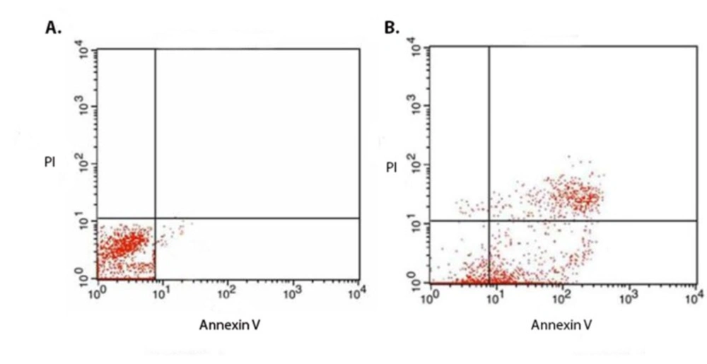
(A) Flow cytometric analysis of the non-treated and (B) treated cells for evaluation of the mechanism of the chimeric protein induced cell death. The MCF7 cells were treated with IC50 (1.88 µM) of the chimeric protein for 4 h. Annexin V and PI staining analysis of the MCF7 cells showed the induction of apoptosis in the treated cells. In case of untreated control cells, result showed 98.0% non- stained cells. However, for the treated MCF7 cells, the amount of the non-stained cells was 15.26% while the annexin V stained cells (early apoptotic cell) accounted for about 43% ± 6.36% (n = 3)
| Pro- Apoptotic | Fold increase/decrease | Anti-Apoptotic | Fold increase/decrease |
|---|
| AIF | 4.26 | Bcl2 | -2.18 |
| Bax | 2.4 | | |
| FAS | 0.9 | | |
| DR4 | 1.1 | | |
| Casp3 | 2.3 | | |
Evaluation of cell death mechanism by flow cytometry
To assess the cell death mechanism induced by the chimeric protein using Flow cytometry, MCF7 cells were seeded in wells of six-well plates and treated with the IC50 concentration of the chimeric protein and subjected to staining with the Annexin V/PI according to instructions of the Annexin-V-FLUOS Staining kit (Roche, Germany).
Evaluation of pro- and anti-apoptotic gene transcriptions
In order to further evaluate the mechanism of apoptosis induced by the chimeric protein, Real Time PCR analysis of mRNA expression of some pro- and anti-apoptotic genes was performed using Apoptosis RT Array™ kit (NanoCinna, Iran) according to the manufacturer’s instructions.
The mRNA expression of GAPDH was considered as a housekeeping gene. Briefly, following treatment of MCF7 cells with the IC50 concentration of the chimeric protein, total cellular RNA was isolated using RNeasy™ Mini Kit (Qiagen, Germany) followed by DNAse treatment and subjected to cDNA synthesis using RevertAid™ First Strand cDNA Synthesis Kit (Thermo Scientific, USA). Next, the prepared cDNA was used for Real-time RT-PCR analysis by AB Applied BioSystems (Thermo Fisher Scientific, USA).
Statistical analyses
Data were expressed as a mean ± standard error. The SPSS 20 software was used for statistical analysis. Statistical significance was determined by one-way ANOVA with Tukey′s post-hoc test. Differences were considered statistically significant when p < 0.05.