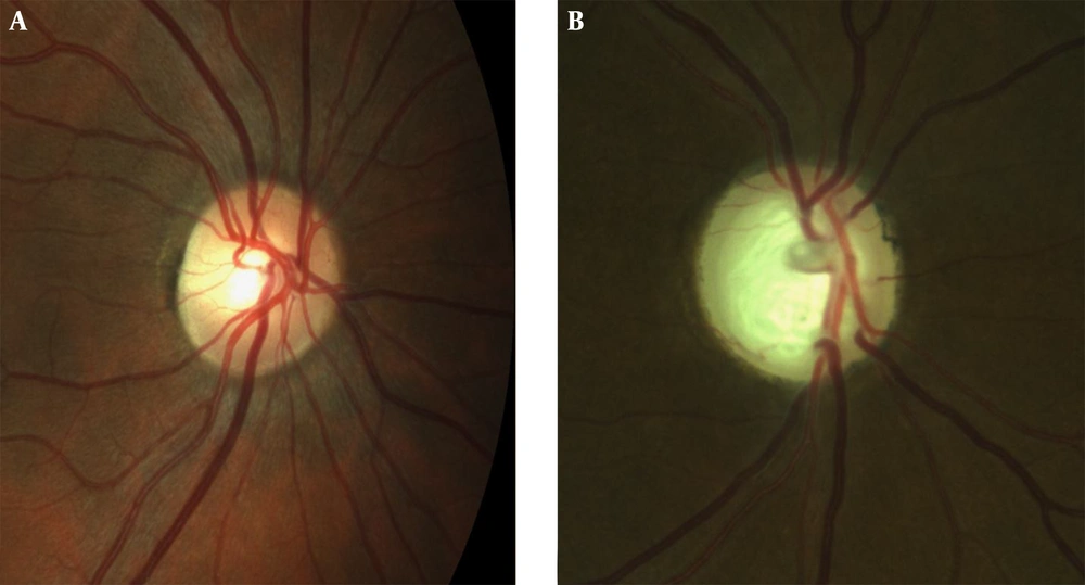
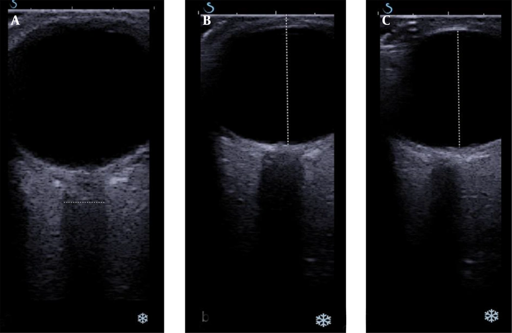
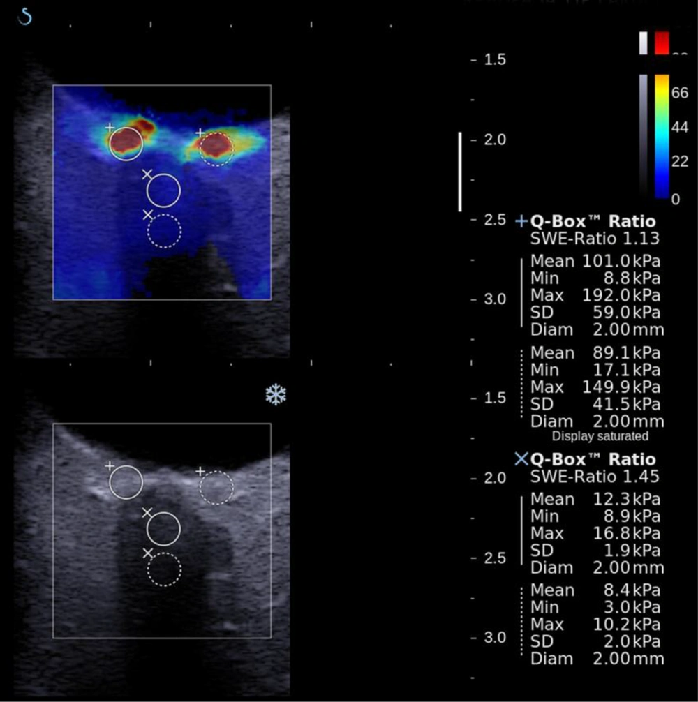
In this study, we demonstrated a novel use of SWE technology to evaluate the biomechanical properties of the ppSC and ON and studied whether these elasticity measurements correlated with any clinical variables in glaucoma patients. As is known, there is currently no available in vivo method to evaluate the biomechanical properties of the ppSC and ON. Evaluations in the study showed that the mean stiffness values were significantly higher in the perineural sclera’s nasal and temporal regions and the ON in glaucomatous eyes compared to healthy controls (P < 0.05).
Chronic high intraocular pressure may increase the structural stiffness of the ppSC and directly lead to LC deformation (
21). It is well known that the deformative changes of the ppSC develop under the strain and stress of the disease course and may have potential roles in glaucoma pathogenesis. Chronic intolerably high IOP may cause biomechanical changes in the ppSC and it may affect the susceptibility of ganglion cell axons. Hence, it is important to show ppSC changes when attempting to understand the effect of IOP on the ON head.
Changes that occur in the ON are the most prominent features of glaucomatous damage. Studies on the morphology of the ON head in monkeys (
22,
23) and ON head biomechanics in humans (
8,
24) have increased our knowledge about the biomechanics of the ON head and peripapillary structures in glaucoma. These studies all underline the significant deformative/degenerative changes in the material properties and architecture of the ppSC as well as the ON, hence aiding the understanding of the impact of glaucomatous disease. Higher stiffness values among glaucoma patients in the current study may be a reflection of direct/indirect effects of the intolerably high IOP on the target tissues. Higher elasticity may also be a predisposing risk factor in glaucoma development and vice versa. However, we showed no histopathological correlation with SWE findings.
The stiffness values were correlated inversely with the mean thickness of the ON, which was lower in glaucomatous patients (3 mm) than in normal eyes (3.5 mm; P < 0.05). Furthermore, the C/D ratio, which is an important ophthalmoscopy finding, was well correlated with stiffness values. We did not find a correlation between visual field (i.e., MD and PSD) and elastography parameters. We have two possible explanations for this finding. First, our sample size was not large and most of the glaucoma patients included in the study had mild to moderate glaucoma. Therefore, their functional damage may be associated with less anatomic and morphologic damage. Visual field parameters reflect the functional status of the ON and functional damage has been reported to be affected relatively late, compared to anatomical changes, in the course of glaucoma. Second, peripapillary stiffness increases in glaucomatous patients may be a preliminary indication of glaucoma rather than a result of glaucomatous damage. As a potential prognostic factor, stiffness increases might be discovered in the early stages of the disease.
Herein, we investigated the usefulness of SWE in in vivo evaluation of the ON and peripapillary structures in glaucomatous eyes and healthy control eyes. There were significant differences in the stiffness values of the ON and peripapillary structures in glaucomatous and healthy eyes with SWE. Also, our results obtained from SWE were correlated with the C/D ratio, which indicates the severity of glaucoma and IOP, which is the main parameter of follow up for ophthalmologists. The correlations between the C/D ratio, ON diameter, and higher SWE measures suggested that SWE has the potential to aid in the diagnosis of glaucoma. Interestingly, beyond a glaucomatous impact, SWE did not indicate a high correlation with IOP increases directly. The results were correlated with the degenerative changes in the ON and ppSC.
The optic nerve color coding in SWE imaging is a bit slower than the peripapillary sclera. Gennison et al. published a study on SWE imaging that investigates the effect of muscle fiber orientation on SWE imaging (
8,
24). A significant difference of SWE values was revealed in different orientations of probe, parallel or perpendicular, on the muscle. However, there was no way to image the optic nerve in parallel orientation with ultrasonography probe in our study. In all cases, we obtained SWE values of the optic nerve regardless of the patient characteristics. Hence, the study depicts the glaucoma effect on the optic nerve and does not intend to suggest a diagnostic threshold level.
In a recent study, Agladioglu et al. investigated the elasticity index and strain ratio of orbital and retro-orbital structures in glaucomatous patients by strain elastography imaging (
25). According to this study, there were no statistical differences in the strain elasticity index of these structures between glaucomatous patients and healthy controls. For instance, there was a statistical difference in the strain ratio of anterior and posterior vitreous among glaucomatous eyes and the healthy controls. First, semi-quantitative methodology of the strain elastography might be the cause of insignificant results. Secondly, investigating such a small area with the strain elastography is subjective regarding SWE, which can evaluate the 2 mm thin areas quantitatively by small ROI calibrating. Furthermore, fluid content of the vitreous might have an insufficient inductive role of mechanical compression due to the elastic nature of vitreous.
Although considerable differences were noted between glaucomatous and normal eyes in general, an overlap of stiffness between patients with glaucoma and healthy controls was noticed. This may be due to other factors playing a role in the pathogenesis of glaucoma. Two main limitations of the current study include a small sample size and relatively small number of patients with advanced glaucoma. As this is the first in vivo study, further studies with a larger patient sample size are needed.
Initial findings suggest that the method has potential use in the disease. The representation of glaucomatous damage should deliver new insights for patient evaluation. Patients with restricted ophthalmologic evaluation due to red eye, leucoria, and cataract disease are potential subjects of the SWE imaging. Long-term evaluation of glaucoma damage to optic nerve could be accomplished before field of view injury develops.
In conclusion, this manuscript discusses evaluating the biomechanical properties of peripapillary tissue in glaucoma in vivo. The study compared elasticity parameters in healthy subjects and glaucomatous patients. We found that the SWE technique is potentially a valuable tool for evaluation of the peripapillary region, particularly in glaucoma. We observed stiffer ppSC and ON SWE values in eyes with glaucoma and the elastography values were correlated with anatomical parameters. Further studies on how best to use SWE for the diagnosis and risk evaluation of glaucoma and other optic neuropathy diseases are needed.