The FNAB plays an essential role in the evaluation of euthyroid patients with thyroid nodules, while the TBSRTC assigns a malignancy risk to each cytological category. In our study, the term “indeterminate” was used to identify “follicular neoplasm/suspicious for a follicular neoplasm” and “suspicious for malignancy” results of the TBSRTC. The distinction between follicular thyroid carcinoma and follicular adenoma cannot be made using an FNAB, and surgery is required to distinguish the benign nodules from the malignant ones. In addition, when an FNAB is suspicious for malignancy, nearly all physicians recommend surgery to exclude it. The introduction of a diagnostic test which helps to decrease the number of unnecessary surgical resections of benign nodules could have an important positive impact on patients management.
Malignant cells exhibit a number of aberrant characteristics when compared to cells in normal tissues, and these characteristics can, potentially, be used to image tumors. A PET scan with FDG relies upon the increased glucose metabolism found in a wide range of malignancies, using it to detect both primary lesions and their metastases. However, the usefulness of an FDG PET/CT in the presurgical evaluation of indeterminate thyroid nodules is conflicting, due to the contradictory results found in the literature (
16-
19).
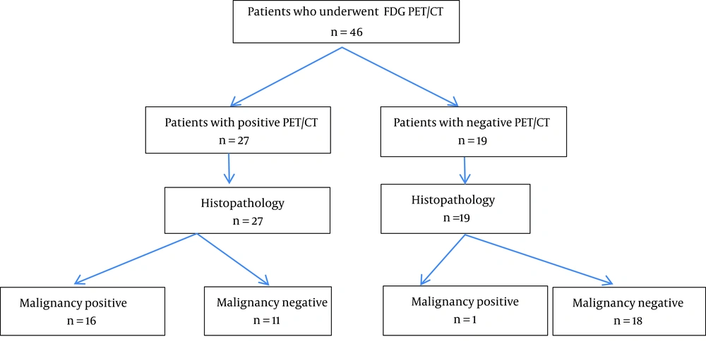
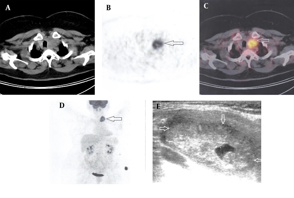
In our study, 37% (17/46) of the patients demonstrated thyroid carcinomas in their final histopathological reports. The FDG PET/CT detected 16/17 malignant lesions, with a very good sensitivity of 94%. Previously, Vriens et al. (
17) and Wang et al. (
16) meta-analyzed the usefulness of the FDG PET in evaluating thyroid nodules with indeterminate FNAB results. Sensitivity and specificity of our study similar to those found in their studies. In the study by Vriens et al. (
17), it was reported that a false negative PET result was only found in those nodules with a maximum histological dimension of ≤ 1.5 cm. In contrast, the study by Wang et al. (
16) found 8 false negative cases, and 5 of them had dimensions ≥ 2 cm. In our study, there was 1 FDG negative malignant lesion (papillary cancer), with a dimension of 2.5 cm, and 6 FDG positive malignant lesions (4 papillary, 1 follicular, and 1 medullary cancer) with dimensions ≤ 1.5 cm. Therefore, as suggested by Wang et al. (
16), it is unlikely that the size of the lesion is the most important factor for explaining the false negative phenomena in our study.
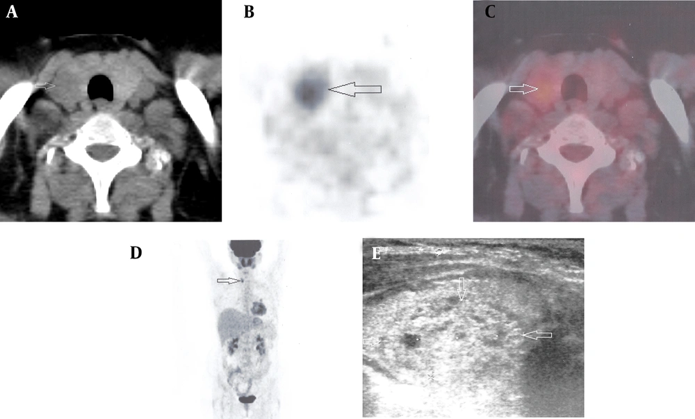
In the present study, the FDG was not found to be negative in all of the benign nodules, and the FDG accumulation was detected in 11 of the 29 benign tumors. The specificity, and the positive predictive and negative predictive values were 62%, 59%, and 95%, respectively. In the SUV analysis, no significant difference was found between the benign and malignant nodules: however, there was a considerable overlap between the groups. In the previously published literature, the reports disagreed about the usefulness of the quantification of the FDG uptake, measured in terms of the SUV, for the characterization of the thyroid nodules. While some of the studies showed no significant differences between the SUVs of the benign and malignant nodules (
20,
21), others suggested that the SUVs can be used to differentiate malignant from benign nodules(
16,
22-
24). However, different cutoffs for the SUVs were found in these studies and, for the most part, there was considerable overlap in their glucose metabolic activities (
16,
22-
24). SUVs rely on the reconstruction, acquisition, and region-of-interest parameters, and the variability found in the SUV methodology makes it difficult to compare the results obtained from different centers. Therefore, additional standardized large multicenter studies may be necessary to generalize the role of the SUV measurements in the characterization of indeterminate thyroid nodules.
For all of these reasons, regardless of the SUV measurements, we recommend further examinations on any FDG positive nodule with indeterminate cytology. According to our analysis, despite there being some false positive cases, over half of the patients could have avoided unnecessary surgeries (62.1%, 18/29). However, the FDG PET/CT is not available in some regions, and it is expensive, which could, for the time being, limit its routine use. In those patients on whom surgery cannot be performed, close regular follow-ups, including repeated FNABs and thyroid ultrasonography, should be performed. Overall, the FDG PET/CT may provide physicians with additional confidence when deciding to observe lesions, rather than to proceed immediately to surgical resection. The careful selection of those patients who could benefit most from the additional information provided by this imaging method is important, and the FDG PET/CT scan could be more useful for patients with a high risk of surgical morbidity and mortality.
In summary, our study revealed that the FDG PET/CT has high sensitivity and a negative predictive value for identifying malignancies in thyroid nodules with indeterminate cytological findings. Therefore, the FDG PET/CT may be a helpful tool in the clinical management of these nodules.