Ultrasound is the preferred method for the evaluation of DDH in infants between 6 weeks and 6 months of age. Alpha angle measurement using the Graf method is the accepted diagnostic method, however, variations in probe orientation can result in significant changes in the measured alpha angle. Thus, it is possible to generate many acceptable images with different alpha angles, which can result in different Graf categories for the same hip (
3).
To our knowledge, there is no study in the literature investigating CT findings for the primary diagnosis of DDH. Most of the studies focusing on DDH and CT are related to the post-reduction assessment of hips (
7). We investigated CT for its possible supportive role in the diagnosis of DDH.
Acetabular cortical thicknesses did not reveal a significant difference between left and right hips in the control group. Acetabular margins (rim) were sharp edged or had a beak-like appearance in normal hips and there was no acetabular surface irregularity and shallowing in the control group.
Most of the patients with DDH were girls in our study (88%), which is consistent with previous studies. In a study by LeBa et al., DDH was found to be more common in females (83%) (
8).
Bilateral DDH was more common than unilateral DDH in our study. This finding is consistent with some previous studies, (
9) but the bilateral occurrence rate is not clear since previous studies have reported varying results (
10).
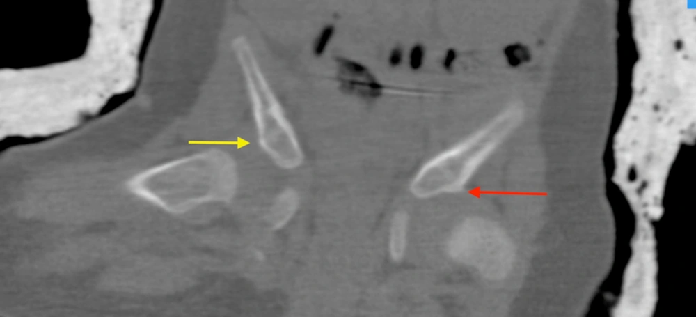
All the unilateral DDH patients showed acetabular bony margin irregularity and flattening. In addition, 94% of bilateral DDH patients showed either focal or diffuse acetabular bony margin irregularity and flattening. Hence, acetabular marginal irregularity and flattening was the most frequent and reliable finding in the diagnosis of DDH in our study. This part of hip is also visible on ultrasonography, so it can be a reliable finding in the diagnosis of DDH. Marginal irregularity and flattening were seen even in 2-month-old babies. Therefore, acetabular margin evaluation might be an important part of ultrasonography screening in babies at risk. A sharp angled or beak-like acetabular rim is highly suggestive of a normal hip.
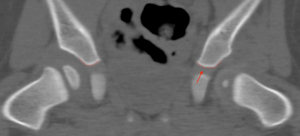
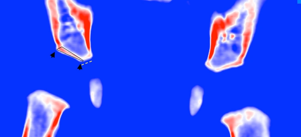
Another frequent finding was seen in the inferior part of the acetabulum in patients with unilateral DDH. A shallow acetabulum, caused by a thicker ilium compared to the normal side in these patients, was seen in 93% of the dysplastic hips. There was also a smooth angulation on the inferior margin compared to the normal side. This finding cannot be evaluated with ultrasonography; therefore CT imaging might be needed for patients suspected of having this condition. To our knowledge, this finding was not reported before in literature.
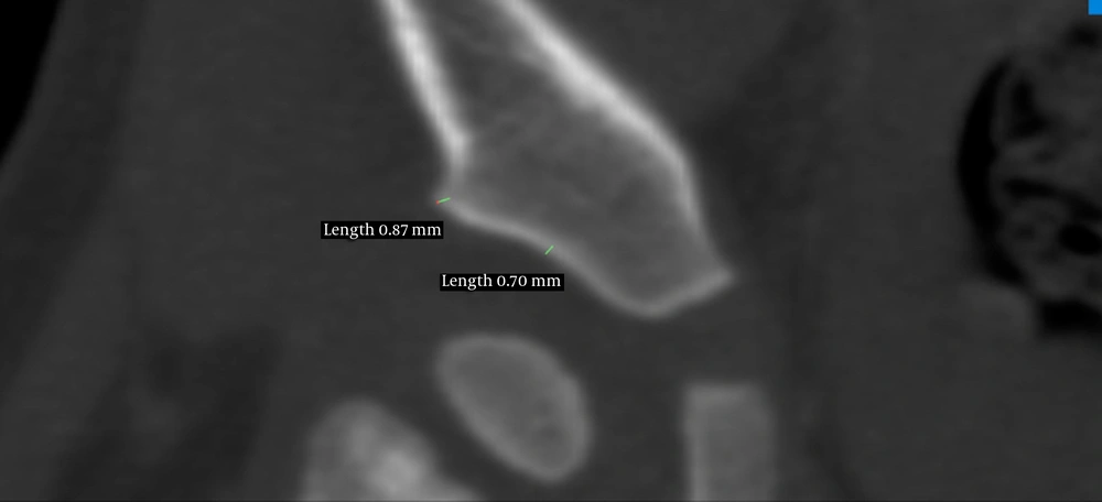
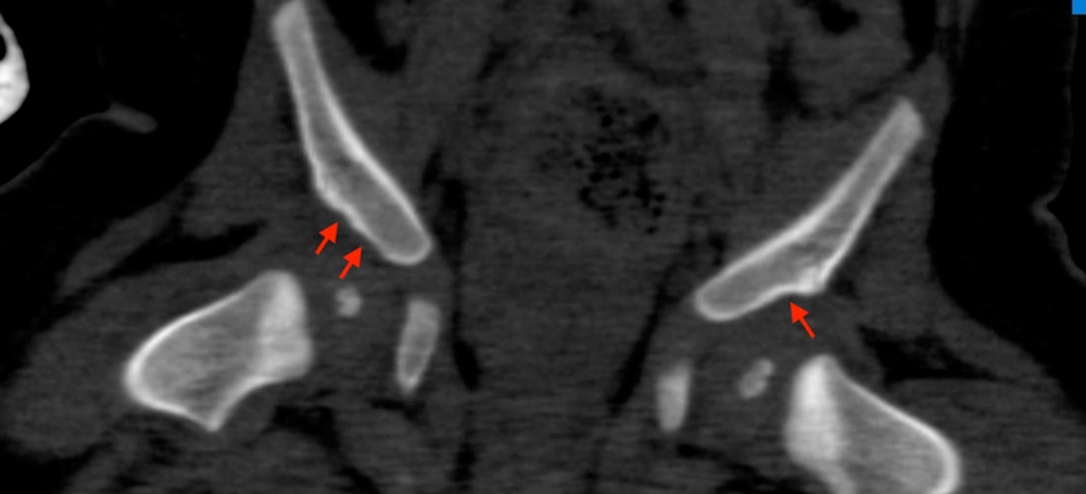
In normal hips, a triangular shaped acetabular cortex without any irregularity was seen. The acetabular cortex was thicker in the superolateral aspect and thinner in the inferomedial aspect of the acetabulum in normal hips. The lateral aspect of normal hips showed a sharp beak-like margin (
Figure 6). In dysplastic hips, the triangular shaped acetabular cortex was not seen in most cases, rather, the cortex was irregular with focal thinning, and the superolateral aspect of the acetabulum was irregular and flattened in the coronal reformatted images.
Coronal reformatted CT image. Color clut image of normal hips of a 3 month-old baby. Normal hips with a sharp angled or beak like acetabular margin is seen (arrows). Acetabular cortex with triangular shape, thickest at superolateral aspect and thinnest at inferomedial aspect showing gradual decrease in thickness (black lines).
In patients with unilateral DDH, the mean acetabular cortical thickness of the dysplastic hips was significantly lower than that of the normal side in the central part and the acetabular margin. Cortical thinning might be related to an insufficient vascular supply of the iliac bone on the dysplastic hip side. In a study by Meng at al., decreased femoral head perfusion was showed in children with DDH (
11). In patients with bilateral DDH, acetabular cortical thickness was somewhat lower in the most severe dysplastic side, however, this difference was not significant. Cortical thinning was most prominent in the acetabular margins of the dysplastic hips.
Acetabular surface irregularity was seen in 82% of the dysplastic side in patients with unilateral DDH, and 91% in bilateral DDH. The youngest baby with surface irregularity was 2 months old. In the control group, most of the hips had a regular acetabular surface, although a minimal smooth undulation was present in a small number of them. Sometimes dysplastic hips showed minimal irregularity, which can be difficult to view on ultrasonography. Further studies are needed to evaluate the visibility of surface irregularities with ultrasound examination.
The relationship between a shallow acetabulum and an underdeveloped femoral head in the development of hip dysplasia is well known. In our study, a shallow acetabulum was seen in 84% of the unilateral dysplastic hips and in 86% of the bilateral dysplastic hips. It is difficult to identify a shallow acetabulum in 2-month-old babies because of physiologic dysplasia; in these age group, the acetabula of normal hips are also shallow in appearance.
Subcortical sclerosis was not a frequent finding in our study, and we saw no cases of it in 2 or 3-month-old babies. The youngest baby with subcortical sclerosis was 4 months old.
In conclusion, our study demonstrates that acetabular marginal irregularity and flattening is an important finding in the diagnosis of DDH when combined with bone thickening in the inferior part of ilium. CT cannot be recommended as an initial modality in the diagnosis of DDH, but extra attention should be given to marginal irregularity of acetabulum during ultrasound imaging. To know the changes related to DDH in the acetabulum will help in the diagnosis of DDH in patients older than 6 months old with a pelvic CT scan due to diseases other than DDH.