
Enhancement of Anti-MRSA Activity in Endophytic Streptomyces drozdowiczii Ca2- 5 Through OSMAC and Light-Regime Optimization

Author(s):
Maryam BeiranvandMaryam BeiranvandMaryam Beiranvand ORCID1, Saeid AfsharSaeid AfsharSaeid Afshar ORCID1, 2, Hashemi Shahraki AbdolrazaghHashemi Shahraki AbdolrazaghHashemi Shahraki Abdolrazagh ORCID3, Sajad YaghoubiSajad YaghoubiSajad Yaghoubi ORCID4,*, Hadi SamadiyanHadi SamadiyanHadi Samadiyan ORCID5,**
1Department of Medical Biotechnology, School of Advanced Medical Sciences and Technologies, Hamadan University of Medical Sciences, Hamadan, Iran
2Cancer Research Center, Institute of Cancer, Hamadan University of Medical Sciences, Hamadan, Iran
3Division of Laboratory Services , Centers for Disease Control and Prevention (CDC), 630 Hart Lane, Nashville, TN37216, USA
4Department of Basic Medical Sciences, Neyshabur University of Medical Sciences, Neyshabur, Iran
5Research Center for Molecular Medicine, Institute of Cancer, Avicenna Health Research Institute, Hamadan University of Medical Sciences, Hamadan, Iran
Corresponding Authors:
*Corresponding Author: Department of Basic Medical Sciences, Neyshabur University of Medical Sciences, Neyshabur, Iran. Email: [email protected]
**Corresponding Author: Research Center for Molecular Medicine, Institute of Cancer, Avicenna Health Research Institute, Hamadan University of Medical Sciences, Hamadan, Iran. Email: [email protected]

Jundishapur Journal of Microbiology:Vol. 19, issue 6; e172325
Published online:Jun 30, 2026
Article type:Research Article
Received:Apr 27, 2026
Accepted:Jun 02, 2026
How to Cite:Beiranvand M, Afshar S, Shahraki Abdolrazagh H, Yaghoubi S, Samadiyan H. Enhancement of Anti-MRSA Activity in Endophytic Streptomyces drozdowiczii Ca2- 5 Through OSMAC and Light-Regime Optimization. Jundishapur J Microbiol. 2026;19(6):e172325. doi: https://doi.org/10.5812/jjm-172325

Abstract

Background:

Multidrug-resistant bacteria, particularly methicillin-resistant Staphylococcus aureus (MRSA), pose a major global health threat and cause hundreds of thousands of deaths annually. Endophytic Streptomyces strains are promising sources of bioactive metabolites; however, many of their biosynthetic gene clusters remain silent under standard laboratory conditions.

Objectives:

This study aimed to activate silent biosynthetic pathways in an endophytic Streptomyces isolate using the one strain–many compounds (OSMAC) strategy to enhance the production of metabolites with anti-MRSA activity.

Methods:

An endophytic Streptomyces strain was isolated from medicinal plants in Iran. The OSMAC approach was applied by systematically varying key culture parameters, including nutrient composition, pH, aeration, incubation time, and the physicochemical conditions of the medium. Light regimes were also optimized to better simulate the bacterium’s natural habitat. The antibacterial activity of the extracts was evaluated against MRSA ATCC 43300 using the standard agar well diffusion method.

Results:

Optimization of culture conditions significantly increased the production of secondary metabolites with potent anti-MRSA activity. The largest inhibition zones were achieved under specific combinations of nutrient limitation, a slightly acidic pH, and defined light exposure, compared with standard laboratory conditions.

Conclusions:

This study highlights the OSMAC strategy as an effective and cost-efficient method for activating silent biosynthetic pathways in endophytic Streptomyces by manipulating culture conditions. The optimized approach provides a valuable framework for discovering novel anti-MRSA compounds from actinobacteria.

1. Background

Multidrug-resistant bacteria cause infections that are increasingly difficult to treat with existing antibiotics (1). Among these bacteria, methicillin-resistant Staphylococcus aureus (MRSA) is of particular concern and poses a serious risk to vulnerable populations, including older adults, children, and immunocompromised individuals (1-4). According to the Centers for Disease Control and Prevention (CDC), antimicrobial resistance was responsible for more than 1.27 million deaths globally in 2019, with MRSA contributing to a substantial proportion of these cases each year (5, 6). These alarming statistics underscore the urgent need to discover and develop new antimicrobial agents (7, 8).
Endophytic actinomycetes, particularly members of the genus Streptomyces, are prolific producers of bioactive compounds, including antibiotics, antifungals, anticancer agents, and immunosuppressants (9-11). Despite their remarkable biosynthetic potential, many of their secondary metabolite gene clusters remain silent under standard laboratory conditions (12, 13). Various strategies have been used to activate these cryptic biosynthetic gene clusters (BGCs), including genetic engineering (14-16), co-cultivation with other microbes (17, 18), and the OSMAC approach. The OSMAC strategy relies on altering culture conditions, such as nutrient sources and physical parameters, thereby enabling a single strain to produce diverse secondary metabolites without genetic manipulation.

2. Objectives

This study aimed to isolate endophytic Streptomyces from Iranian medicinal plants and optimize key physicochemical parameters, including nutrient sources, aeration, incubation time, and light regime, to maximize anti-MRSA activity.

3. Methods

3.1. Sample Collection

In May 2024, medicinal plants, including Zataria multiflora [IPNI: 461954 - 1] and Allium sativum [IPNI: 528796 - 1], which are well known for their traditional use in the treatment of MRSA-associated infections (19, 20), were collected from different altitudes across Mount Damavand (coordinates: 35°57′04″N, 52°06′32″E), Iran. Plant samples were immediately placed in sterile plastic bags, kept under cold conditions, and transported to the Microbiology Laboratory of the Pasteur Institute of Iran for the isolation of endophytic Streptomyces and further processing (21).

3.2. Isolation of Streptomyces From Plant Samples

Plant samples were surface-sterilized using a previously described multistep protocol (22). Sterilized root and stem tissues were aseptically cut into approximately 4 × 4 mm segments and plated onto selective media, including R3A (22), ISP4, and GMA. Plates were incubated at 28 °C for 4 days, and microbial growth was monitored daily. Colonies exhibiting typical Streptomyces-like morphology were purified by repeated subculturing, grouped according to similar morphological characteristics, and screened for antimicrobial activity.

3.3. Initial Antimicrobial Activity Assay

A single colony of each isolate was inoculated into 100 mL of Mannitol Soya Flour (MSF) broth (23), containing 2 g mannitol, 2 g soya flour, and 10 mM CaC2. Cultures were incubated at 28 °C for 7 days on a rotary shaker at 200 rpm. After incubation, cultures were centrifuged at 4000 rpm for 20 minutes using a Gyrozen 1524 centrifuge to separate the biomass from the supernatant. Both fractions were tested for antimicrobial activity using the agar well diffusion method against a lawn of Staphylococcus aureus (ATCC 43300). The inoculum was standardized to a 0.5 McFarland turbidity standard. Briefly, 25 µL of each sample was pipetted into 6-mm wells in agar plates, followed by overnight incubation at 37 °C. Several isolates showed notable antimicrobial activity, with one isolate (Ca2 - 5) exhibiting particularly strong activity against MRSA. Notably, the cell-free supernatant displayed stronger inhibitory activity than the biomass, indicating that it was a promising candidate for further study.

3.4. OSMAC-Based Optimization of Antimicrobial Activity

To explore the metabolic potential of the selected Streptomyces strain, the OSMAC approach was applied using 96 different culture media formulations (Table 1 in Supplementary File) to evaluate antimicrobial activity. Each 100 mL culture contained 2 g of nitrogen sources (soybean meal, peptone, or yeast extract) and 2 g of carbon sources (mannitol, glucose, starch, or chitin), supplemented with trace elements, including 10 mM CaCl2. Cultivation conditions were diversified by varying shaking speed (0 - 200 rpm), incubation temperature, and light exposure, with dark conditions achieved by wrapping flasks in aluminum foil. After incubation, cultures were centrifuged at 4000 rpm for 20 minutes, and the antimicrobial activity of the supernatants was assessed against MRSA (ATCC 43300) using the agar well diffusion method with a 0.5 McFarland inoculum standard. Inhibition zones (mm) were measured after overnight incubation at 37 °C using a Scan 4000 scanner. All 96 conditions were initially screened once, and active conditions were retested in two independent experiments to confirm the reproducibility and consistency of antimicrobial activity.

3.5. Molecular Characterization

3.5.1. Genomic DNA Extraction

Genomic DNA was extracted using the boiling method (24). Briefly, bacterial cultures grown in Tryptic Soy Broth (TSB) were centrifuged at 15,000 × g for 10 minutes. The pellet was washed once with sterile water, resuspended in sterile water, and boiled at 100 °C for 10 minutes. After centrifugation, the supernatant containing DNA was collected and stored at -20 °C until use.

3.5.2. PCR Amplification of the 16S rRNA Gene

The 16S rRNA gene was amplified using the universal bacterial primers 27F (5′-AGAGTTTGATCCTGGCTCAG-3′) and 1525R (5′-AAGGAGGTGATCCAGCCGCA-3′), purchased from Bioneer (USA). PCR was performed in an Eppendorf 96 AG thermal cycler (model 22331, Hamburg, Germany) under the following conditions: initial denaturation at 94 °C for 3 minutes, followed by 35 cycles of 94 °C for 30 seconds, 60 °C for 30 seconds, and 68 °C for 1 minute, with a final extension at 68 °C for 5 minutes. PCR products were visualized using SYBR Safe DNA Gel Stain after electrophoresis on a 1% agarose gel at 90 V for 1 hour and purified using the GeneJET PCR Purification Kit.

3.5.3. Sequencing of the 16S rRNA Gene

Sequencing was performed using the ABI PRISM Dye Terminator Cycle Sequencing Ready Reaction Kit (Applied Biosystems, USA) on an ABI 310 DNA sequencer, according to the manufacturer's instructions. Both forward (27F) and reverse (1525R) primers were used. The obtained sequences were analyzed and edited using CLC Main Workbench software (version 7.0.3). Consensus sequences were exported in FASTA format and compared with sequences in the NCBI GenBank database using BLASTn to determine sequence similarity.

3.5.4. Phylogenetic Tree Analysis

Sequences showing high similarity were retrieved and aligned using MEGA X software. A maximum-likelihood phylogenetic tree was constructed to illustrate evolutionary relationships, with bootstrap analysis based on 200 replicates.

3.6. Statistical Analysis

All assays were performed in seven technical replicates, and results were expressed as mean ± SD. A value of 6 mm was considered the baseline (no inhibition). Data normality was assessed using the Shapiro-Wilk test. Differences among groups were analyzed using one-way or two-way analysis of variance, whereas nonparametric data were evaluated using the Kruskal-Wallis test. Associations between cultivation conditions and antimicrobial activity (active vs inactive) were analyzed using the chi-square test. Statistical significance was set at P < 0.05.

4. Results

4.1. Antimicrobial Activity of Endophytic Streptomyces Isolates

A total of 20 endophytic Streptomyces strains were successfully isolated from surface-sterilized root and stem tissues of medicinal plants collected from Mount Damavand, Iran. The isolates were initially characterized based on morphological features, including colony color, size, and texture, and were subsequently screened for antimicrobial activity.
The 20 isolates were cultured in ISP4 medium and then inoculated into 100 mL of MSF broth in Erlenmeyer flasks. Cultures were incubated at 30 °C with shaking at 200 rpm for 7 days. Three isolates (Ca2 - 1, Ca2 - 4, and Ca2 - 5) exhibited significant antibacterial activity against MRSA. Isolates with no activity against the test organisms were excluded from further experiments. Isolate Ca2 - 5 was selected for OSMAC optimization to enhance its anti-MRSA activity.

4.2. Effects of OSMAC Conditions on Anti-MRSA Activity

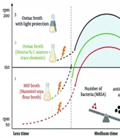
Antimicrobial activity was evaluated using the agar well diffusion method. Inhibition zone diameters were classified as follows: <6 mm, no activity; 6 - 10 mm, weak activity; 11 - 15 mm, moderate activity; 16 - 20 mm, strong activity; and > 20 mm, very strong activity. Isolate Ca2 - 5 was cultured under 96 different OSMAC conditions by systematically varying carbon and nitrogen sources, trace elements, and physical parameters, including shaking speed, incubation time, and light conditions, as shown in Table 1 in Supplementary File. These modifications resulted in significant differences in anti-MRSA activity, with the statistical analysis presented separately. The highest anti-MRSA activity was observed with soybean-mannitol, peptone-mannitol, and yeast extract-mannitol combinations. Mannitol was the most effective carbon source, whereas yeast extract was the most potent nitrogen source, particularly when combined with mannitol, starch, or glucose (Figures 1 and 2). Optimization of shaking speed indicated that 200 rpm produced the highest antimicrobial activity (Figure 3). Peak activity was observed between days 3 and 5 of incubation (Figure 1). In addition, dark incubation, achieved by wrapping flasks with aluminum foil, significantly enhanced anti-MRSA activity compared with that under light conditions (Figures 1 and 2), likely through regulation of secondary metabolite biosynthesis. The overall OSMAC optimization workflow is summarized in Figure 4.
Distribution of nitrogen sources (soybean, peptone, and yeast extract) and carbon sources (mannitol, starch, glucose, and chitin) across different incubation conditions for isolate Ca2 - 5. The pie chart shows the breakdown of sources into three primary groups, each divided into seven subgroups corresponding to incubation periods from day 1 to day 7. These subgroups are categorized under light and dark conditions at four shaking speeds (50, 100, 150, and 200 rpm).
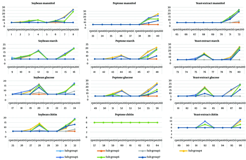
Figure 1.

Distribution of nitrogen sources (soybean, peptone, and yeast extract) and carbon sources (mannitol, starch, glucose, and chitin) across different incubation conditions for isolate Ca2 - 5. The pie chart shows the breakdown of sources into three primary groups, each divided into seven subgroups corresponding to incubation periods from day 1 to day 7. These subgroups are categorized under light and dark conditions at four shaking speeds (50, 100, 150, and 200 rpm).

Antimicrobial activity of isolate Ca2 - 5 against MRSA on day 4 at 200 rpm under light and dark conditions. Panel A shows results under light incubation, and panel B shows results under dark incubation. Numbers 1 - 12 represent different nitrogen and carbon source combinations: 1) soybean-starch, 2) soybean-glucose, 3) soybean-mannitol, 4) soybean-chitin, 5) peptone-starch, 6) peptone-mannitol, 7) peptone-glucose, 8) peptone-chitin, 9) yeast extract-mannitol, 10) yeast extract-chitin, 11) yeast extract-glucose, and 12) yeast extract-starch.
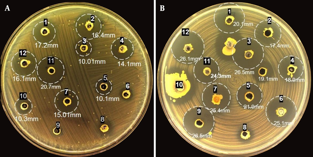
Figure 2.

Antimicrobial activity of isolate Ca2 - 5 against MRSA on day 4 at 200 rpm under light and dark conditions. Panel A shows results under light incubation, and panel B shows results under dark incubation. Numbers 1 - 12 represent different nitrogen and carbon source combinations: 1) soybean-starch, 2) soybean-glucose, 3) soybean-mannitol, 4) soybean-chitin, 5) peptone-starch, 6) peptone-mannitol, 7) peptone-glucose, 8) peptone-chitin, 9) yeast extract-mannitol, 10) yeast extract-chitin, 11) yeast extract-glucose, and 12) yeast extract-starch.
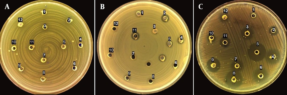
Comparative analysis of the antibacterial activity of isolate ID Ca2 - 5 against MRSA at varying agitation speeds: A) 50 rpm, B) 100 rpm, and C) 150 rpm. Within each panel, specific configurations are outlined, representing various combinations of nitrogen and carbon sources: 1) soybean-starch, 2) soybean-glucose, 3) soybean-mannitol, 4) soybean-chitin, 5) peptone-starch, 6) peptone-mannitol, 7) peptone-glucose, 8) peptone-chitin, 9) yeast extract-mannitol, 10) yeast extract-chitin, 11) yeast extract-glucose, and 12) yeast extract-starch.
Figure 3.

Comparative analysis of the antibacterial activity of isolate ID Ca2 - 5 against MRSA at varying agitation speeds: A) 50 rpm, B) 100 rpm, and C) 150 rpm. Within each panel, specific configurations are outlined, representing various combinations of nitrogen and carbon sources: 1) soybean-starch, 2) soybean-glucose, 3) soybean-mannitol, 4) soybean-chitin, 5) peptone-starch, 6) peptone-mannitol, 7) peptone-glucose, 8) peptone-chitin, 9) yeast extract-mannitol, 10) yeast extract-chitin, 11) yeast extract-glucose, and 12) yeast extract-starch.

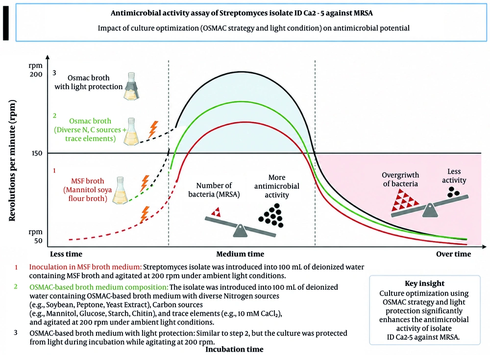
Graphical abstract illustrating the effect of culture optimization on 3 the antimicrobial activity of <i>Streptomyces</i> isolate ID Ca2 - 5 against MRSA.
Figure 4.

Graphical abstract illustrating the effect of culture optimization on 3 the antimicrobial activity of Streptomyces isolate ID Ca2 - 5 against MRSA.

4.3. Molecular Identification of Streptomyces Strains

Molecular identification was performed based on 16S rRNA gene sequencing (Figure 5). The sequence of isolate Ca2 - 5 was most closely related to Streptomyces drozdowiczii NBRC 101007T (GenBank accession AB249957), showing 100% identity with 98% query coverage over an approximate sequence length of 601 bp. Isolate Ca2 - 1 was closely related to Streptomyces poonensis JCM 481, showing 98% identity with 95% query coverage over an approximate sequence length of 695 bp. Isolate Ca2 - 4 was most closely related to Streptomyces flavovirens NBRC 3716T (GenBank accession AB184834), showing 99.44% identity with 94% query coverage over an approximate sequence length of 669 bp. The 16S rRNA gene sequences were deposited in GenBank under accession numbers PV998000.1 (Ca2 - 1), PQ773515 (Ca2 - 4), and PQ773516 (Ca2 - 5).
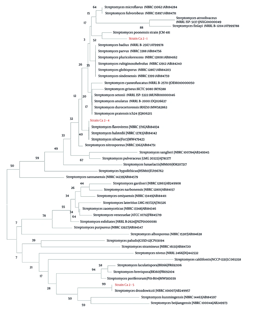
Phylogenetic analysis of <i>Streptomyces</i> isolates based on DNA sequencing. The genetic sequences of the isolates are detailed below. Isolate Ca2 - 5 showed 100% sequence identity with <i>Streptomyces drozdowiczii</i> NBRC 101007(T) (GenBank: AB249957). Isolate Ca2 - 1 showed the highest sequence similarity to <i>Streptomyces poonensis</i> JCM 481, while Ca2 - 4 showed 99.44% similarity to <i>Streptomyces flavovirens</i> NBRC 3716 (GenBank: AB184834).
Figure 5.

Phylogenetic analysis of Streptomyces isolates based on DNA sequencing. The genetic sequences of the isolates are detailed below. Isolate Ca2 - 5 showed 100% sequence identity with Streptomyces drozdowiczii NBRC 101007(T) (GenBank: AB249957). Isolate Ca2 - 1 showed the highest sequence similarity to Streptomyces poonensis JCM 481, while Ca2 - 4 showed 99.44% similarity to Streptomyces flavovirens NBRC 3716 (GenBank: AB184834).

4.4. Statistical Analysis

Multifactorial statistical analysis revealed that shaking speed and carbon source were the most significant determinants of antimicrobial activity (P < 0.001), whereas nitrogen source had a moderate effect (P < 0.05). Light conditions had a minor or context-dependent influence. A strong interaction between rpm and carbon source was observed, indicating that metabolite production in Streptomyces Ca2 - 5 is highly condition dependent. Overall, 43.7% of OSMAC conditions exhibited detectable antimicrobial activity against MRSA ATCC 43300, confirming the successful activation of cryptic biosynthetic pathways.

5. Discussion

Alarming projections suggest that, without rapid intervention, approximately 10 million lives could be lost due to antimicrobial resistance (AMR) by 2050 (25). Despite the encouraging discovery of approximately 1600 new natural products (NPs) annually over the past two decades (26), the repeated identification of known compounds remains a substantial impediment in the field. Advances in genomic analysis have revealed a disparity between the number of gene clusters identified through bioinformatics analyses as being involved in the production of secondary metabolites and the number of chemically characterized secondary metabolites (27, 28). BGCs typically comprise co-localized genes encoding enzymes responsible for secondary metabolite production (29, 30). Genome sequencing has revealed cryptic BGCs in microbes that are not associated with chemically characterized molecules (31). These BGCs are believed to be dormant or inactive under laboratory conditions (30).
The production of secondary metabolites (SMs) requires cellular energy and resources, making it a regulated process activated in response to specific environmental conditions (3, 13). Various approaches, such as modifying growth conditions without genetic manipulation, can trigger global physiological changes and enhance SM production (32). These approaches, derived from fermentation optimization practices, have shown that altering cultivation parameters, such as nutrients, trace elements, physical factors (pH and temperature), and chemical elicitors (sublethal antibiotic concentrations and communication molecules), can affect SM production in microorganisms. The influence of nutrient composition on secondary metabolite production varies substantially among microbial species.
Enhanced production of antimicrobial metabolites was achieved using the OSMAC approach, with culture conditions designed to mimic the natural plant-associated habitat. The results demonstrated that shaking speed, incubation time, carbon and nitrogen sources, and particularly light conditions, had a significant effect on anti-MRSA activity. Increased agitation speeds (150 and 200 rpm) markedly enhanced antimicrobial activity by improving oxygen transfer, nutrient distribution, and the expression of secondary metabolite biosynthetic genes, whereas low agitation (50 rpm) resulted in oxygen limitation and reduced metabolite production. The optimal incubation period was 4 - 6 days, corresponding to the stationary growth phase; shorter periods limited biomass formation, whereas longer periods led to nutrient depletion and the accumulation of toxic by-products.
Among the tested substrates, soybean meal showed the strongest effect, especially when combined with mannitol or glucose at higher shaking speeds. Yeast extract also performed well, whereas chitin exhibited weak activity due to its poor degradability. These findings highlight the critical role of the carbon-to-nitrogen (C/N) ratio and strain-specific substrate utilization in the OSMAC strategy. Moreover, visible light exerted an inhibitory effect on antibiotic production, with dark incubation significantly enhancing anti-MRSA activity compared with light conditions. Statistical analysis indicated that shaking speed was the most significant factor influencing antimicrobial production (P < 0.001), whereas light conditions had a weaker, context-dependent effect. These results demonstrate the strong dependence of metabolite production on culture conditions in Streptomyces Ca2 - 5 and are consistent with the natural ecology of Streptomyces, which inhabits dark environments such as soil and plant tissues (33).

5.1. Study Limitations

Despite promising anti-MRSA activity, this study has limitations. The identification of isolate Ca2 - 5 was based solely on 16S rRNA analysis, and genome-based methods are required for definitive species-level classification. In addition, although OSMAC conditions enhanced bioactivity, the active metabolites were not isolated or characterized, indicating that enhanced bioactivity was observed rather than the discovery of novel antimicrobial compounds.

5.2. Conclusions

This study demonstrates the potential of endophytic Streptomyces as a promising source of antimicrobial metabolites. By optimizing environmental and cultivation parameters, including nutrient sources, rotation speed, and light exposure, we enhanced antimicrobial metabolite production. Key findings demonstrated that soybean and mannitol significantly boosted antimicrobial activity, whereas dark incubation may better mimic the low-light conditions encountered in plant-associated microenvironments. These optimizations provide a framework for advancing bioactivity-enhancement efforts and addressing the growing global challenge posed by multidrug-resistant pathogens.
Acknowledgments

Footnotes

  • AI Use Disclosure:The authors declare that no generative AI tools were used in the creation of this article.

  • Authors' Contribution:M. B. contributed to methodology, investigation, data curation, formal analysis, and original draft preparation. A. H. S. contributed to methodology, formal analysis, software, validation, and manuscript review and editing. S. A. contributed to methodology, formal analysis, validation, and manuscript review and editing. H. S. R. contributed to conceptualization, project administration, validation, supervision, and manuscript review and editing. S. Y. contributed to conceptualization, validation, supervision, and manuscript review and editing.

  • Conflict of Interests Statement:The authors declare that they have no known financial or personal conflicts of interest that could have influenced the work reported in this paper.

  • Data Availability:The datasets supporting the findings of this study are available from the corresponding author upon reasonable request. Furthermore, the sequence data generated during this research have been deposited in the National Center for Biotechnology Information (NCBI) repository and can be accessed at [https://www.ncbi.nlm.nih.gov/](https://www.ncbi.nlm.nih.gov/) under accession numbers PQ773515, PQ773516 and PV998000.1.

  • Ethical Approval:This study involved Streptomyces samples collected from natural aquatic environments under academic approval from Hamadan University of Medical Sciences (IR.UMSHA.REC.1404.472: https://ethics.research.ac.ir/IR.UMSHA.REC.1404.472).

  • Funding/Support:This research was financially supported by Hamadan University of Medical Sciences [project code: 43347980].

References

  • 1.
    Morehead MS, Scarbrough C. Emergence of global antibiotic resistance. J Primary Care Clin Office Pract. 2018;45(3):467-484. [PubMed ID: 30115335]. https://doi.org/10.1016/j.pop.2018.05.006.
  • 2.
    Cassat JE, Thomsen I. Staphylococcus aureus infections in children. J Curr Opin Infect Dis. 2021;34(5):510-518. [PubMed ID: 34524201]. [PubMed Central ID: PMC8630804]. https://doi.org/10.1097/QCO.0000000000000752.
  • 3.
    Sharma R, Hammerschlag MR. Treatment of methicillin-resistant Staphylococcus aureus (MRSA) infections in children: a reappraisal of vancomycin. J Curr Infect Dis Rep. 2019;21(10). 37. [PubMed ID: 31486979]. https://doi.org/10.1007/s11908-019-0695-4.
  • 4.
    Band VI, Satola SW, Burd EM, Farley MM, Jacob JT, Weiss DS. Carbapenem-resistant Klebsiella pneumoniae exhibiting clinically undetected colistin heteroresistance leads to treatment failure in a murine model of infection. J mBio. 2018;9(2). e02448 - 17. [PubMed ID: 29511071]. [PubMed Central ID: PMC5844991]. https://doi.org/10.1128/mbio.02448-17.
  • 5.
    Lewis PO, Heil EL, Covert KL, Cluck DB, therapeutics. Treatment strategies for persistent methicillin-resistant Staphylococcus aureus bacteraemia. J J Clin Pharm. 2018;43(5):614-625. [PubMed ID: 30003555]. https://doi.org/10.1111/jcpt.12743.
  • 6.
    Mahjabeen F, Saha U, Mostafa MN, Siddique F, Ahsan E, Fathma S, et al. An update on treatment options for methicillin-resistant Staphylococcus aureus (MRSA) bacteremia: a systematic review. J Cureus. 2022;14(11):e31486. [PubMed ID: 36523711]. [PubMed Central ID: PMC9749567]. https://doi.org/10.7759/cureus.31486.
  • 7.
    Bai AD, Lo CKL, Komorowski AS, Suresh M, Guo K, Garg A, et al. Staphylococcus aureus bacteraemia mortality: a systematic review and meta-analysis. J Clin Microbiol Infect. 2022;28(8):1076-1084. [PubMed ID: 35339678]. [PubMed Central ID: PMC10328498]. https://doi.org/10.1016/j.cmi.2022.03.015.
  • 8.
    Kourtis AP, Hatfield K, Baggs J, Mu Y, See I, Epson E, et al. Vital signs: epidemiology and recent trends in methicillin-resistant and methicillin-susceptible Staphylococcus aureus bloodstream infections-United States. J Morb Mortal Wkly Rep. 2019;68(9):214-219. [PubMed ID: 30845118]. [PubMed Central ID: PMC6421967]. https://doi.org/10.15585/mmwr.mm6809e1.
  • 9.
    Tiwari P, Thakkar S, Dufossé L. Antimicrobials from endophytes as novel therapeutics to counter drug-resistant pathogens. Crit Rev Biotechnol. 2024;45(1):164-190. [PubMed ID: 38710617]. https://doi.org/10.1080/07388551.2024.2342979.
  • 10.
    Chamekh A, Kharbech O, Driss-Limam R, Fersi C, Khouatmeya M, Chouari R. Evidences for antioxidant response and biosorption potential of Bacillus simplex strain 115 against lead. World J Microbiol Biotechnol. 2021;37(3). 44. [PubMed ID: 33547493]. https://doi.org/10.1007/s11274-021-03009-2.
  • 11.
    Gyawali R, Ibrahim SA. Natural products as antimicrobial agents. Food Control. 2014;46:412-429. https://doi.org/10.1016/j.foodcont.2014.05.047.
  • 12.
    Pinedo-Rivilla C, Aleu J, Durán-Patrón R. Cryptic metabolites from marine-derived microorganisms using OSMAC and epigenetic approaches. J Mar Drugs. 2022;20(2):84. [PubMed ID: 35200614]. [PubMed Central ID: PMC8879561]. https://doi.org/10.3390/md20020084.
  • 13.
    Romano S, Jackson SA, Patry S, Dobson ADW. Stefano JSA, P. Sloane, D. Alan. Extending the "one strain many compounds" (OSMAC) principle to marine microorganisms. J Mar Drugs. 2018;16(7):244. [PubMed ID: 30041461]. [PubMed Central ID: PMC6070831]. https://doi.org/10.3390/md16070244.
  • 14.
    Chen C, Zhang Y, Chen R, Liu K, Wu H, Qiao J, et al. Development of a pre-modification strategy to overcome restriction-modification barriers and enhance genetic engineering in Lactococcus lactis for nisin biosynthesis. Int J Mol Sci. 2025;26(5):2200. [PubMed ID: 40076820]. [PubMed Central ID: PMC11900431]. https://doi.org/10.3390/ijms26052200.
  • 15.
    Racharaks R, Arnold W, Peccia J. Development of CRISPR-Cas9 knock-in tools for free fatty acid production using the fast-growing cyanobacterial strain Synechococcus elongatus UTEX 2973. J Microbiol Methods. 2021;189. 106315. [PubMed ID: 34454980]. https://doi.org/10.1016/j.mimet.2021.106315.
  • 16.
    Müller M, Bialas E, Sturm I, Sood U, Lal R, Bechthold A. Genomic modifications for enhanced antibiotic production in rifamycin derivative-producing Amycolatopsis mediterranei S699 strains: focusing on rifQ and rifO genes. Front Antibiot. 2024;3. 1399139. [PubMed ID: 39816246]. [PubMed Central ID: PMC11732027]. https://doi.org/10.3389/frabi.2024.1399139.
  • 17.
    Buijs Y, Zhang SD, Jørgensen KM, Isbrandt T, Larsen TO, Gram L. Enhancement of antibiotic production by co-cultivation of two antibiotic producing marine Vibrionaceae strains. FEMS Microbiol Ecol. 2021;97(4). fiab041. [PubMed ID: 33693627]. https://doi.org/10.1093/femsec/fiab041.
  • 18.
    Hua E, Zhang Y, Yun K, Pan W, Liu Y, Li S, et al. Whole-cell biosensor and producer co-cultivation-based microfludic platform for screening Saccharopolyspora erythraea with hyper erythromycin production. ACS Synth Biol. 2022;11(8):2697-2708. [PubMed ID: 35561342]. https://doi.org/10.1021/acssynbio.2c00102.
  • 19.
    Samadi Z, Mousavi Ghahfarrokhi SS, Jafari H, Nikrou S, Vazirian M, Samadi N. Rosmarinus officinalis and Zataria multiflora extracts enhanced antibacterial activity of ciprofloxacin against methicillin-resistant Staphylococcus aureus. Nat Prod Res. 2025;39(24):7156-7166. [PubMed ID: 39351970]. https://doi.org/10.1080/14786419.2024.2409982.
  • 20.
    Swangsri T, Reamtong O, Saralamba S, Rakthong P, Thaenkham U, Saralamba N. Exploring the antimicrobial potential of crude peptide extracts from Allium sativum and Allium oschaninii against antibiotic-resistant bacterial strains. Pharm Biol. 2024;62(1):666-675. [PubMed ID: 39205473]. [PubMed Central ID: PMC11363733]. https://doi.org/10.1080/13880209.2024.2395517.
  • 21.
    Hameş-Kocabaş EE, Uzel A. Isolation strategies of marine-derived actinomycetes from sponge and sediment samples. J Microbiol Methods. 2012;88(3):342-347. [PubMed ID: 22285852]. https://doi.org/10.1016/j.mimet.2012.01.010.
  • 22.
    Beiranvand M, Amin M, Hashemi-Shahraki A, Romani B, Yaghoubi S, Sadeghi P. Antimicrobial activity of endophytic bacterial populations isolated from medical plants of Iran. J Iran J Microbiol. 2017;9(1):11-18. [PubMed ID: 28775818]. [PubMed Central ID: PMC5533999].
  • 23.
    Sivalingam P, Hong K, Pote J, Prabakar K. Extreme environment Streptomyces: potential sources for new antibacterial and anticancer drug leads? J Int J Microbiol. 2019;2019(1):5283948-20. [PubMed ID: 31354829]. [PubMed Central ID: PMC6636559]. https://doi.org/10.1155/2019/5283948.
  • 24.
    Loberiza IR, Falk FE, Green TJ, Loudon AH. Efficacy of different "boiling" methods for bacterial DNA extraction. J Microbiol Methods. 2025;237. 107245. [PubMed ID: 40889580]. https://doi.org/10.1016/j.mimet.2025.107245.
  • 25.
    Utt E, Wells C. The global response to the threat of antimicrobial resistance and the important role of vaccines. Pharm Policy Law. 2016;18(1 - 4):179-197. https://doi.org/10.3233/PPL-160442.
  • 26.
    Pye CR, Bertin MJ, Lokey RS, Gerwick WH, Linington RG. Retrospective analysis of natural products provides insights for future discovery trends. Proc Natl Acad Sci U S A. 2017;104(22):5601-5606. [PubMed ID: 28461474]. [PubMed Central ID: PMC5465889]. https://doi.org/10.1073/pnas.1614680114.
  • 27.
    Hautbergue T, Jamin EL, Debrauwer L, Puel O, Oswald IP. From genomics to metabolomics, moving toward an integrated strategy for the discovery of fungal secondary metabolites. J Nat Prod Rep. 2018;35(2):147-173. [PubMed ID: 29384544]. https://doi.org/10.1039/C7NP00032D.
  • 28.
    Nielsen JC, Grijseels S, Prigent S, Ji B, Dainat J, Nielsen KF, et al. Global analysis of biosynthetic gene clusters reveals vast potential of secondary metabolite production in Penicillium species. J Nat Microbiol. 2017;2(6). 17044. [PubMed ID: 28368369]. https://doi.org/10.1038/nmicrobiol.2017.44.
  • 29.
    Xu Z, Park TJ, Cao H. Advances in mining and expressing microbial biosynthetic gene clusters. J Crit Rev Microbiol. 2023;49(1):18-37. [PubMed ID: 35166616]. https://doi.org/10.1080/1040841X.2022.2036099.
  • 30.
    Lee N, Hwang S, Lee Y, Cho S, Palsson B, Cho BK. Synthetic biology tools for novel secondary metabolite discovery in Streptomyces. J Microbiol Biotechnol. 2019;29(5):667-686. [PubMed ID: 31091862]. https://doi.org/10.4014/jmb.1904.04015.
  • 31.
    Liu C, Kakeya H. Cryptic chemical communication: secondary metabolic responses revealed by microbial co-culture. J Chem Asian J. 2020;15(3):327-337. [PubMed ID: 31957936]. https://doi.org/10.1002/asia.201901505.
  • 32.
    Nguyen J, Fernandez V, Pontrelli S, Sauer U, Ackermann M, Stocker R. A distinct growth physiology enhances bacterial growth under rapid nutrient fluctuations. J Nat Commun. 2021;12(1). 3662. [PubMed ID: 34135315]. [PubMed Central ID: PMC8209047]. https://doi.org/10.1038/s41467-021-23439-8.
  • 33.
    Liu H, Li J, Singh BK. Harnessing co-evolutionary interactions between plants and Streptomyces to combat drought stress. Nat Plants. 2024;10(8):1159-1171. [PubMed ID: 39048724]. https://doi.org/10.1038/s41477-024-01749-1.

Copyright

Copyright © 2026, Beiranvand et al. This open-access article is available under the Creative Commons Attribution 4.0 (CC BY 4.0) International License (https://creativecommons.org/licenses/by/4.0/), which allows for unrestricted use, distribution, and reproduction in any medium, provided that the original work is properly cited.

Similar Articles

31
May
2015

Anti-Methicillin Resistant Staphylococcus aureus Activity and Optimal Culture Condition of Streptomyces sp. SUK 25

Ahmad Siti Junaidah,
Sudi Suhaini,
Hasidah Mohd Sidek,
Dayang Fredalina Basri,
Noraziah Mohamad Zin

Siti Junaidah A, Suhaini S, Mohd Sidek H, Basri DF, Zin NM. Anti-Methicillin Resistant Staphylococcus aureus Activity and Optimal Culture Condition of Streptomyces sp. SUK 25. Jundishapur J Microbiol. 2015;8(5):e16784. doi: https://doi.org/10.5812/jjm.16784

21
Nov
2016

Isolation and Identification of a New Cold and Alkaline Tolerant Antibiotic Producing Streptomyces sp. From Soil

Sadaf Sadat Rafati,
Bahar Shahnavaz,
Ali Makhdoumi

Rafati SS, Shahnavaz B, Makhdoumi A. Isolation and Identification of a New Cold and Alkaline Tolerant Antibiotic Producing Streptomyces sp. From Soil. Jundishapur J Microbiol. 2016;9(12):e39692. doi: https://doi.org/10.5812/jjm.39692

30
Jan
2024
Jundishapur J Nat Pharm Prod

Screening, Identification, and Pigment Characteristics of Actinomycin D-Producing Actinomycetes from Purple Soil in the Sichuan Basin

Xin Wan,
Rui Liu,
Peng Jiang,
LiHuan Li,
JingPing Chen,
HongFu Wei
,et al.

Wan X, Liu R, Jiang P, Li L, Chen J, et al. Screening, Identification, and Pigment Characteristics of Actinomycin D-Producing Actinomycetes from Purple Soil in the Sichuan Basin. Jundishapur J Nat Pharm Prod. 2024;19(1):e142936. doi: https://doi.org/10.5812/jjnpp-142936

21
Aug
2017

Antibacterial Activity and Composition of the Fermentation Broth of Streptomyces Parvus 33

Wenjuan Zhang,
Saijian Ma,
Chao An,
Wenjiao Xue

Zhang W, Ma S, An C, Xue W. Antibacterial Activity and Composition of the Fermentation Broth of Streptomyces Parvus 33. Jundishapur J Microbiol. 2017;10(8):e12677. doi: https://doi.org/10.5812/jjm.12677

31
Oct
2018

Antibacterial and Antitumor Potential of Actinomycetes Isolated from Mangrove Soil in the Maowei Sea of the Southern Coast of China

Bin Gong,
Shuang Chen,
Wenwen Lan,
Yanmin Huang,
Xiangcheng Zhu

Gong B, Chen S, Lan W, Huang Y, Zhu X. Antibacterial and Antitumor Potential of Actinomycetes Isolated from Mangrove Soil in the Maowei Sea of the Southern Coast of China. Iran J Pharm Res. 2018;17(4):e124876. doi: https://doi.org/10.22037/ijpr.2018.2280

Download PDF2.53 MB

Crossmark

Crossmark

Checking

Share on
Cited by
Metrics

Ordering Reprints

Articles are published under the Creative Commons license stated on each article. No permission or royalty fee is required for uses permitted by that license. CCC handles optional bulk and customized reprint orders. Any quotation covers production and delivery services only, not copyright permission. > Request Reprints from CCC 

Search Relations

Author(s):

Related Articles