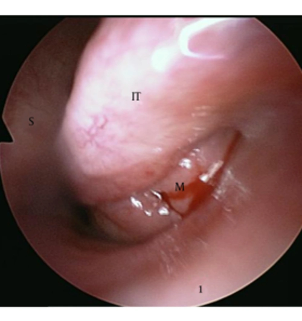
CEH can occur in a variety of locations. Although most hematomas resolve spontaneously, a few can persist for long periods forming slowly expanding space-occupying masses (
1). This expanding process is attributable to the irritant effects of blood and the products of its breakdown, which induce repeated episodes of bleeding from the capillaries in the granulation tissue (
10). Sinonasal organizing (organized) hematoma is a rare type of CEH originating from the paranasal sinuses whose clinical characteristics lead to be misdiagnosed as a malignant or locally aggressive neoplasm (
3). The number of reported cases has dramatically risen since 2005 because of elucidation of the characteristic imaging findings and the specific pathologic findings (
11-
15). Because the maxillary sinus is the largest paranasal sinus that allows negative pressure, significant leakage of blood may develop in the nasal cavity into the sinus through the ostium (
15). Therefore, organizing hematoma most commonly affects the maxillary sinus and is known as OHMS (
11).
Although diagnostic criteria for correct identification of OHMS are not well known (
3), corrective preoperative diagnosis is important to avoid unnecessary extensive surgery because this condition is curative with a simple, conservative endoscopic approach and rarely recurs (
11).
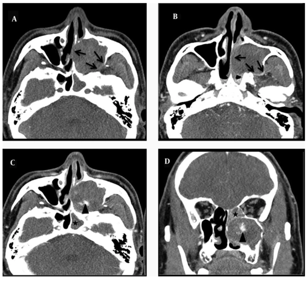
The reported CT findings of organizing hematoma include an expansive, clearly demarcated and compressive mass causing bony erosion without a destructive feature (
14). After administration of contrast, heterogeneous enhancement in a patchy distribution is usually found within the lesion (
15,
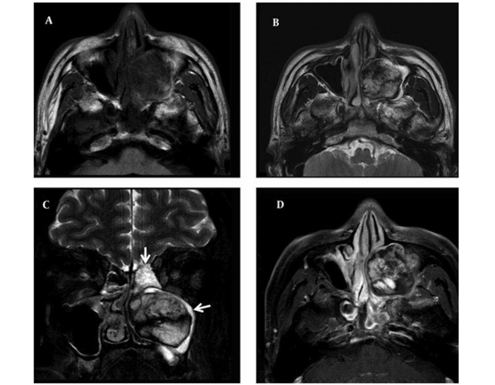
16). MRI is superior to CT for determining the margin and extent of the lesion. In MR images, it has biphasic appearance in which the central part of the lesion presents as low intensity on T1-weighted images and high intensity on T2-weighted images, corresponding with areas of fresh hemorrhage and the peripheral part is less enhanced with a zone of fibrosis, especially called as “shells” of T2 hypointensity which is the most diagnostic finding on MRI (
3,
14,
17). However, if it is not possible to differentiate benign tumors from malignancy by CT and MRI, or if the patient cannot perform MRI scan because of severe claustrophobia, definite preoperative diagnosis is difficult.
CEHs can be misdiagnosed as malignant tumors because of their large size and slow, progressive enlargement (
5,
7). Although
18FDG-PET images of CEH are not widely available,
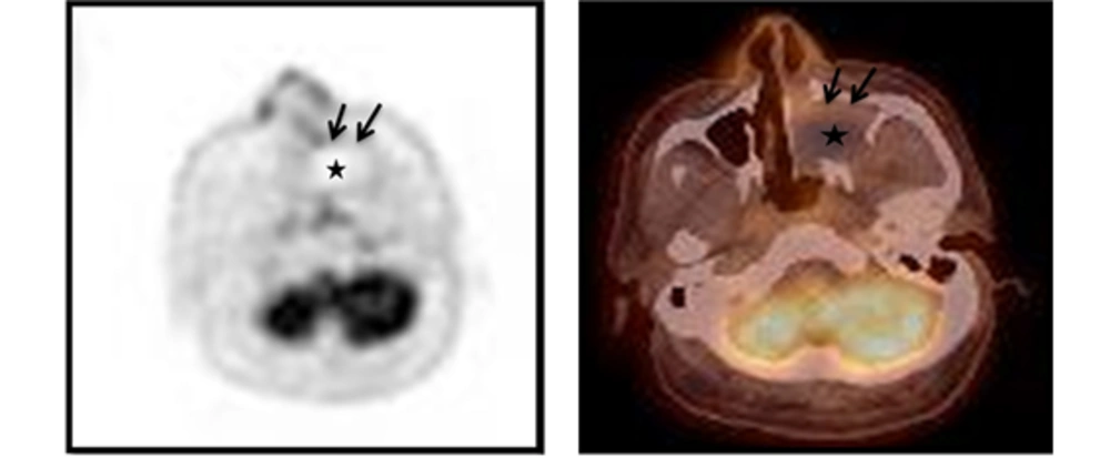
18FDG-PET shows moderate FDG uptake, the maximum standardized uptake value (SUVmax) of 3.1 to 5.5, only at the periphery of the mass with central photon defects that is probably characteristic of CEH, suggesting chronic inflammation or granulation tissue in the fibrous wall (
5-
9). If the SUVmax were set to a cut-off point of 3.0 to distinguish between benign and malignant tumors, these lesions could be misdiagnosed as malignant tumors (
5). However, the use of FDG-PET imaging for tumor diagnosis is limited by the fact that FDG, a glucose analog, is taken up not only by tumor cells but also by macrophages and immature granulation tissue containing fibroblasts and inflammation (
18). CEH has abundant granulation tissue with neovascularization, macrophages and inflammation, which corresponds to organizing processes. These pathological changes are considered to have resulted in increased FDG uptake within the organizing hematoma (
5-
9).
In our patient,
18FDG-PET images of the lesion revealed central photon defects with moderately increased FDG uptake in the peripheral rim of the space-occupying mass in the left maxillary sinus. The SUVmax of the lesion was 3.8. This is consistent with previous reports of
18FDG-PET imaging of CEH. Although the FDG uptake pattern might be seen if a malignant tumor has a tendency of central necrosis, the characteristics of FDG-PET images of OHMS and CEH, including FDG uptake in the peripheral rim with central photon defect of the mass as a result of inflammation should be recognized as a potential interpretive pitfall that mimics a malignant tumor (
5).
In summary, we have presented 18FDG-PET findings of OHMS that showed an increased FDG uptake in the peripheral rim of the mass with central photopenia. To our knowledge, this is the first report in literature reporting 18FDG-PET findings of OHMS with SUVmax values suggestive of a malignancy. Careful interpretation of metabolic (FDG-PET/CT) and anatomic (CT and MRI) images should be performed to accurately characterize the expansile lesion of the maxillary sinus in order to increase specificity and reduce equivocal findings significantly.