


1. Background
2. Objectives
3. Patients and Methods
3.1. Case Selection
3.2. Imaging Examination
3.3. Post-Processing
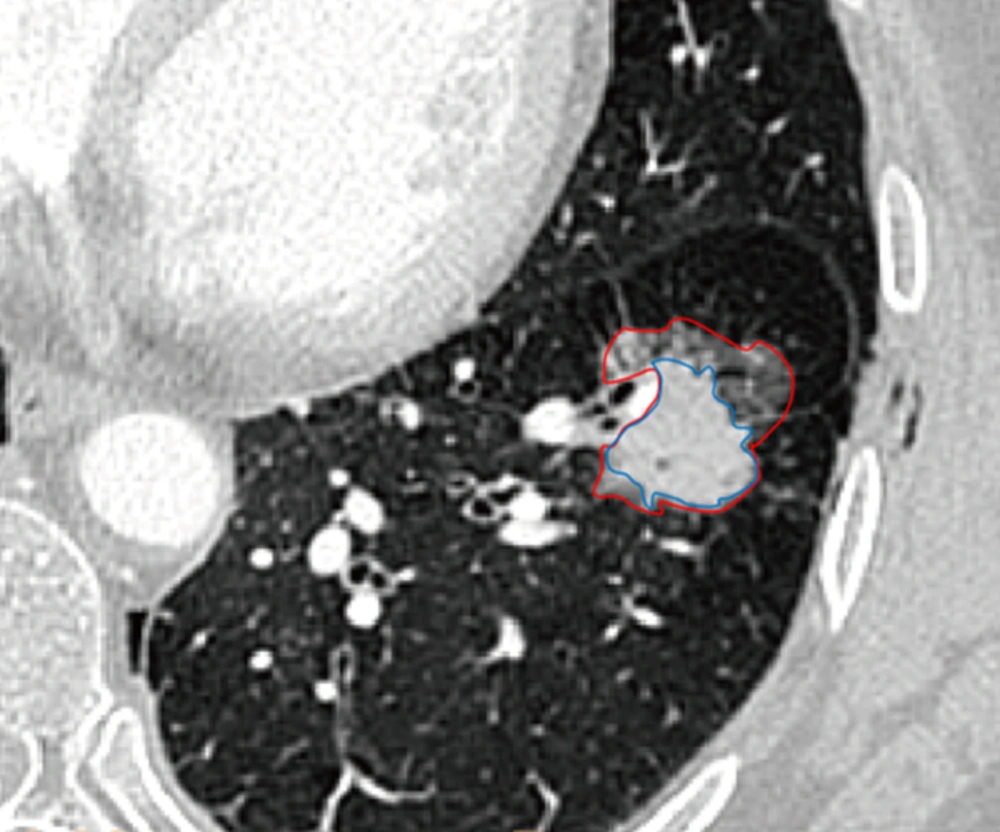
Semiautomated segmentation and volume measurement of sub-solid nodule (SSN) in a 63-year-old woman. Segmentation of solid portion and ground-glass portion on lung window (red line: SSN contour; blue line: the contour of the inner solid portion).
![Semiautomated segmentation and volume measurement of subsolid nodule (SSN) in a 63-year-old woman. Corresponding nodule segmentation on volume tissue segmentation software (purple: solid portion in SSN; pink: ground-glass portion in SSN). Thus, the ground glass opacity (GGO) ratio of this invasive adenocarcinoma was 44.75% (Volume <sub>GGO</sub>/Volume<sub>SSN</sub>: [4571.5 - 2525.7]/4571.5).](https://brieflands.com/journals/ijradiology/articles/22027/figures/iranjradiol-In_Press-In_Press-22027-i002-preview-preview.webp "Semiautomated segmentation and volume measurement of subsolid nodule (SSN) in a 63-year-old woman. Corresponding nodule segmentation on volume tissue segmentation software (purple: solid portion in SSN; pink: ground-glass portion in SSN). Thus, the ground glass opacity (GGO) ratio of this invasive adenocarcinoma was 44.75% (Volume <sub>GGO</sub>/Volume<sub>SSN</sub>: [4571.5 - 2525.7]/4571.5).")
Semiautomated segmentation and volume measurement of subsolid nodule (SSN) in a 63-year-old woman. Corresponding nodule segmentation on volume tissue segmentation software (purple: solid portion in SSN; pink: ground-glass portion in SSN). Thus, the ground glass opacity (GGO) ratio of this invasive adenocarcinoma was 44.75% (Volume GGO/VolumeSSN: [4571.5 - 2525.7]/4571.5).
3.4. Pathological Measurement
3.5. Statistical Analysis
4. Results
| Measurement Method | Minimum, cm | Maximum, cm | Mean, cm | SD |
|---|---|---|---|---|
| Manual | 1.00 | 4.30 | 2.23 | 0.62 |
| Semi-automatic | 1.10 | 4.40 | 2.41 | 0.64 |
| Pathological | 0.50 | 3.00 | 1.79 | 0.64 |
Abbreviations: ICC, intraclass correlation coefficient ; SD, Standard deviation.
| Measurement Method | Inter-Observer Difference, cm | t | ICC (95% Confidence Interval) | |
|---|---|---|---|---|
| Mean ± SD | 95% limits of agreement (%) | |||
| Manual | 0.06182 ± 0.15810 | (-10.7 , 16.1) | 2.900 | 0.9826 (0.9702 - 0.9898) |
| Semi-automatic | -0.03636 ± 0.18794 | (-17.1 , 14.3) | -1.435 | 0.9818 (0.9688 - 0.9894) |
Abbreviations: ICC,intraclass correlation coefficient ; SD, Standard deviation.
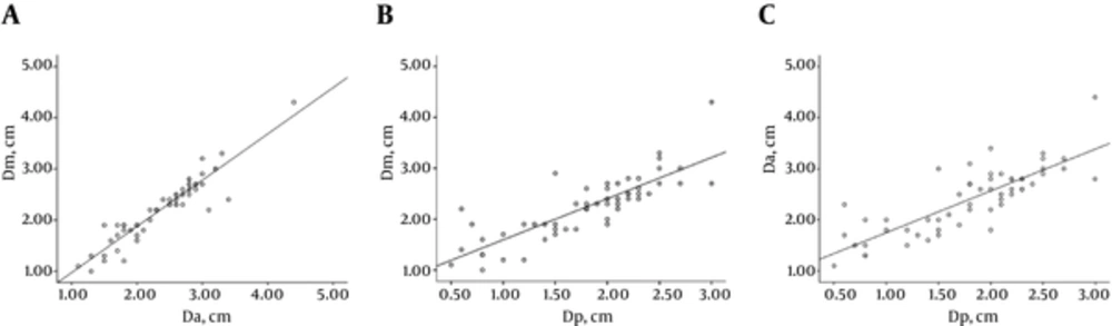
A - C, There was a positive correlation between manual (Dm) and semi-automatic (Da) measurements (r = 0.935), between manual (Dm) and pathological measurements (Dp; r = 0.833) and between semi-automatic (Da) and pathological measurements (Dp; r = 0.813) of the largest tumor diameter.
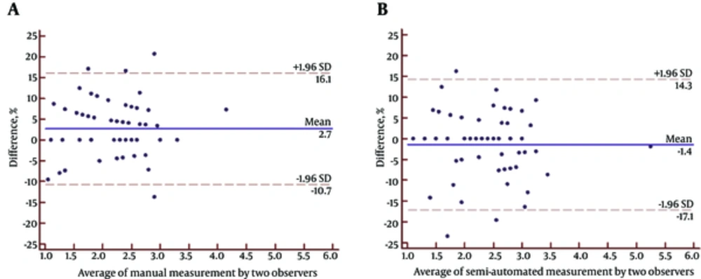
A - B. Bland-Altman plots used to measure agreement with repeated measures between manual and semi-automatic measurements. The mean difference between the two measurements is shown with a solid line; the lower and upper 95% limits of agreement are the dashed lines at (-%10.7 , %16.1) for A and (-%17.1 , %14.3) for B. The magnitude of the difference between manual and semi-automatic measurements regarding average tumor size does not seem to differ since there is no apparent pattern on the plot.
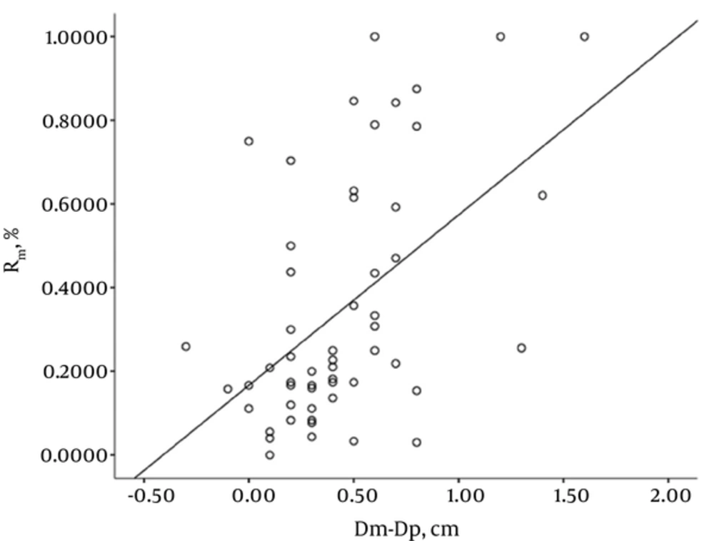
Correlation of ground glass opacity ratio with the discrepancy between manual measurement (Dm) and pathological measurement (Dp) of the largest diameter of a tumor (r = 0.474).
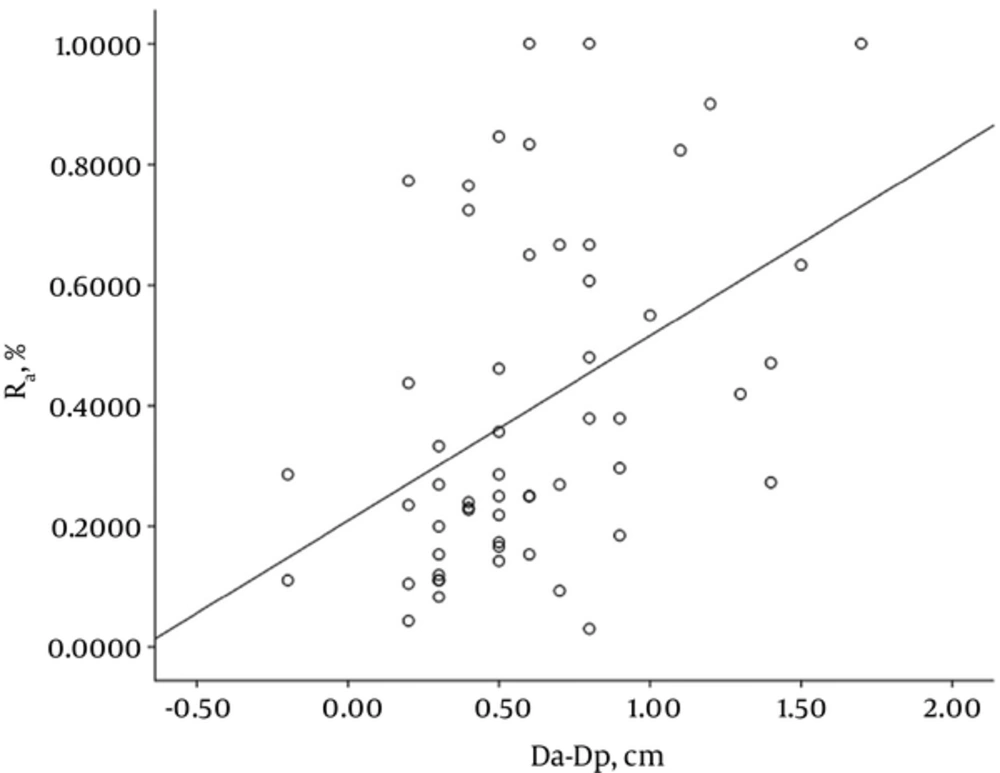
Correlation of ground glass opacity ratio with the discrepancy between semi-automatic measurement (Da) and pathological measurement (Dp) of the largest tumor diameter (r = 0.457).




